 Movie
Movie Controller


+ Open data

- Basic information
| Entry | Database: PDB / ID: 6v0v | ||||||||||||||||||||||||||||||||||||||||||
|---|---|---|---|---|---|---|---|---|---|---|---|---|---|---|---|---|---|---|---|---|---|---|---|---|---|---|---|---|---|---|---|---|---|---|---|---|---|---|---|---|---|---|---|
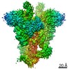
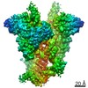
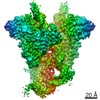
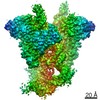
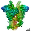
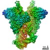
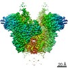
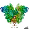
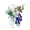
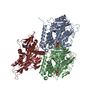
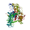
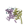
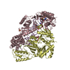
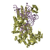
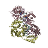
| Title | Cryo-EM structure of mouse WT RAG1/2 NFC complex (DNA0) | ||||||||||||||||||||||||||||||||||||||||||
 Components Components |
| ||||||||||||||||||||||||||||||||||||||||||
 Keywords Keywords | RECOMBINATION/DNA / V(D)J recombination / RAG / SCID / RECOMBINATION / RECOMBINATION-DNA complex | ||||||||||||||||||||||||||||||||||||||||||
| Function / homology |  Function and homology information Function and homology informationmature B cell differentiation involved in immune response / DNA recombinase complex / B cell homeostatic proliferation / endodeoxyribonuclease complex / negative regulation of T cell differentiation in thymus / pre-B cell allelic exclusion / positive regulation of organ growth / V(D)J recombination / negative regulation of T cell apoptotic process / negative regulation of thymocyte apoptotic process ...mature B cell differentiation involved in immune response / DNA recombinase complex / B cell homeostatic proliferation / endodeoxyribonuclease complex / negative regulation of T cell differentiation in thymus / pre-B cell allelic exclusion / positive regulation of organ growth / V(D)J recombination / negative regulation of T cell apoptotic process / negative regulation of thymocyte apoptotic process / phosphatidylinositol-3,4-bisphosphate binding / regulation of behavioral fear response / histone H3K4me3 reader activity / phosphatidylinositol-3,5-bisphosphate binding / regulation of T cell differentiation / positive regulation of T cell differentiation / T cell lineage commitment / B cell lineage commitment / T cell homeostasis / phosphatidylinositol-3,4,5-trisphosphate binding / T cell differentiation / protein autoubiquitination / phosphatidylinositol-4,5-bisphosphate binding / phosphatidylinositol binding / B cell differentiation / thymus development / RING-type E3 ubiquitin transferase / visual learning / ubiquitin-protein transferase activity / T cell differentiation in thymus / ubiquitin protein ligase activity / chromatin organization / endonuclease activity / DNA recombination / histone binding / sequence-specific DNA binding / Hydrolases; Acting on ester bonds / adaptive immune response / defense response to bacterium / hydrolase activity / chromatin binding / protein homodimerization activity / zinc ion binding / nucleoplasm / metal ion binding / identical protein binding / nucleus Similarity search - Function | ||||||||||||||||||||||||||||||||||||||||||
| Biological species |   | ||||||||||||||||||||||||||||||||||||||||||
| Method | ELECTRON MICROSCOPY / single particle reconstruction / cryo EM / Resolution: 3.61 Å | ||||||||||||||||||||||||||||||||||||||||||
 Authors Authors | Chen, X. / Yang, W. / Gellert, M. | ||||||||||||||||||||||||||||||||||||||||||
| Funding support |  United States, 1items United States, 1items
| ||||||||||||||||||||||||||||||||||||||||||
 Citation Citation | Journal: Nat Struct Mol Biol / Year: 2020 Title: Cutting antiparallel DNA strands in a single active site. Authors: Xuemin Chen / Yanxiang Cui / Robert B Best / Huaibin Wang / Z Hong Zhou / Wei Yang / Martin Gellert / Abstract: A single enzyme active site that catalyzes multiple reactions is a well-established biochemical theme, but how one nuclease site cleaves both DNA strands of a double helix has not been well ...A single enzyme active site that catalyzes multiple reactions is a well-established biochemical theme, but how one nuclease site cleaves both DNA strands of a double helix has not been well understood. In analyzing site-specific DNA cleavage by the mammalian RAG1-RAG2 recombinase, which initiates V(D)J recombination, we find that the active site is reconfigured for the two consecutive reactions and the DNA double helix adopts drastically different structures. For initial nicking of the DNA, a locally unwound and unpaired DNA duplex forms a zipper via alternating interstrand base stacking, rather than melting as generally thought. The second strand cleavage and formation of a hairpin-DNA product requires a global scissor-like movement of protein and DNA, delivering the scissile phosphate into the rearranged active site. | ||||||||||||||||||||||||||||||||||||||||||
| History |
|
- Structure visualization
| Movie |
Movie viewer |
|---|---|
| Structure viewer | Molecule: MolmilJmol/JSmol |
- Downloads & links
-Download
| PDBx/mmCIF format | 6v0v.cif.gz | 211.5 KB | Display | PDBx/mmCIF format |
|---|---|---|---|---|
| PDB format | pdb6v0v.ent.gz | 154 KB | Display | PDB format |
| PDBx/mmJSON format | 6v0v.json.gz | Tree view | PDBx/mmJSON format | |
| Others |  Other downloads Other downloads |
-Validation report
| Arichive directory | https://data.pdbj.org/pub/pdb/validation_reports/v0/6v0vftp://data.pdbj.org/pub/pdb/validation_reports/v0/6v0v | HTTPS FTP |
|---|
-Related structure data
| Related structure data | 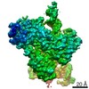 21003MC 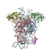 6oemC 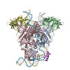 6oenC 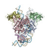 6oeoC 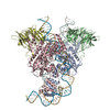 6oepC 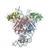 6oeqC 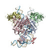 6oerC C: citing same article ( M: map data used to model this data |
|---|---|
| Similar structure data |


-Links
 PDBj
PDBj


- Assembly
| Deposited unit | 
|
|---|---|
| 1 |
|
-Components
-V(D)J recombination-activating protein ... , 2 types, 2 molecules AB
| #1: Protein | Mass: 88524.625 Da / Num. of mol.: 1 Source method: isolated from a genetically manipulated source Source: (gene. exp.)  Homo sapiens (human) Homo sapiens (human)References: UniProt: P15919, Hydrolases; Acting on ester bonds, RING-type E3 ubiquitin transferase |
|---|---|
| #2: Protein | Mass: 58158.254 Da / Num. of mol.: 1 Source method: isolated from a genetically manipulated source Source: (gene. exp.) Homo sapiens (human) / References: UniProt: P21784 |
-DNA chain , 2 types, 2 molecules FI
| #3: DNA chain | Mass: 14268.144 Da / Num. of mol.: 1 / Source method: obtained synthetically / Source: (synth.) |
|---|---|
| #4: DNA chain | Mass: 14064.058 Da / Num. of mol.: 1 / Source method: obtained synthetically / Source: (synth.) |
-Non-polymers , 2 types, 3 molecules 

| #5: Chemical |  Mass: 40.078 Da / Num. of mol.: 2 / Source method: obtained synthetically / Formula: Ca / Feature type: SUBJECT OF INVESTIGATION Mass: 40.078 Da / Num. of mol.: 2 / Source method: obtained synthetically / Formula: Ca / Feature type: SUBJECT OF INVESTIGATION#6: Chemical | ChemComp-ZN / |  Mass: 65.409 Da / Num. of mol.: 1 / Source method: obtained synthetically / Formula: Zn Mass: 65.409 Da / Num. of mol.: 1 / Source method: obtained synthetically / Formula: Zn |
|---|
-Details
| Has ligand of interest | Y |
|---|---|
| Has protein modification | N |
-Experimental details
-Experiment
| Experiment | Method: ELECTRON MICROSCOPY |
|---|---|
| EM experiment | Aggregation state: PARTICLE / 3D reconstruction method: single particle reconstruction |
- Sample preparation
| Component | Name: RAG1/2 Nick-forming complex (DNA0) / Type: COMPLEX / Entity ID: #1-#4 / Source: MULTIPLE SOURCES |
|---|---|
| Source (natural) | Organism: |
| Buffer solution | pH: 7.4 |
| Specimen | Embedding applied: NO / Shadowing applied: NO / Staining applied: NO / Vitrification applied: YES |
| Vitrification | Cryogen name: ETHANE |
- Electron microscopy imaging
| Experimental equipment |  Model: Titan Krios / Image courtesy: FEI Company |
|---|---|
| Microscopy | Model: FEI TITAN KRIOS |
| Electron gun | Electron source:  FIELD EMISSION GUN / Accelerating voltage: 300 kV / Illumination mode: FLOOD BEAM FIELD EMISSION GUN / Accelerating voltage: 300 kV / Illumination mode: FLOOD BEAM |
| Electron lens | Mode: BRIGHT FIELD |
| Image recording | Electron dose: 45 e/Å2 / Film or detector model: GATAN K2 SUMMIT (4k x 4k) |
- Processing
| Software | Name: PHENIX / Version: 1.14_3260: / Classification: refinement | ||||||||||||||||||||||||
|---|---|---|---|---|---|---|---|---|---|---|---|---|---|---|---|---|---|---|---|---|---|---|---|---|---|
| EM software | Name: PHENIX / Category: model refinement | ||||||||||||||||||||||||
| CTF correction | Type: PHASE FLIPPING AND AMPLITUDE CORRECTION | ||||||||||||||||||||||||
| 3D reconstruction | Resolution: 3.61 Å / Resolution method: FSC 0.143 CUT-OFF / Num. of particles: 111362 / Symmetry type: POINT | ||||||||||||||||||||||||
| Refine LS restraints |
|
