 Movie
Movie Controller

[English] 日本語
 Yorodumi
- EMDB-20033: Cryo-EM structure of mouse RAG1/2 12RSS-NFC/23RSS-PRC complex (DNA1) -
+ Open data

- Basic information
| Entry | Database: EMDB / ID: EMD-20033 | |||||||||
|---|---|---|---|---|---|---|---|---|---|---|
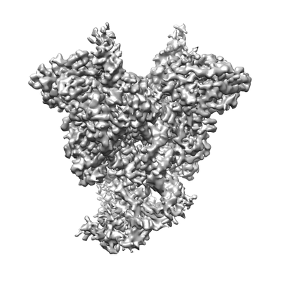
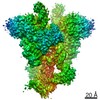
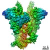
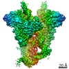
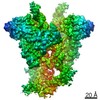
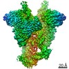
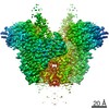
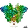
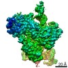
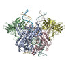
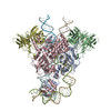
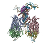
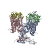
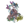
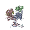
| Title | Cryo-EM structure of mouse RAG1/2 12RSS-NFC/23RSS-PRC complex (DNA1) | |||||||||
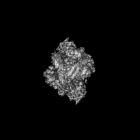
 Map data Map data | RAG1/2 12RSS-NFC/23RSS-PRC | |||||||||
 Sample Sample |
| |||||||||
 Keywords Keywords | V(D)J recombination / DNA Transposition / RAG / SCID / RECOMBINATION / RECOMBINATION-DNA complex | |||||||||
| Function / homology |  Function and homology information Function and homology informationmature B cell differentiation involved in immune response / DNA recombinase complex / B cell homeostatic proliferation / endodeoxyribonuclease complex / negative regulation of T cell differentiation in thymus / pre-B cell allelic exclusion / positive regulation of organ growth / V(D)J recombination / negative regulation of T cell apoptotic process / negative regulation of thymocyte apoptotic process ...mature B cell differentiation involved in immune response / DNA recombinase complex / B cell homeostatic proliferation / endodeoxyribonuclease complex / negative regulation of T cell differentiation in thymus / pre-B cell allelic exclusion / positive regulation of organ growth / V(D)J recombination / negative regulation of T cell apoptotic process / negative regulation of thymocyte apoptotic process / phosphatidylinositol-3,4-bisphosphate binding / regulation of behavioral fear response / histone H3K4me3 reader activity / phosphatidylinositol-3,5-bisphosphate binding / regulation of T cell differentiation / positive regulation of T cell differentiation / T cell lineage commitment / B cell lineage commitment / T cell homeostasis / phosphatidylinositol-3,4,5-trisphosphate binding / T cell differentiation / protein autoubiquitination / phosphatidylinositol-4,5-bisphosphate binding / phosphatidylinositol binding / B cell differentiation / thymus development / RING-type E3 ubiquitin transferase / visual learning / ubiquitin-protein transferase activity / T cell differentiation in thymus / ubiquitin protein ligase activity / chromatin organization / endonuclease activity / DNA recombination / histone binding / sequence-specific DNA binding / Hydrolases; Acting on ester bonds / adaptive immune response / defense response to bacterium / hydrolase activity / chromatin binding / protein homodimerization activity / zinc ion binding / nucleoplasm / metal ion binding / identical protein binding / nucleus Similarity search - Function | |||||||||
| Biological species |   | |||||||||
| Method | single particle reconstruction / cryo EM / Resolution: 3.7 Å | |||||||||
 Authors Authors | Chen X / Cui Y / Zhou ZH / Yang W / Gellert M | |||||||||
| Funding support |  United States, 1 items United States, 1 items
| |||||||||
 Citation Citation | Journal: Nat Struct Mol Biol / Year: 2020 Title: Cutting antiparallel DNA strands in a single active site. Authors: Xuemin Chen / Yanxiang Cui / Robert B Best / Huaibin Wang / Z Hong Zhou / Wei Yang / Martin Gellert / Abstract: A single enzyme active site that catalyzes multiple reactions is a well-established biochemical theme, but how one nuclease site cleaves both DNA strands of a double helix has not been well ...A single enzyme active site that catalyzes multiple reactions is a well-established biochemical theme, but how one nuclease site cleaves both DNA strands of a double helix has not been well understood. In analyzing site-specific DNA cleavage by the mammalian RAG1-RAG2 recombinase, which initiates V(D)J recombination, we find that the active site is reconfigured for the two consecutive reactions and the DNA double helix adopts drastically different structures. For initial nicking of the DNA, a locally unwound and unpaired DNA duplex forms a zipper via alternating interstrand base stacking, rather than melting as generally thought. The second strand cleavage and formation of a hairpin-DNA product requires a global scissor-like movement of protein and DNA, delivering the scissile phosphate into the rearranged active site. | |||||||||
| History |
|
- Structure visualization
| Movie |
Movie viewer |
|---|---|
| Structure viewer | EM map: SurfViewMolmilJmol/JSmol |
| Supplemental images |
- Downloads & links
-EMDB archive
| Map data | emd_20033.map.gz | 77.2 MB | EMDB map data format | |
|---|---|---|---|---|
| Header (meta data) | emd-20033-v30.xmlemd-20033.xml | 23.1 KB 23.1 KB | Display Display | EMDB header |
| Images |  emd_20033.png emd_20033.png | 57.2 KB | ||
| Filedesc metadata | emd-20033.cif.gz | 7.9 KB | ||
| Archive directory |  http://ftp.pdbj.org/pub/emdb/structures/EMD-20033ftp://ftp.pdbj.org/pub/emdb/structures/EMD-20033 http://ftp.pdbj.org/pub/emdb/structures/EMD-20033ftp://ftp.pdbj.org/pub/emdb/structures/EMD-20033 | HTTPS FTP |
-Related structure data
| Related structure data |  6oepMC  6oemC  6oenC  6oeoC  6oeqC  6oerC  6v0vC C: citing same article ( M: atomic model generated by this map |
|---|---|
| Similar structure data |


-Links
| EMDB pages | EMDB (EBI/PDBe) / EMDataResource |
|---|---|
| Related items in Molecule of the Month |


-Map
| File | Download / File: emd_20033.map.gz / Format: CCP4 / Size: 83.7 MB / Type: IMAGE STORED AS FLOATING POINT NUMBER (4 BYTES) | ||||||||||||||||||||||||||||||||||||||||||||||||||||||||||||
|---|---|---|---|---|---|---|---|---|---|---|---|---|---|---|---|---|---|---|---|---|---|---|---|---|---|---|---|---|---|---|---|---|---|---|---|---|---|---|---|---|---|---|---|---|---|---|---|---|---|---|---|---|---|---|---|---|---|---|---|---|---|
| Annotation | RAG1/2 12RSS-NFC/23RSS-PRC | ||||||||||||||||||||||||||||||||||||||||||||||||||||||||||||
| Projections & slices | Image control
Images are generated by Spider. | ||||||||||||||||||||||||||||||||||||||||||||||||||||||||||||
| Voxel size | X=Y=Z: 1.07 Å | ||||||||||||||||||||||||||||||||||||||||||||||||||||||||||||
| Density |
| ||||||||||||||||||||||||||||||||||||||||||||||||||||||||||||
| Symmetry | Space group: 1 | ||||||||||||||||||||||||||||||||||||||||||||||||||||||||||||
| Details | EMDB XML:
CCP4 map header:
| ||||||||||||||||||||||||||||||||||||||||||||||||||||||||||||
 Z (Sec.)
 Y (Row.)
 X (Col.)


-Supplemental data
- Sample components
-Entire : mouse RAG1/2 12RSS-NFC/23RSS-PRC complex (DNA1)
| Entire | Name: mouse RAG1/2 12RSS-NFC/23RSS-PRC complex (DNA1) |
|---|---|
| Components |
|
-Supramolecule #1: mouse RAG1/2 12RSS-NFC/23RSS-PRC complex (DNA1)
| Supramolecule | Name: mouse RAG1/2 12RSS-NFC/23RSS-PRC complex (DNA1) / type: complex / ID: 1 / Parent: 0 / Macromolecule list: #1-#6 |
|---|---|
| Source (natural) | Organism: |
-Macromolecule #1: V(D)J recombination-activating protein 1
| Macromolecule | Name: V(D)J recombination-activating protein 1 / type: protein_or_peptide / ID: 1 / Number of copies: 2 / Enantiomer: LEVO / EC number: Hydrolases; Acting on ester bonds |
|---|---|
| Source (natural) | Organism: |
| Molecular weight | Theoretical: 119.388352 KDa |
| Recombinant expression | Organism:  Homo sapiens (human) Homo sapiens (human) |
| Sequence | String: MAASLPSTLS FSSAPDEIQH PQIKFSEWKF KLFRVRSFEK APEEAQKEKD SSEGKPYLEQ SPVVPEKPGG QNSILTQRAL KLHPKFSKK FHADGKSSDK AVHQARLRHF CRICGNRFKS DGHSRRYPVH GPVDAKTQSL FRKKEKRVTS WPDLIARIFR I DVKADVDS ...String: MAASLPSTLS FSSAPDEIQH PQIKFSEWKF KLFRVRSFEK APEEAQKEKD SSEGKPYLEQ SPVVPEKPGG QNSILTQRAL KLHPKFSKK FHADGKSSDK AVHQARLRHF CRICGNRFKS DGHSRRYPVH GPVDAKTQSL FRKKEKRVTS WPDLIARIFR I DVKADVDS IHPTEFCHDC WSIMHRKFSS SHSQVYFPRK VTVEWHPHTP SCDICFTAHR GLKRKRHQPN VQLSKKLKTV LN HARRDRR KRTQARVSSK EVLKKISNCS KIHLSTKLLA VDFPAHFVKS ISCQICEHIL ADPVETSCKH LFCRICILRC LKV MGSYCP SCRYPCFPTD LESPVKSFLN ILNSLMVKCP AQDCNEEVSL EKYNHHVSSH KESKETLVHI NKGGRPRQHL LSLT RRAQK HRLRELKIQV KEFADKEEGG DVKAVCLTLF LLALRARNEH RQADELEAIM QGRGSGLQPA VCLAIRVNTF LSCSQ YHKM YRTVKAITGR QIFQPLHALR NAEKVLLPGY HPFEWQPPLK NVSSRTDVGI IDGLSGLASS VDEYPVDTIA KRFRYD SAL VSALMDMEED ILEGMRSQDL DDYLNGPFTV VVKESCDGMG DVSEKHGSGP AVPEKAVRFS FTVMRITIEH GSQNVKV FE EPKPNSELCC KPLCLMLADE SDHETLTAIL SPLIAEREAM KSSELTLEMG GIPRTFKFIF RGTGYDEKLV REVEGLEA S GSVYICTLCD TTRLEASQNL VFHSITRSHA ENLQRYEVWR SNPYHESVEE LRDRVKGVSA KPFIETVPSI DALHCDIGN AAEFYKIFQL EIGEVYKHPN ASKEERKRWQ ATLDKHLRKR MNLKPIMRMN GNFARKLMTQ ETVDAVCELI PSEERHEALR ELMDLYLKM KPVWRSSCPA KECPESLCQY SFNSQRFAEL LSTKFKYRYE GKITNYFHKT LAHVPEIIER DGSIGAWASE G NQSGNKLF RRFRKMNARQ SKCYEMEDVL KHHWLYTSKY LQKFMNAHNA LKSSGFTMNS KETLGDPLGI EDSLESQDSM EF UniProtKB: V(D)J recombination-activating protein 1 |
-Macromolecule #2: V(D)J recombination-activating protein 2
| Macromolecule | Name: V(D)J recombination-activating protein 2 / type: protein_or_peptide / ID: 2 / Number of copies: 2 / Enantiomer: LEVO |
|---|---|
| Source (natural) | Organism: |
| Molecular weight | Theoretical: 59.13841 KDa |
| Recombinant expression | Organism: Homo sapiens (human) |
| Sequence | String: MSLQMVTVGH NIALIQPGFS LMNFDGQVFF FGQKGWPKRS CPTGVFHFDI KQNHLKLKPA IFSKDSCYLP PLRYPATCSY KGSIDSDKH QYIIHGGKTP NNELSDKIYI MSVACKNNKK VTFRCTEKDL VGDVPEPRYG HSIDVVYSRG KSMGVLFGGR S YMPSTQRT ...String: MSLQMVTVGH NIALIQPGFS LMNFDGQVFF FGQKGWPKRS CPTGVFHFDI KQNHLKLKPA IFSKDSCYLP PLRYPATCSY KGSIDSDKH QYIIHGGKTP NNELSDKIYI MSVACKNNKK VTFRCTEKDL VGDVPEPRYG HSIDVVYSRG KSMGVLFGGR S YMPSTQRT TEKWNSVADC LPHVFLIDFE FGCATSYILP ELQDGLSFHV SIARNDTVYI LGGHSLASNI RPANLYRIRV DL PLGTPAV NCTVLPGGIS VSSAILTQTN NDEFVIVGGY QLENQKRMVC SLVSLGDNTI EISEMETPDW TSDIKHSKIW FGS NMGNGT IFLGIPGDNK QAMSEAFYFY TLRCSEEDLS EDQKIVSNSQ TSTEDPGDST PFEDSEEFCF SAEATSFDGD DEFD TYNED DEDDESVTGY WITCCPTCDV DINTWVPFYS TELNKPAMIY CSHGDGHWVH AQCMDLEERT LIHLSEGSNK YYCNE HVQI ARALQTPKRN PPLQKPPMKS LHKKGSGKVL TPAKKSFLRR LFD UniProtKB: V(D)J recombination-activating protein 2 |
-Macromolecule #3: DNA (46-MER)
| Macromolecule | Name: DNA (46-MER) / type: dna / ID: 3 / Number of copies: 1 / Classification: DNA |
|---|---|
| Source (natural) | Organism: |
| Molecular weight | Theoretical: 15.528942 KDa |
| Sequence | String: (DC)(DG)(DG)(DG)(DT)(DT)(DT)(DT)(DT)(DG) (DT)(DT)(DA)(DA)(DG)(DG)(DG)(DC)(DT)(DG) (DT)(DA)(DT)(DC)(DA)(DC)(DT)(DG)(DT) (DG)(DT)(DA)(DA)(DG)(DA)(DC)(DA)(DG)(DG) (DC) (DC)(DA)(DG)(DA)(DT)(DC)(DC)(DA) (DG)(DG) |
-Macromolecule #4: DNA (46-MER)
| Macromolecule | Name: DNA (46-MER) / type: dna / ID: 4 / Number of copies: 1 / Classification: DNA |
|---|---|
| Source (natural) | Organism: |
| Molecular weight | Theoretical: 15.275817 KDa |
| Sequence | String: (DC)(DC)(DT)(DG)(DG)(DA)(DT)(DC)(DT)(DG) (DG)(DC)(DC)(DT)(DG)(DT)(DC)(DT)(DT)(DA) (DC)(DA)(DC)(DA)(DG)(DT)(DG)(DA)(DT) (DA)(DC)(DA)(DG)(DC)(DC)(DC)(DT)(DT)(DA) (DA) (DC)(DA)(DA)(DA)(DA)(DA)(DC)(DC) (DC)(DG) |
-Macromolecule #5: DNA (57-MER)
| Macromolecule | Name: DNA (57-MER) / type: dna / ID: 5 / Number of copies: 1 / Classification: DNA |
|---|---|
| Source (natural) | Organism: |
| Molecular weight | Theoretical: 18.809023 KDa |
| Sequence | String: (DC)(DG)(DG)(DG)(DT)(DT)(DT)(DT)(DT)(DG) (DT)(DC)(DT)(DG)(DG)(DC)(DT)(DT)(DC)(DA) (DC)(DA)(DC)(DT)(DT)(DG)(DA)(DT)(DT) (DT)(DG)(DC)(DA)(DT)(DC)(DA)(DC)(DT)(DG) (DT) (DG)(DT)(DA)(DA)(DG)(DA) ...String: (DC)(DG)(DG)(DG)(DT)(DT)(DT)(DT)(DT)(DG) (DT)(DC)(DT)(DG)(DG)(DC)(DT)(DT)(DC)(DA) (DC)(DA)(DC)(DT)(DT)(DG)(DA)(DT)(DT) (DT)(DG)(DC)(DA)(DT)(DC)(DA)(DC)(DT)(DG) (DT) (DG)(DT)(DA)(DA)(DG)(DA)(DC)(DA) (DG)(DG)(DC)(DC)(DA)(DG)(DA)(DT)(DC)(DC) (DA)(DG) (DG) |
-Macromolecule #6: DNA (57-MER)
| Macromolecule | Name: DNA (57-MER) / type: dna / ID: 6 / Number of copies: 1 / Classification: DNA |
|---|---|
| Source (natural) | Organism: |
| Molecular weight | Theoretical: 18.792076 KDa |
| Sequence | String: (DC)(DC)(DT)(DG)(DG)(DA)(DT)(DC)(DT)(DG) (DG)(DC)(DC)(DT)(DG)(DT)(DC)(DT)(DT)(DA) (DC)(DA)(DC)(DA)(DG)(DT)(DG)(DA)(DT) (DG)(DC)(DA)(DA)(DA)(DT)(DC)(DA)(DA)(DG) (DT) (DG)(DT)(DG)(DA)(DA)(DG) ...String: (DC)(DC)(DT)(DG)(DG)(DA)(DT)(DC)(DT)(DG) (DG)(DC)(DC)(DT)(DG)(DT)(DC)(DT)(DT)(DA) (DC)(DA)(DC)(DA)(DG)(DT)(DG)(DA)(DT) (DG)(DC)(DA)(DA)(DA)(DT)(DC)(DA)(DA)(DG) (DT) (DG)(DT)(DG)(DA)(DA)(DG)(DC)(DC) (DA)(DG)(DA)(DC)(DA)(DA)(DA)(DA)(DA)(DC) (DC)(DC) (DG) |
-Macromolecule #7: CALCIUM ION
| Macromolecule | Name: CALCIUM ION / type: ligand / ID: 7 / Number of copies: 3 / Formula: CA |
|---|---|
| Molecular weight | Theoretical: 40.078 Da |
-Macromolecule #8: ZINC ION
| Macromolecule | Name: ZINC ION / type: ligand / ID: 8 / Number of copies: 2 / Formula: ZN |
|---|---|
| Molecular weight | Theoretical: 65.409 Da |
-Experimental details
-Structure determination
| Method | cryo EM |
|---|---|
 Processing Processing | single particle reconstruction |
| Aggregation state | particle |
-Sample preparation
| Buffer | pH: 7.4 |
|---|---|
| Grid | Details: unspecified |
| Vitrification | Cryogen name: ETHANE |
- Electron microscopy
| Microscope | FEI TITAN KRIOS |
|---|---|
| Image recording | Film or detector model: GATAN K2 SUMMIT (4k x 4k) / Average electron dose: 42.0 e/Å2 |
| Electron beam | Acceleration voltage: 300 kV / Electron source:  FIELD EMISSION GUN FIELD EMISSION GUN |
| Electron optics | Illumination mode: FLOOD BEAM / Imaging mode: BRIGHT FIELD |
| Experimental equipment |  Model: Titan Krios / Image courtesy: FEI Company |
