 Movie
Movie Controller

[English] 日本語
 Yorodumi
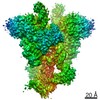
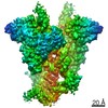
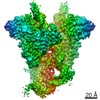
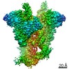
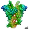
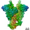
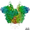
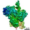
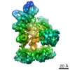
- EMDB-20039: Cryo-EM structure of mouse RAG1/2 NFC complex (DNA1) with C2 symmetry -
+ Open data

- Basic information
| Entry | Database: EMDB / ID: EMD-20039 | |||||||||
|---|---|---|---|---|---|---|---|---|---|---|
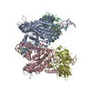
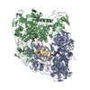
| Title | Cryo-EM structure of mouse RAG1/2 NFC complex (DNA1) with C2 symmetry | |||||||||
 Map data Map data | Mouse RAG1/2 NFC complex | |||||||||
 Sample Sample |
| |||||||||
| Biological species |  | |||||||||
| Method | single particle reconstruction / cryo EM / Resolution: 3.44 Å | |||||||||
 Authors Authors | Chen X / Cui Y / Zhou ZH / Yang W / Gellert M | |||||||||
| Funding support |  United States, 1 items United States, 1 items
| |||||||||
 Citation Citation | Journal: Nat Struct Mol Biol / Year: 2020 Title: Cutting antiparallel DNA strands in a single active site. Authors: Xuemin Chen / Yanxiang Cui / Robert B Best / Huaibin Wang / Z Hong Zhou / Wei Yang / Martin Gellert / Abstract: A single enzyme active site that catalyzes multiple reactions is a well-established biochemical theme, but how one nuclease site cleaves both DNA strands of a double helix has not been well ...A single enzyme active site that catalyzes multiple reactions is a well-established biochemical theme, but how one nuclease site cleaves both DNA strands of a double helix has not been well understood. In analyzing site-specific DNA cleavage by the mammalian RAG1-RAG2 recombinase, which initiates V(D)J recombination, we find that the active site is reconfigured for the two consecutive reactions and the DNA double helix adopts drastically different structures. For initial nicking of the DNA, a locally unwound and unpaired DNA duplex forms a zipper via alternating interstrand base stacking, rather than melting as generally thought. The second strand cleavage and formation of a hairpin-DNA product requires a global scissor-like movement of protein and DNA, delivering the scissile phosphate into the rearranged active site. | |||||||||
| History |
|
- Structure visualization
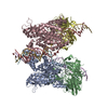
| Movie |
 Movie viewer Movie viewer |
|---|---|
| Structure viewer | EM map: SurfViewMolmilJmol/JSmol |
| Supplemental images |

- Downloads & links
-EMDB archive
| Map data | emd_20039.map.gz | 5.3 MB | EMDB map data format | |
|---|---|---|---|---|
| Header (meta data) | emd-20039-v30.xmlemd-20039.xml | 10.7 KB 10.7 KB | Display Display | EMDB header |
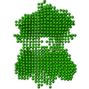
| Images | 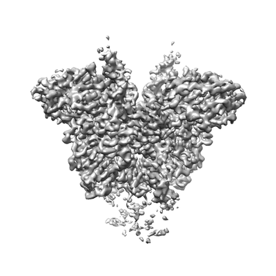 emd_20039.png emd_20039.png | 48.1 KB | ||
| Archive directory |  http://ftp.pdbj.org/pub/emdb/structures/EMD-20039ftp://ftp.pdbj.org/pub/emdb/structures/EMD-20039 http://ftp.pdbj.org/pub/emdb/structures/EMD-20039ftp://ftp.pdbj.org/pub/emdb/structures/EMD-20039 | HTTPS FTP |
-Related structure data
| Related structure data | 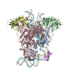 6oemC 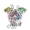 6oenC 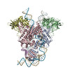 6oeoC 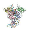 6oepC 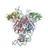 6oeqC 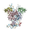 6oerC  6v0vC C: citing same article ( |
|---|---|
| Similar structure data |


-Links
| EMDB pages | EMDB (EBI/PDBe) / EMDataResource |
|---|
-Map
| File | Download / File: emd_20039.map.gz / Format: CCP4 / Size: 83.7 MB / Type: IMAGE STORED AS FLOATING POINT NUMBER (4 BYTES) | ||||||||||||||||||||||||||||||||||||||||||||||||||||||||||||
|---|---|---|---|---|---|---|---|---|---|---|---|---|---|---|---|---|---|---|---|---|---|---|---|---|---|---|---|---|---|---|---|---|---|---|---|---|---|---|---|---|---|---|---|---|---|---|---|---|---|---|---|---|---|---|---|---|---|---|---|---|---|
| Annotation | Mouse RAG1/2 NFC complex | ||||||||||||||||||||||||||||||||||||||||||||||||||||||||||||
| Projections & slices | Image control
Images are generated by Spider. | ||||||||||||||||||||||||||||||||||||||||||||||||||||||||||||
| Voxel size | X=Y=Z: 1.07 Å | ||||||||||||||||||||||||||||||||||||||||||||||||||||||||||||
| Density |
| ||||||||||||||||||||||||||||||||||||||||||||||||||||||||||||
| Symmetry | Space group: 1 | ||||||||||||||||||||||||||||||||||||||||||||||||||||||||||||
| Details | EMDB XML:
CCP4 map header:
| ||||||||||||||||||||||||||||||||||||||||||||||||||||||||||||
 Z (Sec.)
 Y (Row.)
 X (Col.)


-Supplemental data
- Sample components
-Entire : RAG1/2 Nick-forming complex (DNA1)
| Entire | Name: RAG1/2 Nick-forming complex (DNA1) |
|---|---|
| Components |
|
-Supramolecule #1: RAG1/2 Nick-forming complex (DNA1)
| Supramolecule | Name: RAG1/2 Nick-forming complex (DNA1) / type: complex / ID: 1 / Parent: 0 / Macromolecule list: #1-#7 |
|---|---|
| Source (natural) | Organism: |
| Recombinant expression | Organism:  Homo sapiens (human) Homo sapiens (human) |
-Experimental details
-Structure determination
| Method | cryo EM |
|---|---|
 Processing Processing | single particle reconstruction |
| Aggregation state | particle |
-Sample preparation
| Buffer | pH: 7.4 |
|---|---|
| Grid | Details: unspecified |
| Vitrification | Cryogen name: ETHANE |
| Details | mouse RAG1/2 NFC complex (DNA2) |
- Electron microscopy
| Microscope | FEI TITAN KRIOS |
|---|---|
| Image recording | Film or detector model: GATAN K2 SUMMIT (4k x 4k) / Average electron dose: 42.0 e/Å2 |
| Electron beam | Acceleration voltage: 300 kV / Electron source:  FIELD EMISSION GUN FIELD EMISSION GUN |
| Electron optics | Illumination mode: FLOOD BEAM / Imaging mode: BRIGHT FIELD |
| Experimental equipment |  Model: Titan Krios / Image courtesy: FEI Company |
-Image processing
| Final reconstruction | Resolution.type: BY AUTHOR / Resolution: 3.44 Å / Resolution method: FSC 0.143 CUT-OFF / Number images used: 109388 |
|---|---|
| Initial angle assignment | Type: NOT APPLICABLE |
| Final angle assignment | Type: NOT APPLICABLE |
