 Movie
Movie Controller


+ Open data

- Basic information
| Entry | Database: EMDB / ID: EMD-24941 | ||||||||||||||||||
|---|---|---|---|---|---|---|---|---|---|---|---|---|---|---|---|---|---|---|---|
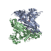
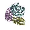
| Title | Helicobacter Hepaticus CcsBA Open Conformation | ||||||||||||||||||
 Map data Map data | |||||||||||||||||||
 Sample Sample |
| ||||||||||||||||||
 Keywords Keywords | Cytochrome c biogenesis / Heme transporter / Heme lyase / MEMBRANE PROTEIN | ||||||||||||||||||
| Function / homology | ResB-like domain / ResB-like family / Cytochrome c-type biogenesis protein CcsA/CcmC / Cytochrome c assembly protein / Cytochrome C assembly protein / cytochrome complex assembly / heme binding / plasma membrane / Cytochrome c biogenesis protein Function and homology information Function and homology information | ||||||||||||||||||
| Biological species |  Helicobacter hepaticus (bacteria) Helicobacter hepaticus (bacteria) | ||||||||||||||||||
| Method | single particle reconstruction / cryo EM / Resolution: 3.56 Å | ||||||||||||||||||
 Authors Authors | Mendez DL / Lowder EP | ||||||||||||||||||
| Funding support | 5 items
| ||||||||||||||||||
 Citation Citation | Journal: Nat Chem Biol / Year: 2022 Title: Cryo-EM of CcsBA reveals the basis for cytochrome c biogenesis and heme transport. Authors: Deanna L Mendez / Ethan P Lowder / Dustin E Tillman / Molly C Sutherland / Andrea L Collier / Michael J Rau / James A J Fitzpatrick / Robert G Kranz /  Abstract: Although the individual structures and respiratory functions of cytochromes are well studied, the structural basis for their assembly, including transport of heme for attachment, are unknown. We ...Although the individual structures and respiratory functions of cytochromes are well studied, the structural basis for their assembly, including transport of heme for attachment, are unknown. We describe cryo-electron microscopy (cryo-EM) structures of CcsBA, a bifunctional heme transporter and cytochrome c (cyt c) synthase. Models built from the cryo-EM densities show that CcsBA is trapped with heme in two conformations, herein termed the closed and open states. The closed state has heme located solely at a transmembrane (TM) site, with a large periplasmic domain oriented such that access of heme to the cytochrome acceptor is denied. The open conformation contains two heme moieties, one in the TM-heme site and another in an external site (P-heme site). The presence of heme in the periplasmic site at the base of a chamber induces a large conformational shift that exposes the heme for reaction with apocytochrome c (apocyt c). Consistent with these structures, in vivo and in vitro cyt c synthase studies suggest a mechanism for transfer of the periplasmic heme to cytochrome. | ||||||||||||||||||
| History |
|
- Structure visualization
| Movie |
Movie viewer |
|---|---|
| Structure viewer | EM map: SurfViewMolmilJmol/JSmol |
| Supplemental images |

- Downloads & links
-EMDB archive
| Map data | emd_24941.map.gz | 59.6 MB | EMDB map data format | |
|---|---|---|---|---|
| Header (meta data) | emd-24941-v30.xmlemd-24941.xml | 17.5 KB 17.5 KB | Display Display | EMDB header |
| Images | 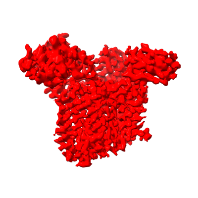 emd_24941.png emd_24941.png | 78.6 KB | ||
| Filedesc metadata | emd-24941.cif.gz | 7.2 KB | ||
| Archive directory |  http://ftp.pdbj.org/pub/emdb/structures/EMD-24941ftp://ftp.pdbj.org/pub/emdb/structures/EMD-24941 http://ftp.pdbj.org/pub/emdb/structures/EMD-24941ftp://ftp.pdbj.org/pub/emdb/structures/EMD-24941 | HTTPS FTP |
-Related structure data
| Related structure data | 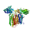 7s9yMC  7s9zC M: atomic model generated by this map C: citing same article ( |
|---|---|
| Similar structure data |


-Links
| EMDB pages | EMDB (EBI/PDBe) / EMDataResource |
|---|
-Map
| File | Download / File: emd_24941.map.gz / Format: CCP4 / Size: 64 MB / Type: IMAGE STORED AS FLOATING POINT NUMBER (4 BYTES) | ||||||||||||||||||||||||||||||||||||||||||||||||||||||||||||
|---|---|---|---|---|---|---|---|---|---|---|---|---|---|---|---|---|---|---|---|---|---|---|---|---|---|---|---|---|---|---|---|---|---|---|---|---|---|---|---|---|---|---|---|---|---|---|---|---|---|---|---|---|---|---|---|---|---|---|---|---|---|
| Projections & slices | Image control
Images are generated by Spider. | ||||||||||||||||||||||||||||||||||||||||||||||||||||||||||||
| Voxel size | X=Y=Z: 1.1 Å | ||||||||||||||||||||||||||||||||||||||||||||||||||||||||||||
| Density |
| ||||||||||||||||||||||||||||||||||||||||||||||||||||||||||||
| Symmetry | Space group: 1 | ||||||||||||||||||||||||||||||||||||||||||||||||||||||||||||
| Details | EMDB XML:
CCP4 map header:
| ||||||||||||||||||||||||||||||||||||||||||||||||||||||||||||
 Z (Sec.)
 Y (Row.)
 X (Col.)


-Supplemental data
- Sample components
-Entire : Complex of CcsBA with two hemes present
| Entire | Name: Complex of CcsBA with two hemes present |
|---|---|
| Components |
|
-Supramolecule #1: Complex of CcsBA with two hemes present
| Supramolecule | Name: Complex of CcsBA with two hemes present / type: complex / ID: 1 / Parent: 0 / Macromolecule list: #1 / Details: Purified from E. coli in DDM |
|---|---|
| Source (natural) | Organism: Helicobacter hepaticus (bacteria) |
-Macromolecule #1: Cytochrome c biogenesis protein
| Macromolecule | Name: Cytochrome c biogenesis protein / type: protein_or_peptide / ID: 1 / Number of copies: 1 / Enantiomer: LEVO |
|---|---|
| Source (natural) | Organism: Helicobacter hepaticus (bacteria) |
| Molecular weight | Theoretical: 107.291469 KDa |
| Recombinant expression | Organism: |
| Sequence | String: MMNIIKTLFC SMKMVLLLIG IYATACGIAT FIEKYEGTLA ARLWVYDAFW FEILHIWLVA CLIGCFITSK AWQRKKYASL LLHASFIVI IIGAGITRYY GFEGLMNLRE GQSVNFISTN THYIFIQIKN PQGDVESVRI PTYIDEKVNH KINQHLTFFG K PLTLHTEE ...String: MMNIIKTLFC SMKMVLLLIG IYATACGIAT FIEKYEGTLA ARLWVYDAFW FEILHIWLVA CLIGCFITSK AWQRKKYASL LLHASFIVI IIGAGITRYY GFEGLMNLRE GQSVNFISTN THYIFIQIKN PQGDVESVRI PTYIDEKVNH KINQHLTFFG K PLTLHTEE FTAKQVNMSE LFILNASIDF LGKNEKTLIM RDGNNAPTKE NITMLEIEGY KIFLAWGIDN IALPFSIKLK KF ELERYPG SNSPASYTSE VEVLDGQNPP LPFRIFMNNV LDYGGYRFFQ SSYHPDEKGS ILSVNNDPGK TPTYIGYAML ILG VIWLLF DKNGRFATLG RFLKTQKFFS LMLCSALCYA LSSPQIAYAS TQSQTDFQPL SENEIPPLQD IPSMIKALAD TSSL TNDFD RILVQDFGGR IKPMHTLANE YIHKLTQQRT FKGLNPSQVF LGMLFYPQEW QSIQMIATKS PKLRQILGLD ENQKH IAYI DVFTPQGQYI LQNYVEAANL KSPSLRDTFE KDVISVDERI NYAFLIYTGQ VLRIFPDNKS PNNQWLYPLQ AISSAV AQD DTKKAKELMQ IYKKFAQGMQ QGINTHNWQE AAQATRDIRT FQQNNGGSLL ISPAKVDSEI WLNLYNPFYQ LTYPYIF IS IVLFIIVLVG ILKNTPTRPL IHKVFYILLF ALFILHTCGL GLRWYVSEHA PWSNAYESML YIAWAAILSG VVFFRRSN L ALCASSFLAG MTLFVANLGD MDPQIGNLMP VLKSYWLNIH VSVITASYGF LGLCFMLGLI TLIMFLLRNE KRSQVDCSI LSLSALNEMS MILGLFLLSV GNFLGGIWAN ESWGRYWGWD SKETWALISI GVYAIILHLR FVVPKNFPFI FASASVIGFF SVLMTYFGV NYYLTGMHSY AAGEAEPVPL WVELMVAGII LLIIIASRKR VLDMPHLHHH HHH UniProtKB: Cytochrome c biogenesis protein |
-Macromolecule #2: HEME B/C
| Macromolecule | Name: HEME B/C / type: ligand / ID: 2 / Number of copies: 2 / Formula: HEB |
|---|---|
| Molecular weight | Theoretical: 618.503 Da |
| Chemical component information |  ChemComp-HEB: |
-Macromolecule #3: PHOSPHATIDYLETHANOLAMINE
| Macromolecule | Name: PHOSPHATIDYLETHANOLAMINE / type: ligand / ID: 3 / Number of copies: 1 / Formula: PTY |
|---|---|
| Molecular weight | Theoretical: 734.039 Da |
| Chemical component information |  ChemComp-PTY: |
-Experimental details
-Structure determination
| Method | cryo EM |
|---|---|
 Processing Processing | single particle reconstruction |
| Aggregation state | particle |
-Sample preparation
| Concentration | 2.8 mg/mL | ||||||||||||
|---|---|---|---|---|---|---|---|---|---|---|---|---|---|
| Buffer | pH: 8 Component:
| ||||||||||||
| Grid | Model: Quantifoil R2/2 / Material: COPPER / Mesh: 300 / Pretreatment - Type: PLASMA CLEANING / Pretreatment - Time: 60 sec. / Pretreatment - Atmosphere: OTHER / Pretreatment - Pressure: 0.009300000000000001 kPa | ||||||||||||
| Vitrification | Cryogen name: ETHANE / Chamber humidity: 100 % / Chamber temperature: 277.15 K / Instrument: FEI VITROBOT MARK IV Details: Blot for 2 seconds at a blot force of -1 and plunge frozen. |
- Electron microscopy
| Microscope | TFS KRIOS |
|---|---|
| Temperature | Min: 82.0 K / Max: 84.0 K |
| Specialist optics | Spherical aberration corrector: Microscope is outfitted with a Cs image corrector with two hexapole elements. Energy filter - Name: GIF Bioquantum / Energy filter - Slit width: 20 eV / Details: Specific energy filter was a Gatan BioQuantum 968. |
| Details | Preliminary grid screening was performed manually. |
| Image recording | Film or detector model: GATAN K2 QUANTUM (4k x 4k) / Detector mode: COUNTING / Digitization - Dimensions - Width: 3832 pixel / Digitization - Dimensions - Height: 3704 pixel / Number grids imaged: 1 / Number real images: 8676 / Average exposure time: 8.0 sec. / Average electron dose: 66.0 e/Å2 |
| Electron beam | Acceleration voltage: 300 kV / Electron source:  FIELD EMISSION GUN FIELD EMISSION GUN |
| Electron optics | C2 aperture diameter: 150.0 µm / Calibrated defocus max: 2.5 µm / Calibrated defocus min: 1.0 µm / Calibrated magnification: 105000 / Illumination mode: FLOOD BEAM / Imaging mode: BRIGHT FIELD / Cs: 0.01 mm / Nominal defocus max: 2.5 µm / Nominal defocus min: 1.0 µm / Nominal magnification: 105000 |
| Sample stage | Specimen holder model: FEI TITAN KRIOS AUTOGRID HOLDER / Cooling holder cryogen: NITROGEN |
| Experimental equipment |  Model: Titan Krios / Image courtesy: FEI Company |
+Image processing
-Atomic model buiding 1
| Details | De novo modelling was undertaken using COOT. Both Phenix and ISOLDE were used for refinement |
|---|---|
| Refinement | Space: REAL / Protocol: FLEXIBLE FIT |
| Output model | PDB-7s9y: |
