 Movie
Movie Controller


+ Open data

- Basic information
| Entry | Database: PDB / ID: 7s9z | ||||||||||||||||||
|---|---|---|---|---|---|---|---|---|---|---|---|---|---|---|---|---|---|---|---|
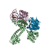
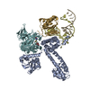
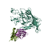
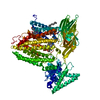
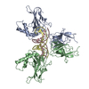
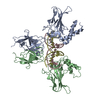
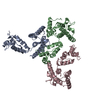
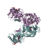
| Title | Helicobacter Hepaticus CcsBA Closed Conformation | ||||||||||||||||||
 Components Components | Cytochrome c biogenesis protein | ||||||||||||||||||
 Keywords Keywords | MEMBRANE PROTEIN / Cytochrome c biogenesis / Heme transporter / Heme lyase | ||||||||||||||||||
| Function / homology |  Function and homology information Function and homology information | ||||||||||||||||||
| Biological species |  Helicobacter hepaticus (bacteria) Helicobacter hepaticus (bacteria) | ||||||||||||||||||
| Method | ELECTRON MICROSCOPY / single particle reconstruction / cryo EM / Resolution: 4.14 Å | ||||||||||||||||||
 Authors Authors | Mendez, D.L. / Lowder, E.P. / Tillman, D.E. / Sutherland, M.C. / Collier, A.L. / Rau, M.J. / Fitzpatrick, J.A. / Kranz, R.G. | ||||||||||||||||||
| Funding support | 5items
| ||||||||||||||||||
 Citation Citation | Journal: Nat Chem Biol / Year: 2022 Title: Cryo-EM of CcsBA reveals the basis for cytochrome c biogenesis and heme transport. Authors: Deanna L Mendez / Ethan P Lowder / Dustin E Tillman / Molly C Sutherland / Andrea L Collier / Michael J Rau / James A J Fitzpatrick / Robert G Kranz /  Abstract: Although the individual structures and respiratory functions of cytochromes are well studied, the structural basis for their assembly, including transport of heme for attachment, are unknown. We ...Although the individual structures and respiratory functions of cytochromes are well studied, the structural basis for their assembly, including transport of heme for attachment, are unknown. We describe cryo-electron microscopy (cryo-EM) structures of CcsBA, a bifunctional heme transporter and cytochrome c (cyt c) synthase. Models built from the cryo-EM densities show that CcsBA is trapped with heme in two conformations, herein termed the closed and open states. The closed state has heme located solely at a transmembrane (TM) site, with a large periplasmic domain oriented such that access of heme to the cytochrome acceptor is denied. The open conformation contains two heme moieties, one in the TM-heme site and another in an external site (P-heme site). The presence of heme in the periplasmic site at the base of a chamber induces a large conformational shift that exposes the heme for reaction with apocytochrome c (apocyt c). Consistent with these structures, in vivo and in vitro cyt c synthase studies suggest a mechanism for transfer of the periplasmic heme to cytochrome. | ||||||||||||||||||
| History |
|
- Structure visualization
| Movie |
Movie viewer |
|---|---|
| Structure viewer | Molecule: MolmilJmol/JSmol |
- Downloads & links
-Download
| PDBx/mmCIF format | 7s9z.cif.gz | 176.3 KB | Display | PDBx/mmCIF format |
|---|---|---|---|---|
| PDB format | pdb7s9z.ent.gz | 135.9 KB | Display | PDB format |
| PDBx/mmJSON format | 7s9z.json.gz | Tree view | PDBx/mmJSON format | |
| Others |  Other downloads Other downloads |
-Validation report
| Arichive directory | https://data.pdbj.org/pub/pdb/validation_reports/s9/7s9zftp://data.pdbj.org/pub/pdb/validation_reports/s9/7s9z | HTTPS FTP |
|---|
-Related structure data
| Related structure data |  24942MC 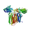 7s9yC M: map data used to model this data C: citing same article ( |
|---|---|
| Similar structure data |


-Links
 PDBj
PDBj
- Assembly
| Deposited unit | 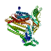
|
|---|---|
| 1 |
|
-Components
| #1: Protein | Mass: 107291.469 Da / Num. of mol.: 1 Source method: isolated from a genetically manipulated source Source: (gene. exp.) Helicobacter hepaticus (bacteria) / Gene: ccsBA / Production host: |
|---|---|
| #2: Chemical | ChemComp-PTY /   Mass: 734.039 Da / Num. of mol.: 1 / Source method: obtained synthetically / Formula: C40H80NO8P / Comment: phospholipid*YM Mass: 734.039 Da / Num. of mol.: 1 / Source method: obtained synthetically / Formula: C40H80NO8P / Comment: phospholipid*YM |
| #3: Chemical | ChemComp-HEB /   Mass: 618.503 Da / Num. of mol.: 1 / Source method: obtained synthetically / Formula: C34H34FeN4O4 / Feature type: SUBJECT OF INVESTIGATION Mass: 618.503 Da / Num. of mol.: 1 / Source method: obtained synthetically / Formula: C34H34FeN4O4 / Feature type: SUBJECT OF INVESTIGATION |
| Has ligand of interest | Y |
| Has protein modification | Y |
-Experimental details
-Experiment
| Experiment | Method: ELECTRON MICROSCOPY |
|---|---|
| EM experiment | Aggregation state: PARTICLE / 3D reconstruction method: single particle reconstruction |
- Sample preparation
| Component | Name: Complex of CcsBA with one heme present / Type: COMPLEX / Details: Purified from E. coli in DDM / Entity ID: #1 / Source: RECOMBINANT | ||||||||||||||||||||
|---|---|---|---|---|---|---|---|---|---|---|---|---|---|---|---|---|---|---|---|---|---|
| Molecular weight | Experimental value: NO | ||||||||||||||||||||
| Source (natural) | Organism: Helicobacter hepaticus (bacteria) | ||||||||||||||||||||
| Source (recombinant) | Organism: | ||||||||||||||||||||
| Buffer solution | pH: 8 | ||||||||||||||||||||
| Buffer component |
| ||||||||||||||||||||
| Specimen | Conc.: 2.8 mg/ml / Embedding applied: NO / Shadowing applied: NO / Staining applied: NO / Vitrification applied: YES | ||||||||||||||||||||
| Specimen support | Grid material: COPPER / Grid mesh size: 300 divisions/in. / Grid type: Quantifoil R2/2 | ||||||||||||||||||||
| Vitrification | Instrument: FEI VITROBOT MARK IV / Cryogen name: ETHANE / Humidity: 100 % / Chamber temperature: 277.15 K Details: Blot for 2 seconds at a blot force of -1 and plunge frozen |
- Electron microscopy imaging
| Experimental equipment |  Model: Titan Krios / Image courtesy: FEI Company |
|---|---|
| Microscopy | Model: TFS KRIOS / Details: Preliminary grid screening was performed manually. |
| Electron gun | Electron source:  FIELD EMISSION GUN / Accelerating voltage: 300 kV / Illumination mode: FLOOD BEAM FIELD EMISSION GUN / Accelerating voltage: 300 kV / Illumination mode: FLOOD BEAM |
| Electron lens | Mode: BRIGHT FIELD / Nominal magnification: 105000 X / Calibrated magnification: 105000 X / Nominal defocus max: 2500 nm / Nominal defocus min: 1000 nm / Calibrated defocus min: 1000 nm / Calibrated defocus max: 2500 nm / Cs: 0.01 mm / C2 aperture diameter: 150 µm / Alignment procedure: ZEMLIN TABLEAU |
| Specimen holder | Cryogen: NITROGEN / Specimen holder model: FEI TITAN KRIOS AUTOGRID HOLDER / Temperature (max): 84 K / Temperature (min): 82 K |
| Image recording | Average exposure time: 8 sec. / Electron dose: 66 e/Å2 / Detector mode: COUNTING / Film or detector model: GATAN K2 QUANTUM (4k x 4k) / Num. of grids imaged: 1 / Num. of real images: 8676 |
| EM imaging optics | Energyfilter name: GIF Bioquantum / Details: Specific energy filter was a Gatan BioQuantum 968. / Energyfilter slit width: 20 eV Spherical aberration corrector: Microscope is outfitted with a Cs image corrector with two hexapole elements. |
| Image scans | Width: 3832 / Height: 3704 / Movie frames/image: 40 |
- Processing
| EM software |
| ||||||||||||||||||||||||||||||||||||||||||||||||||
|---|---|---|---|---|---|---|---|---|---|---|---|---|---|---|---|---|---|---|---|---|---|---|---|---|---|---|---|---|---|---|---|---|---|---|---|---|---|---|---|---|---|---|---|---|---|---|---|---|---|---|---|
| CTF correction | Type: PHASE FLIPPING AND AMPLITUDE CORRECTION | ||||||||||||||||||||||||||||||||||||||||||||||||||
| Particle selection | Num. of particles selected: 1778157 Details: Initial particle picking was facilitated using the blob picker in cryoSPARC. Picked particles were then subjected to 2D classification. Once converged, a sub-set of 2D classes were manually ...Details: Initial particle picking was facilitated using the blob picker in cryoSPARC. Picked particles were then subjected to 2D classification. Once converged, a sub-set of 2D classes were manually picked and reclassified using a smaller number of classes. | ||||||||||||||||||||||||||||||||||||||||||||||||||
| Symmetry | Point symmetry: C1 (asymmetric) | ||||||||||||||||||||||||||||||||||||||||||||||||||
| 3D reconstruction | Resolution: 4.14 Å / Resolution method: FSC 0.143 CUT-OFF / Num. of particles: 117488 / Algorithm: FOURIER SPACE / Num. of class averages: 1 / Symmetry type: POINT | ||||||||||||||||||||||||||||||||||||||||||||||||||
| Atomic model building | Protocol: FLEXIBLE FIT / Space: REAL Details: Model was built de novo into density map using COOT. Both Phenix and ISOLDE were used for refinement |
