 Movie
Movie Controller


+ Open data

- Basic information
| Entry | Database: PDB / ID: 3vjb | ||||||
|---|---|---|---|---|---|---|---|
| Title | Crystal structure of the human squalene synthase | ||||||
 Components Components | Squalene synthase | ||||||
 Keywords Keywords | TRANSFERASE / Farnesyl-diphosphate farnesyltransferase / Head-to-head synthases / Cholesterol biosynthesis / Oxidoreductase | ||||||
| Function / homology |  Function and homology information Function and homology informationsqualene synthase / farnesyl diphosphate metabolic process / Cholesterol biosynthesis / squalene synthase [NAD(P)H] activity / steroid biosynthetic process / cholesterol biosynthetic process / Activation of gene expression by SREBF (SREBP) / PPARA activates gene expression / endoplasmic reticulum membrane / endoplasmic reticulum ...squalene synthase / farnesyl diphosphate metabolic process / Cholesterol biosynthesis / squalene synthase [NAD(P)H] activity / steroid biosynthetic process / cholesterol biosynthetic process / Activation of gene expression by SREBF (SREBP) / PPARA activates gene expression / endoplasmic reticulum membrane / endoplasmic reticulum / membrane / metal ion binding Similarity search - Function | ||||||
| Biological species |  Homo sapiens (human) Homo sapiens (human) | ||||||
| Method |  X-RAY DIFFRACTION / SYNCHROTRON / MOLECULAR REPLACEMENT / Resolution: 2.05 Å X-RAY DIFFRACTION / SYNCHROTRON / MOLECULAR REPLACEMENT / Resolution: 2.05 Å | ||||||
 Authors Authors | Liu, C.I. / Jeng, W.Y. / Chang, W.J. / Wang, A.H.J. | ||||||
 Citation Citation | Journal: J.Biol.Chem. / Year: 2012 Title: Binding modes of zaragozic acid A to human squalene synthase and staphylococcal dehydrosqualene synthase Authors: Liu, C.I. / Jeng, W.Y. / Chang, W.J. / Ko, T.P. / Wang, A.H.J. | ||||||
| History |
|
- Structure visualization
| Structure viewer | Molecule: MolmilJmol/JSmol |
|---|
- Downloads & links
-Download
| PDBx/mmCIF format | 3vjb.cif.gz | 830.3 KB | Display | PDBx/mmCIF format |
|---|---|---|---|---|
| PDB format | pdb3vjb.ent.gz | 695.8 KB | Display | PDB format |
| PDBx/mmJSON format | 3vjb.json.gz | Tree view | PDBx/mmJSON format | |
| Others |  Other downloads Other downloads |
-Validation report
| Arichive directory | https://data.pdbj.org/pub/pdb/validation_reports/vj/3vjbftp://data.pdbj.org/pub/pdb/validation_reports/vj/3vjb | HTTPS FTP |
|---|
-Related structure data
| Related structure data | 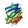 3vj8C 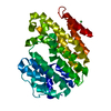 3vj9C 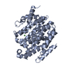 3vjaC 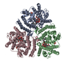 3vjcC 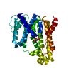 3vjdC 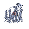 3vjeC  1ezfS C: citing same article ( S: Starting model for refinement |
|---|---|
| Similar structure data |


-Links
 PDBj
PDBj

- Assembly
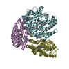
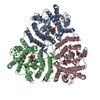
| Deposited unit | 
| ||||||||
|---|---|---|---|---|---|---|---|---|---|
| 1 | 
| ||||||||
| 2 | 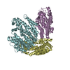
| ||||||||
| Unit cell |
|
-Components
| #1: Protein | Mass: 39448.969 Da / Num. of mol.: 6 / Fragment: UNP residues 31-370 Source method: isolated from a genetically manipulated source Source: (gene. exp.) Homo sapiens (human) / Gene: FDFT1 / Plasmid: pET-28a / Production host:  #2: Water | ChemComp-HOH / |  Mass: 18.015 Da / Num. of mol.: 926 / Source method: isolated from a natural source / Formula: H2O Mass: 18.015 Da / Num. of mol.: 926 / Source method: isolated from a natural source / Formula: H2O |
|---|
-Experimental details
-Experiment
| Experiment | Method: X-RAY DIFFRACTION / Number of used crystals: 1 |
|---|
- Sample preparation
| Crystal | Density Matthews: 2.55 Å3/Da / Density % sol: 51.68 % |
|---|---|
| Crystal grow | Temperature: 298 K / Method: vapor diffusion, hanging drop / pH: 7.5 Details: 1.4M sodium citrate tribasic dihydrate, 0.1M HEPES sodium, pH 7.5, VAPOR DIFFUSION, HANGING DROP, temperature 298K |
-Data collection
| Diffraction | Mean temperature: 100 K |
|---|---|
| Diffraction source | Source: SYNCHROTRON / Site: NSRRC  / Beamline: BL13B1 / Wavelength: 1 Å / Beamline: BL13B1 / Wavelength: 1 Å |
| Detector | Type: ADSC QUANTUM 315 / Detector: CCD / Date: Jul 19, 2007 Details: Vertically Collimating Premirror, Toroidal Focusing Mirror |
| Radiation | Monochromator: LN2-Cooled Fixed-Exit Double Crystal Si(111) Monochromator Protocol: SINGLE WAVELENGTH / Monochromatic (M) / Laue (L): M / Scattering type: x-ray |
| Radiation wavelength | Wavelength: 1 Å / Relative weight: 1 |
| Reflection | Resolution: 2.05→30 Å / Num. all: 147063 / Num. obs: 144874 / % possible obs: 98.5 % / Observed criterion σ(F): 0 / Observed criterion σ(I): 1 / Redundancy: 3.9 % / Biso Wilson estimate: 41.8 Å2 / Rmerge(I) obs: 0.077 / Net I/σ(I): 20.8 |
| Reflection shell | Resolution: 2.05→2.12 Å / Redundancy: 3.7 % / Rmerge(I) obs: 0.402 / Mean I/σ(I) obs: 3.6 / Num. unique all: 14667 / % possible all: 91.4 |
- Processing
| Software |
| |||||||||||||||||||||||||||||||||||||||||||||||||||||||||||||||||||||||||||||||||||||||||||||||||||||||||||||||||||||||||||||||||||||||||||||||||||||||||||||||||||||||||||||||
|---|---|---|---|---|---|---|---|---|---|---|---|---|---|---|---|---|---|---|---|---|---|---|---|---|---|---|---|---|---|---|---|---|---|---|---|---|---|---|---|---|---|---|---|---|---|---|---|---|---|---|---|---|---|---|---|---|---|---|---|---|---|---|---|---|---|---|---|---|---|---|---|---|---|---|---|---|---|---|---|---|---|---|---|---|---|---|---|---|---|---|---|---|---|---|---|---|---|---|---|---|---|---|---|---|---|---|---|---|---|---|---|---|---|---|---|---|---|---|---|---|---|---|---|---|---|---|---|---|---|---|---|---|---|---|---|---|---|---|---|---|---|---|---|---|---|---|---|---|---|---|---|---|---|---|---|---|---|---|---|---|---|---|---|---|---|---|---|---|---|---|---|---|---|---|---|---|
| Refinement | Method to determine structure: MOLECULAR REPLACEMENT Starting model: PDB ENTRY 1EZF Resolution: 2.05→29.4 Å / Cor.coef. Fo:Fc: 0.966 / Cor.coef. Fo:Fc free: 0.941 / SU B: 10.58 / SU ML: 0.128 / Isotropic thermal model: Isotropic with TLS / Cross valid method: THROUGHOUT / ESU R Free: 0.167 / Stereochemistry target values: MAXIMUM LIKELIHOOD / Details: HYDROGENS HAVE BEEN ADDED IN THE RIDING POSITIONS
| |||||||||||||||||||||||||||||||||||||||||||||||||||||||||||||||||||||||||||||||||||||||||||||||||||||||||||||||||||||||||||||||||||||||||||||||||||||||||||||||||||||||||||||||
| Solvent computation | Ion probe radii: 0.8 Å / Shrinkage radii: 0.8 Å / VDW probe radii: 1.4 Å / Solvent model: MASK | |||||||||||||||||||||||||||||||||||||||||||||||||||||||||||||||||||||||||||||||||||||||||||||||||||||||||||||||||||||||||||||||||||||||||||||||||||||||||||||||||||||||||||||||
| Displacement parameters | Biso mean: 54.303 Å2
| |||||||||||||||||||||||||||||||||||||||||||||||||||||||||||||||||||||||||||||||||||||||||||||||||||||||||||||||||||||||||||||||||||||||||||||||||||||||||||||||||||||||||||||||
| Refinement step | Cycle: LAST / Resolution: 2.05→29.4 Å
| |||||||||||||||||||||||||||||||||||||||||||||||||||||||||||||||||||||||||||||||||||||||||||||||||||||||||||||||||||||||||||||||||||||||||||||||||||||||||||||||||||||||||||||||
| Refine LS restraints |
| |||||||||||||||||||||||||||||||||||||||||||||||||||||||||||||||||||||||||||||||||||||||||||||||||||||||||||||||||||||||||||||||||||||||||||||||||||||||||||||||||||||||||||||||
| LS refinement shell | Resolution: 2.052→2.162 Å / Total num. of bins used: 10
| |||||||||||||||||||||||||||||||||||||||||||||||||||||||||||||||||||||||||||||||||||||||||||||||||||||||||||||||||||||||||||||||||||||||||||||||||||||||||||||||||||||||||||||||
| Refinement TLS params. | Method: refined / Refine-ID: X-RAY DIFFRACTION
| |||||||||||||||||||||||||||||||||||||||||||||||||||||||||||||||||||||||||||||||||||||||||||||||||||||||||||||||||||||||||||||||||||||||||||||||||||||||||||||||||||||||||||||||
| Refinement TLS group |
|
