[English] 日本語
Yorodumi
- EMDB-13054: Cryo-EM structure of nonameric EPEC SctV-C -

+
Open data


ID or keywords:

Loading...

-
Basic information

Entry
Database: EMDB / ID: EMD-13054
TitleCryo-EM structure of nonameric EPEC SctV-C
Map dataPost-processed map_masked
Sample
  • Complex: EPEC SctV-C nonamer
    • Protein or peptide: Translocator EscV
KeywordsT3SS / SctV nonamer / export gate / PROTEIN TRANSPORT
Function / homology
Function and homology information


protein secretion / plasma membrane
Similarity search - Function
Type III secretion protein HrcV / FHIPEP conserved site / Bacterial export FHIPEP family signature. / Type III secretion system FHIPEP / FHIPEP, domain 3 / FHIPEP, domain 4 / FHIPEP family
Similarity search - Domain/homology
Biological speciesEscherichia coli O127:H6 str. E2348/69 (bacteria) / Escherichia coli O127:H6 (strain E2348/69 / EPEC) (bacteria)
Methodsingle particle reconstruction / cryo EM / Resolution: 3.1 Å
AuthorsYuan B / Wald J / Fahrenkamp D / Marlovits TC
Funding support Austria, 1 items
OrganizationGrant numberCountry
Austrian Science FundI 2408-B22 Austria
Citation
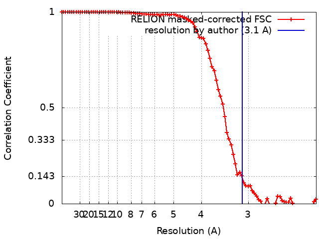
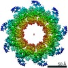
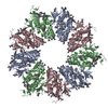
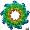
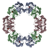
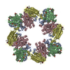
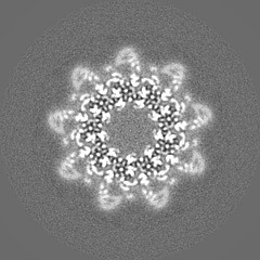
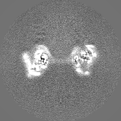
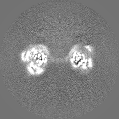
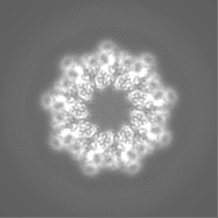
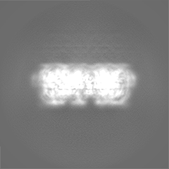
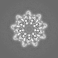
Journal: J Mol Biol / Year: 2021
Title: Structural Dynamics of the Functional Nonameric Type III Translocase Export Gate.
Authors: Biao Yuan / Athina G Portaliou / Rinky Parakra / Jochem H Smit / Jiri Wald / Yichen Li / Bindu Srinivasu / Maria S Loos / Harveer Singh Dhupar / Dirk Fahrenkamp / Charalampos G Kalodimos / ...Authors: Biao Yuan / Athina G Portaliou / Rinky Parakra / Jochem H Smit / Jiri Wald / Yichen Li / Bindu Srinivasu / Maria S Loos / Harveer Singh Dhupar / Dirk Fahrenkamp / Charalampos G Kalodimos / Franck Duong van Hoa / Thorben Cordes / Spyridoula Karamanou / Thomas C Marlovits / Anastassios Economou /
Abstract: Type III protein secretion is widespread in Gram-negative pathogens. It comprises the injectisome with a surface-exposed needle and an inner membrane translocase. The translocase contains the SctRSTU ...Type III protein secretion is widespread in Gram-negative pathogens. It comprises the injectisome with a surface-exposed needle and an inner membrane translocase. The translocase contains the SctRSTU export channel enveloped by the export gate subunit SctV that binds chaperone/exported clients and forms a putative ante-chamber. We probed the assembly, function, structure and dynamics of SctV from enteropathogenic E. coli (EPEC). In both EPEC and E. coli lab strains, SctV forms peripheral oligomeric clusters that are detergent-extracted as homo-nonamers. Membrane-embedded SctV is necessary and sufficient to act as a receptor for different chaperone/exported protein pairs with distinct C-domain binding sites that are essential for secretion. Negative staining electron microscopy revealed that peptidisc-reconstituted His-SctV forms a tripartite particle of ∼22 nm with a N-terminal domain connected by a short linker to a C-domain ring structure with a ∼5 nm-wide inner opening. The isolated C-domain ring was resolved with cryo-EM at 3.1 Å and structurally compared to other SctV homologues. Its four sub-domains undergo a three-stage "pinching" motion. Hydrogen-deuterium exchange mass spectrometry revealed this to involve dynamic and rigid hinges and a hyper-flexible sub-domain that flips out of the ring periphery and binds chaperones on and between adjacent protomers. These motions are coincident with local conformational changes at the pore surface and ring entry mouth that may also be modulated by the ATPase inner stalk. We propose that the intrinsic dynamics of the SctV protomer are modulated by chaperones and the ATPase and could affect allosterically the other subunits of the nonameric ring during secretion.
History
DepositionJun 9, 2021-
Header (metadata) releaseSep 29, 2021-
Map releaseSep 29, 2021-
UpdateJul 17, 2024-
Current statusJul 17, 2024Processing site: PDBe / Status: Released

-
Structure visualization
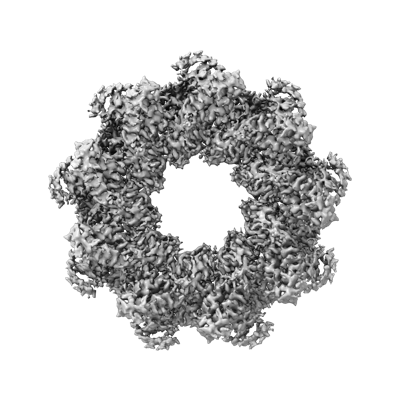
Movie
  • Surface view with section colored by density value
  • Surface level: 0.026
  • Imaged by UCSF Chimera
  • Download
  • Surface view colored by cylindrical radius
  • Surface level: 0.026
  • Imaged by UCSF Chimera
  • Download
  • Surface view with fitted model
  • Atomic models: PDB-7osl
  • Surface level: 0.026
  • Imaged by UCSF Chimera
  • Download
Movie viewer
Structure viewerEM map:
SurfViewMolmilJmol/JSmol
Supplemental images

Downloads & links

-
Map

FileDownload / File: emd_13054.map.gz / Format: CCP4 / Size: 52.7 MB / Type: IMAGE STORED AS FLOATING POINT NUMBER (4 BYTES)
AnnotationPost-processed map_masked
Voxel sizeX=Y=Z: 1.1 Å
Density
Contour LevelBy AUTHOR: 0.0234 / Movie #1: 0.026
Minimum - Maximum-0.1659851 - 0.23508382
Average (Standard dev.)0.0006150173 (±0.0065155383)
SymmetrySpace group: 1
Details

EMDB XML:

Map geometry
Axis orderXYZ
Origin000
Dimensions240240240
Spacing240240240
CellA=B=C: 264.0 Å
α=β=γ: 90.0 °

CCP4 map header:

modeImage stored as Reals
Å/pix. X/Y/Z1.11.11.1
M x/y/z240240240
origin x/y/z0.0000.0000.000
length x/y/z264.000264.000264.000
α/β/γ90.00090.00090.000
MAP C/R/S123
start NC/NR/NS000
NC/NR/NS240240240
D min/max/mean-0.1660.2350.001

-
Supplemental data

-
Mask #1

Fileemd_13054_msk_1.map
Projections & Slices
AxesZYX

Projections

Slices (1/2)
Density Histograms

-
Half map: half-map1 from auto-refinement

Fileemd_13054_half_map_1.map
Annotationhalf-map1 from auto-refinement
Projections & Slices
AxesZYX

Projections

Slices (1/2)
Density Histograms

-
Half map: Half-map 2 from autorefinement

Fileemd_13054_half_map_2.map
AnnotationHalf-map 2 from autorefinement
Projections & Slices
AxesZYX

Projections

Slices (1/2)
Density Histograms

-
Sample components

-
Entire : EPEC SctV-C nonamer

EntireName: EPEC SctV-C nonamer
Components
  • Complex: EPEC SctV-C nonamer
    • Protein or peptide: Translocator EscV

-
Supramolecule #1: EPEC SctV-C nonamer

SupramoleculeName: EPEC SctV-C nonamer / type: complex / ID: 1 / Parent: 0 / Macromolecule list: all
Source (natural)Organism: Escherichia coli O127:H6 str. E2348/69 (bacteria)
Molecular weightTheoretical: 379.2 kDa/nm

-
Macromolecule #1: Translocator EscV

MacromoleculeName: Translocator EscV / type: protein_or_peptide / ID: 1 / Number of copies: 9 / Enantiomer: LEVO
Source (natural)Organism: Escherichia coli O127:H6 (strain E2348/69 / EPEC) (bacteria)
Strain: E2348/69 / EPEC
Molecular weightTheoretical: 37.952457 KDa
Recombinant expressionOrganism: Escherichia coli 'BL21-Gold(DE3)pLysS AG' (bacteria)
SequenceString: NISPGAEPLI LNLSSNIYSS DITQQIEVMR WNFFEESGIP LPKIIVNPVK NNDSAIEFLL YQESIYKDTL IDDTVYFEAG HAEISFEFV QEKLSTNSIV YKTNKTNQQL AHLTGMDVYA TTNDKITFLL KKLVLSNAKE FIGVQETRYL MDIMERKYNE L VKELQRQL ...String:
NISPGAEPLI LNLSSNIYSS DITQQIEVMR WNFFEESGIP LPKIIVNPVK NNDSAIEFLL YQESIYKDTL IDDTVYFEAG HAEISFEFV QEKLSTNSIV YKTNKTNQQL AHLTGMDVYA TTNDKITFLL KKLVLSNAKE FIGVQETRYL MDIMERKYNE L VKELQRQL GLSKIVDILQ RLVEENVSIR DLRTIFETLI FWSTKEKDVV ILCEYVRIAL RRHILGRYSV SGTLLNVWLI GS DIENELR ESIRQTSSGS YLNISPERTE QIIGFLKNIM NPTGNGVILT ALDIRRYVKK MIEGSFPSVP VLSFQEVGNN IEL KVLGTV NDFRA

UniProtKB: Translocator EscV

-
Experimental details

-
Structure determination

Methodcryo EM
Processingsingle particle reconstruction
Aggregation stateparticle

-
Sample preparation

Concentration1.8 mg/mL
BufferpH: 8
Component:
ConcentrationFormulaName
50.0 mMNaClsodium chloride
50.0 mMTris-HClTris hydrochloride
GridModel: Quantifoil R1.2/1.3 / Material: GOLD / Support film - Material: CARBON / Support film - topology: HOLEY / Pretreatment - Type: GLOW DISCHARGE / Pretreatment - Time: 45 sec. / Pretreatment - Atmosphere: AIR
VitrificationCryogen name: ETHANE / Chamber humidity: 100 % / Chamber temperature: 277 K / Instrument: FEI VITROBOT MARK IV
DetailsThe sample was mono-disperse.

-
Electron microscopy

MicroscopeFEI TITAN KRIOS
Image recordingFilm or detector model: GATAN K2 SUMMIT (4k x 4k) / Detector mode: COUNTING / Number real images: 3739 / Average electron dose: 41.25 e/Å2
Electron beamAcceleration voltage: 300 kV / Electron source: FIELD EMISSION GUN
Electron opticsC2 aperture diameter: 70.0 µm / Illumination mode: FLOOD BEAM / Imaging mode: BRIGHT FIELD / Cs: 2.7 mm
Experimental equipment
Model: Titan Krios / Image courtesy: FEI Company

+
Image processing

Particle selectionNumber selected: 660798
Startup modelType of model: NONE
Final reconstructionResolution.type: BY AUTHOR / Resolution: 3.1 Å / Resolution method: FSC 0.143 CUT-OFF / Software - Name: RELION (ver. 3.1) / Number images used: 105670
Initial angle assignmentType: ANGULAR RECONSTITUTION / Software - Name: RELION (ver. 3.1)
Final angle assignmentType: ANGULAR RECONSTITUTION / Software - Name: RELION (ver. 3.1)
Final 3D classificationNumber classes: 4 / Avg.num./class: 72000 / Software - Name: RELION (ver. 3.1)
FSC plot (resolution estimation)

+
About Yorodumi

-
News

-
Feb 9, 2022. New format data for meta-information of EMDB entries

New format data for meta-information of EMDB entries

  • Version 3 of the EMDB header file is now the official format.
  • The previous official version 1.9 will be removed from the archive.

Related info.:EMDB header

External links:wwPDB to switch to version 3 of the EMDB data model

-
Aug 12, 2020. Covid-19 info

Covid-19 info

URL: https://pdbjlvh1.pdbj.org/emnavi/covid19.php

New page: Covid-19 featured information page in EM Navigator.

Related info.:Covid-19 info / Mar 5, 2020. Novel coronavirus structure data

+
Mar 5, 2020. Novel coronavirus structure data

Novel coronavirus structure data

Related info.:Yorodumi Speices / Aug 12, 2020. Covid-19 info

External links:COVID-19 featured content - PDBj / Molecule of the Month (242):Coronavirus Proteases

+
Jan 31, 2019. EMDB accession codes are about to change! (news from PDBe EMDB page)

EMDB accession codes are about to change! (news from PDBe EMDB page)

  • The allocation of 4 digits for EMDB accession codes will soon come to an end. Whilst these codes will remain in use, new EMDB accession codes will include an additional digit and will expand incrementally as the available range of codes is exhausted. The current 4-digit format prefixed with “EMD-” (i.e. EMD-XXXX) will advance to a 5-digit format (i.e. EMD-XXXXX), and so on. It is currently estimated that the 4-digit codes will be depleted around Spring 2019, at which point the 5-digit format will come into force.
  • The EM Navigator/Yorodumi systems omit the EMD- prefix.

Related info.:Q: What is EMD? / ID/Accession-code notation in Yorodumi/EM Navigator

External links:EMDB Accession Codes are Changing Soon! / Contact to PDBj

+
Jul 12, 2017. Major update of PDB

Major update of PDB

  • wwPDB released updated PDB data conforming to the new PDBx/mmCIF dictionary.
  • This is a major update changing the version number from 4 to 5, and with Remediation, in which all the entries are updated.
  • In this update, many items about electron microscopy experimental information are reorganized (e.g. em_software).
  • Now, EM Navigator and Yorodumi are based on the updated data.

External links:wwPDB Remediation / Enriched Model Files Conforming to OneDep Data Standards Now Available in the PDB FTP Archive

-
Yorodumi

Thousand views of thousand structures

  • Yorodumi is a browser for structure data from EMDB, PDB, SASBDB, etc.
  • This page is also the successor to EM Navigator detail page, and also detail information page/front-end page for Omokage search.
  • The word "yorodu" (or yorozu) is an old Japanese word meaning "ten thousand". "mi" (miru) is to see.

Related info.:EMDB / PDB / SASBDB / Comparison of 3 databanks / Yorodumi Search / Aug 31, 2016. New EM Navigator & Yorodumi / Yorodumi Papers / Jmol/JSmol / Function and homology information / Changes in new EM Navigator and Yorodumi

Read more