[English] 日本語
Yorodumi
- EMDB-12661: Respiratory complex I from Escherichia coli - focused refinement ... -

+
Open data


ID or keywords:

Loading...

-
Basic information

Entry
Database: EMDB / ID: EMD-12661
TitleRespiratory complex I from Escherichia coli - focused refinement of cytoplasmic arm
Map data
Sample
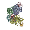
  • Complex: Respiratory complex I from Escherichia coli - focused refinement of cytoplasmic arm
    • Protein or peptide: x 6 types
  • Ligand: x 5 types
Function / homology
Function and homology information


NADH dehydrogenase complex / plasma membrane respiratory chain complex I / Translocases; Catalysing the translocation of protons; Linked to oxidoreductase reactions / NADH:ubiquinone reductase (non-electrogenic) activity / molybdopterin cofactor binding / cellular respiration / respiratory chain complex I / NADH dehydrogenase activity / NADH dehydrogenase (ubiquinone) activity / quinone binding ...NADH dehydrogenase complex / plasma membrane respiratory chain complex I / Translocases; Catalysing the translocation of protons; Linked to oxidoreductase reactions / NADH:ubiquinone reductase (non-electrogenic) activity / molybdopterin cofactor binding / cellular respiration / respiratory chain complex I / NADH dehydrogenase activity / NADH dehydrogenase (ubiquinone) activity / quinone binding / ATP synthesis coupled electron transport / aerobic respiration / respiratory electron transport chain / transmembrane transport / 2 iron, 2 sulfur cluster binding / NAD binding / FMN binding / 4 iron, 4 sulfur cluster binding / iron ion binding / membrane / metal ion binding / plasma membrane / cytoplasm
Similarity search - Function
NADH dehydrogenase, subunit CD / Molybdopterin oxidoreductase Fe4S4 domain / Molybdopterin oxidoreductase Fe4S4 domain / Molybdopterin dinucleotide-binding domain / Molydopterin dinucleotide binding domain / NADH-quinone oxidoreductase, chain I / NAD(P)H-quinone oxidoreductase subunit D/H / NADH:ubiquinone oxidoreductase, 49kDa subunit, conserved site / Respiratory chain NADH dehydrogenase 49 Kd subunit signature. / NADH-quinone oxidoreductase, subunit D ...NADH dehydrogenase, subunit CD / Molybdopterin oxidoreductase Fe4S4 domain / Molybdopterin oxidoreductase Fe4S4 domain / Molybdopterin dinucleotide-binding domain / Molydopterin dinucleotide binding domain / NADH-quinone oxidoreductase, chain I / NAD(P)H-quinone oxidoreductase subunit D/H / NADH:ubiquinone oxidoreductase, 49kDa subunit, conserved site / Respiratory chain NADH dehydrogenase 49 Kd subunit signature. / NADH-quinone oxidoreductase, subunit D / Respiratory-chain NADH dehydrogenase, 49 Kd subunit / NADH:ubiquinone oxidoreductase, subunit G / Respiratory-chain NADH dehydrogenase 75 Kd subunit signature 3. / Respiratory-chain NADH dehydrogenase 75 Kd subunit signature 2. / NADH dehydrogenase, subunit C / NADH ubiquinone oxidoreductase, F subunit / NADH:ubiquinone oxidoreductase, 30kDa subunit, conserved site / Respiratory chain NADH dehydrogenase 30 Kd subunit signature. / 2Fe-2S iron-sulfur cluster binding domain / Respiratory-chain NADH dehydrogenase 75 Kd subunit signature 1. / NADH:ubiquinone oxidoreductase, 75kDa subunit, conserved site / NADH:ubiquinone oxidoreductase, 30kDa subunit / NADH:ubiquinone oxidoreductase, 30kDa subunit superfamily / Respiratory-chain NADH dehydrogenase, 30 Kd subunit / NADH-ubiquinone oxidoreductase-G iron-sulfur binding region / NADH-ubiquinone oxidoreductase-G iron-sulfur binding region / Respiratory-chain NADH dehydrogenase 51 Kd subunit signature 1. / Respiratory-chain NADH dehydrogenase 24 Kd subunit signature. / Aspartate decarboxylase-like domain superfamily / NuoE domain / NADH-quinone oxidoreductase subunit E-like / Thioredoxin-like [2Fe-2S] ferredoxin / NADH-quinone oxidoreductase subunit E, N-terminal / NADH:ubiquinone oxidoreductase, subunit G, iron-sulphur binding / His(Cys)3-ligated-type [4Fe-4S] domain profile. / NADH:ubiquinone oxidoreductase, 51kDa subunit, conserved site / Respiratory-chain NADH dehydrogenase 51 Kd subunit signature 2. / NADH-ubiquinone oxidoreductase 51kDa subunit, FMN-binding domain / NADH-ubiquinone oxidoreductase 51kDa subunit, iron-sulphur binding domain / NADH-ubiquinone oxidoreductase 51kDa subunit, iron-sulphur binding domain superfamily / NADH-ubiquinone oxidoreductase 51kDa subunit, FMN-binding domain superfamily / Respiratory-chain NADH dehydrogenase 51 Kd subunit / NADH-ubiquinone oxidoreductase-F iron-sulfur binding region / NADH-ubiquinone oxidoreductase-F iron-sulfur binding region / Molybdopterin oxidoreductase, 4Fe-4S domain / Prokaryotic molybdopterin oxidoreductases 4Fe-4S domain profile. / Molybdopterin oxidoreductase / Molybdopterin oxidoreductase / [NiFe]-hydrogenase, large subunit / 4Fe-4S dicluster domain / 2Fe-2S ferredoxin-type iron-sulfur binding domain profile. / 2Fe-2S ferredoxin-type iron-sulfur binding domain / 2Fe-2S ferredoxin-like superfamily / 4Fe-4S ferredoxin, iron-sulphur binding, conserved site / 4Fe-4S ferredoxin-type iron-sulfur binding region signature. / 4Fe-4S ferredoxin-type iron-sulfur binding domain profile. / 4Fe-4S ferredoxin-type, iron-sulphur binding domain / Thioredoxin-like superfamily
Similarity search - Domain/homology
NADH-quinone oxidoreductase subunit E / NADH-quinone oxidoreductase subunit I / NADH-quinone oxidoreductase subunit F / NADH-quinone oxidoreductase subunit C/D / NADH-quinone oxidoreductase subunit G
Similarity search - Component
Biological speciesEscherichia coli B (bacteria)
Methodsingle particle reconstruction / cryo EM / Resolution: 2.1 Å
AuthorsKolata P / Efremov RG
Funding support Belgium, 1 items
OrganizationGrant numberCountry
European Research Council (ERC)00281ROEF Belgium
CitationJournal: Elife / Year: 2021
Title: Structure of respiratory complex I reconstituted into lipid nanodiscs reveals an uncoupled conformation.
Authors: Piotr Kolata / Rouslan G Efremov /
Abstract: Respiratory complex I is a multi-subunit membrane protein complex that reversibly couples NADH oxidation and ubiquinone reduction with proton translocation against transmembrane potential. Complex I ...Respiratory complex I is a multi-subunit membrane protein complex that reversibly couples NADH oxidation and ubiquinone reduction with proton translocation against transmembrane potential. Complex I from is among the best functionally characterized complexes, but its structure remains unknown, hindering further studies to understand the enzyme coupling mechanism. Here, we describe the single particle cryo-electron microscopy (cryo-EM) structure of the entire catalytically active complex I reconstituted into lipid nanodiscs. The structure of this mesophilic bacterial complex I displays highly dynamic connection between the peripheral and membrane domains. The peripheral domain assembly is stabilized by unique terminal extensions and an insertion loop. The membrane domain structure reveals novel dynamic features. Unusual conformation of the conserved interface between the peripheral and membrane domains suggests an uncoupled conformation of the complex. Considering constraints imposed by the structural data, we suggest a new simple hypothetical coupling mechanism for the molecular machine.
History
DepositionMar 23, 2021-
Header (metadata) releaseAug 18, 2021-
Map releaseAug 18, 2021-
UpdateAug 18, 2021-
Current statusAug 18, 2021Processing site: PDBe / Status: Released

-
Structure visualization

Movie
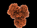
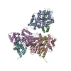
  • Surface view with section colored by density value
  • Surface level: 2.09
  • Imaged by UCSF Chimera
  • Download
  • Surface view colored by radius
  • Surface level: 2.09
  • Imaged by UCSF Chimera
  • Download
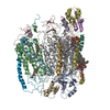
  • Surface view with fitted model
  • Atomic models: PDB-7nz1
  • Surface level: 2.09
  • Imaged by UCSF Chimera
  • Download
Movie viewer
Structure viewerEM map:
SurfViewMolmilJmol/JSmol
Supplemental images

Downloads & links

-
Map

FileDownload / File: emd_12661.map.gz / Format: CCP4 / Size: 21 MB / Type: IMAGE STORED AS FLOATING POINT NUMBER (4 BYTES)
Voxel sizeX=Y=Z: 0.7712 Å
Density
Contour LevelBy AUTHOR: 2.09 / Movie #1: 2.09
Minimum - Maximum-25.512682 - 32.797268
Average (Standard dev.)7.3893955e-12 (±1.0)
SymmetrySpace group: 1
Details

EMDB XML:

Map geometry
Axis orderXYZ
Origin000
Dimensions201167164
Spacing167201164
CellA: 128.7904 Å / B: 155.0112 Å / C: 126.4768 Å
α=β=γ: 90.0 °

CCP4 map header:

modeImage stored as Reals
Å/pix. X/Y/Z0.771197604790420.771199004975120.7712012195122
M x/y/z167201164
origin x/y/z0.0000.0000.000
length x/y/z128.790155.011126.477
α/β/γ90.00090.00090.000
start NX/NY/NZ000
NX/NY/NZ540540540
MAP C/R/S123
start NC/NR/NS000
NC/NR/NS167201164
D min/max/mean-25.51332.7970.000

-
Supplemental data

-
Half map: #2

Fileemd_12661_half_map_1.map
Projections & Slices
AxesZYX

Projections

Slices (1/2)
Density Histograms

-
Half map: #1

Fileemd_12661_half_map_2.map
Projections & Slices
AxesZYX

Projections

Slices (1/2)
Density Histograms

-
Sample components

+
Entire : Respiratory complex I from Escherichia coli - focused refinement ...

EntireName: Respiratory complex I from Escherichia coli - focused refinement of cytoplasmic arm
Components
  • Complex: Respiratory complex I from Escherichia coli - focused refinement of cytoplasmic arm
    • Protein or peptide: NADH-quinone oxidoreductase subunit BNADH dehydrogenase (quinone)
    • Protein or peptide: NADH-quinone oxidoreductase subunit C/DNADH dehydrogenase (quinone)
    • Protein or peptide: NADH-quinone oxidoreductase subunit ENADH dehydrogenase (quinone)
    • Protein or peptide: NADH-quinone oxidoreductase subunit FNADH dehydrogenase (quinone)
    • Protein or peptide: NADH-quinone oxidoreductase subunit GNADH dehydrogenase (quinone)
    • Protein or peptide: NADH-quinone oxidoreductase subunit INADH dehydrogenase (quinone)
  • Ligand: IRON/SULFUR CLUSTERIron–sulfur cluster
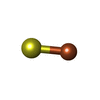
  • Ligand: FE2/S2 (INORGANIC) CLUSTER
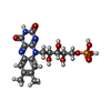
  • Ligand: FLAVIN MONONUCLEOTIDE
  • Ligand: CALCIUM IONCalcium
  • Ligand: water

+
Supramolecule #1: Respiratory complex I from Escherichia coli - focused refinement ...

SupramoleculeName: Respiratory complex I from Escherichia coli - focused refinement of cytoplasmic arm
type: complex / ID: 1 / Parent: 0 / Macromolecule list: #1-#6
Source (natural)Organism: Escherichia coli B (bacteria) / Strain: BL21(AI) / Location in cell: cell membrane
Molecular weightTheoretical: 280 KDa

+
Macromolecule #1: NADH-quinone oxidoreductase subunit B

MacromoleculeName: NADH-quinone oxidoreductase subunit B / type: protein_or_peptide / ID: 1 / Number of copies: 1 / Enantiomer: LEVO / EC number: NADH:ubiquinone reductase (H+-translocating)
Source (natural)Organism: Escherichia coli B (bacteria)
Molecular weightTheoretical: 25.097809 KDa
SequenceString: MDYTLTRIDP NGENDRYPLQ KQEIVTDPLE QEVNKNVFMG KLNDMVNWGR KNSIWPYNFG LSCCYVEMVT SFTAVHDVAR FGAEVLRAS PRQADLMVVA GT(CSX)FTKMAPV IQRLYDQMLE PKWVISMGAC ANSGGMYDIY SVVQGVDKFI PVDVYIP GC PPRPEAYMQA ...String:
MDYTLTRIDP NGENDRYPLQ KQEIVTDPLE QEVNKNVFMG KLNDMVNWGR KNSIWPYNFG LSCCYVEMVT SFTAVHDVAR FGAEVLRAS PRQADLMVVA GT(CSX)FTKMAPV IQRLYDQMLE PKWVISMGAC ANSGGMYDIY SVVQGVDKFI PVDVYIP GC PPRPEAYMQA LMLLQESIGK ERRPLSWVVG DQGVYRANMQ SERERKRGER IAVTNLRTPD EI

+
Macromolecule #2: NADH-quinone oxidoreductase subunit C/D

MacromoleculeName: NADH-quinone oxidoreductase subunit C/D / type: protein_or_peptide / ID: 2 / Number of copies: 1 / Enantiomer: LEVO / EC number: NADH:ubiquinone reductase (H+-translocating)
Source (natural)Organism: Escherichia coli B (bacteria) / Strain: BL21(AI)
Molecular weightTheoretical: 68.321945 KDa
SequenceString: MTDLTAQEPA WQTRDHLDDP VIGELRNRFG PDAFTVQATR TGVPVVWIKR EQLLEVGDFL KKLPKPYVML FDLHGMDERL RTHREGLPA ADFSVFYHLI SIDRNRDIML KVALAENDLH VPTFTKLFPN ANWYERETWD LFGITFDGHP NLRRIMMPQT W KGHPLRKD ...String:
MTDLTAQEPA WQTRDHLDDP VIGELRNRFG PDAFTVQATR TGVPVVWIKR EQLLEVGDFL KKLPKPYVML FDLHGMDERL RTHREGLPA ADFSVFYHLI SIDRNRDIML KVALAENDLH VPTFTKLFPN ANWYERETWD LFGITFDGHP NLRRIMMPQT W KGHPLRKD YPARATEFSP FELTKAKQDL EMEALTFKPE EWGMKRGTEN EDFMFLNLGP NHPSAHGAFR IVLQLDGEEI VD CVPDIGY HHRGAEKMGE RQSWHSYIPY TDRIEYLGGC VNEMPYVLAV EKLAGITVPD RVNVIRVMLS ELFRINSHLL YIS TFIQDV GAMTPVFFAF TDRQKIYDLV EAITGFRMHP AWFRIGGVAH DLPRGWDRLL REFLDWMPKR LASYEKAALQ NTIL KGRSQ GVAAYGAKEA LEWGTTGAGL RATGIDFDVR KARPYSGYEN FDFEIPVGGG VSDCYTRVML KVEELRQSLR ILEQC LNNM PEGPFKADHP LTTPPPKERT LQHIETLITH FLQVSWGPVM PANESFQMIE ATKGINSYYL TSDGSTMSYR TRVRTP SFA HLQQIPAAIR GSLVSDLIVY LGSIDFVMSD VDR

+
Macromolecule #3: NADH-quinone oxidoreductase subunit E

MacromoleculeName: NADH-quinone oxidoreductase subunit E / type: protein_or_peptide / ID: 3 / Number of copies: 1 / Enantiomer: LEVO / EC number: NADH:ubiquinone reductase (H+-translocating)
Source (natural)Organism: Escherichia coli B (bacteria)
Molecular weightTheoretical: 18.630049 KDa
SequenceString:
MHENQQPQTE AFELSAAERE AIEHEMHHYE DPRAASIEAL KIVQKQRGWV PDGAIHAIAD VLGIPASDVE GVATFYSQIF RQPVGRHVI RYCDSVVCHI NGYQGIQAAL EKKLNIKPGQ TTFDGRFTLL PT(CSX)CLGNCDK GPNMMIDEDT HAHLTPE AI PELLERYK

+
Macromolecule #4: NADH-quinone oxidoreductase subunit F

MacromoleculeName: NADH-quinone oxidoreductase subunit F / type: protein_or_peptide / ID: 4 / Number of copies: 1 / Enantiomer: LEVO / EC number: NADH:ubiquinone reductase (H+-translocating)
Source (natural)Organism: Escherichia coli B (bacteria)
Molecular weightTheoretical: 49.368332 KDa
SequenceString: MKNIIRTPET HPLTWRLRDD KQPVWLDEYR SKNGYEGARK ALTGLSPDEI VNQVKDAGLK GRGGAGFSTG LKWSLMPKDE SMNIRYLLC NADEMEPGTY KDRLLMEQLP HLLVEGMLIS AFALKAYRGY IFLRGEYIEA AVNLRRAIAE ATEAGLLGKN I MGTGFDFE ...String:
MKNIIRTPET HPLTWRLRDD KQPVWLDEYR SKNGYEGARK ALTGLSPDEI VNQVKDAGLK GRGGAGFSTG LKWSLMPKDE SMNIRYLLC NADEMEPGTY KDRLLMEQLP HLLVEGMLIS AFALKAYRGY IFLRGEYIEA AVNLRRAIAE ATEAGLLGKN I MGTGFDFE LFVHTGAGRY ICGEETALIN SLEGRRANPR SKPPFPATSG AWGKPT(CSX)VNN VETLCNVPAI LANGVEWY Q NISKSKDAGT KLMGFSGRVK NPGLWELPFG TTAREILEDY AGGMRDGLKF KAWQPGGAGT DFLTEAHLDL PMEFESIGK AGSRLGTALA MAVDHEINMV SLVRNLEEFF ARESCGWCTP CRDGLPWSVK ILRALERGEG QPGDIETLEQ LCRFLGPGKT FCAHAPGAV EPLQSAIKYF REEFEAGIKQ PFSNTHLING IQPNLLKERW

+
Macromolecule #5: NADH-quinone oxidoreductase subunit G

MacromoleculeName: NADH-quinone oxidoreductase subunit G / type: protein_or_peptide / ID: 5 / Number of copies: 1 / Enantiomer: LEVO / EC number: NADH:ubiquinone reductase (H+-translocating)
Source (natural)Organism: Escherichia coli B (bacteria)
Molecular weightTheoretical: 100.419211 KDa
SequenceString: MATIHVDGKE YEVNGADNLL EACLSLGLDI PYFCWHPALG SVGACRQCAV KQYQNAEDTR GRLVMSCMTP ASDGTFISID DEEAKQFRE SVVEWLMTNH PHDCPVCEEG GNCHLQDMTV MTGHSFRRYR FTKRTHRNQD LGPFISHEMN RCIACYRCVR Y YKDYADGT ...String:
MATIHVDGKE YEVNGADNLL EACLSLGLDI PYFCWHPALG SVGACRQCAV KQYQNAEDTR GRLVMSCMTP ASDGTFISID DEEAKQFRE SVVEWLMTNH PHDCPVCEEG GNCHLQDMTV MTGHSFRRYR FTKRTHRNQD LGPFISHEMN RCIACYRCVR Y YKDYADGT DLGVYGAHDN VYFGRPEDGT LESEFSGNLV EICPTGVFTD KTHSERYNRK WDMQFAPSIC QQCSIGCNIS PG ERYGELR RIENRYNGTV NHYFLCDRGR FGYGYVNLKD RPRQPVQRRG DDFITLNAEQ AMQGAADILR QSKKVIGIGS PRA SVESNF ALRELVGEEN FYTGIAHGEQ ERLQLALKVL REGGIYTPAL REIESYDAVL VLGEDVTQTG ARVALAVRQA VKGK AREMA AAQKVADWQI AAILNIGQRA KHPLFVTNVD DTRLDDIAAW TYRAPVEDQA RLGFAIAHAL DNSAPAVDGI EPELQ SKID VIVQALAGAK KPLIISGTNA GSLEVIQAAA NVAKALKGRG ADVGITMIAR SVNSMGLGIM GGGSLEEALT ELETGR ADA VVVLENDLHR HASAIRVNAA LAKAPLVMVV DHQRTAIMEN AHLVLSAASF AESDGTVINN EGRAQRFFQV YDPAYYD SK TVMLESWRWL HSLHSTLLSR EVDWTQLDHV IDAVVAKIPE LAGIKDAAPD ATFRIRGQKL AREPHRYSGR TAMRANIS V HEPRQPQDID TMFTFSMEGN NQPTAHRSQV PFAWAPGWNS PQAWNKFQDE VGGKLRFGDP GVRLFETSEN GLDYFTSVP ARFQPQDGKW RIAPYYHLFG SDELSQRAPV FQSRMPQPYI KLNPADAAKL GVNAGTRVSF SYDGNTVTLP VEIAEGLTAG QVGLPMGMS GIAPVLAGAH LEDLKEAQQ

+
Macromolecule #6: NADH-quinone oxidoreductase subunit I

MacromoleculeName: NADH-quinone oxidoreductase subunit I / type: protein_or_peptide / ID: 6 / Number of copies: 1 / Enantiomer: LEVO / EC number: NADH:ubiquinone reductase (H+-translocating)
Source (natural)Organism: Escherichia coli B (bacteria)
Molecular weightTheoretical: 20.562771 KDa
SequenceString:
MTLKELLVGF GTQVRSIWMI GLHAFAKRET RMYPEEPVYL PPRYRGRIVL TRDPDGEERC VACNLCAVAC PVGCISLQKA ETKDGRWYP EFFRINFSRC IFCGLCEEAC PTTAIQLTPD FEMGEYKRQD LVYEKEDLLI SGPGKYPEYN FYRMAGMAID G KDKGEAEN EAKPIDVKSL LP

+
Macromolecule #7: IRON/SULFUR CLUSTER

MacromoleculeName: IRON/SULFUR CLUSTER / type: ligand / ID: 7 / Number of copies: 7 / Formula: SF4
Molecular weightTheoretical: 351.64 Da
Chemical component information

ChemComp-FS1:
IRON/SULFUR CLUSTER / Iron–sulfur cluster

+
Macromolecule #8: FE2/S2 (INORGANIC) CLUSTER

MacromoleculeName: FE2/S2 (INORGANIC) CLUSTER / type: ligand / ID: 8 / Number of copies: 2 / Formula: FES
Molecular weightTheoretical: 175.82 Da
Chemical component information

ChemComp-FES:
FE2/S2 (INORGANIC) CLUSTER / Iron–sulfur cluster

+
Macromolecule #9: FLAVIN MONONUCLEOTIDE

MacromoleculeName: FLAVIN MONONUCLEOTIDE / type: ligand / ID: 9 / Number of copies: 1 / Formula: FMN
Molecular weightTheoretical: 456.344 Da
Chemical component information

ChemComp-FMN:
FLAVIN MONONUCLEOTIDE / Flavin mononucleotide

+
Macromolecule #10: CALCIUM ION

MacromoleculeName: CALCIUM ION / type: ligand / ID: 10 / Number of copies: 1 / Formula: CA
Molecular weightTheoretical: 40.078 Da

+
Macromolecule #11: water

MacromoleculeName: water / type: ligand / ID: 11 / Number of copies: 1170 / Formula: HOH
Molecular weightTheoretical: 18.015 Da
Chemical component information

ChemComp-HOH:
WATER / Water

-
Experimental details

-
Structure determination

Methodcryo EM
Processingsingle particle reconstruction
Aggregation stateparticle

-
Sample preparation

Concentration0.1 mg/mL
BufferpH: 6.8
Component:
ConcentrationFormulaName
20.0 mMC8H19NO5Bis-TrisBis-tris methane
200.0 mMNaClSodium chlorideSodium chloride
2.0 mMCaCl2Calcium chloride

Details: The buffer was used for gel filtration of protein reconstituted in lipid nanodiscs
GridModel: Quantifoil R0.6/1 / Material: COPPER / Mesh: 300 / Support film - Material: CARBON / Support film - topology: HOLEY ARRAY / Pretreatment - Type: GLOW DISCHARGE / Pretreatment - Atmosphere: AIR / Pretreatment - Pressure: 0.028 kPa
VitrificationCryogen name: ETHANE / Chamber humidity: 97 % / Chamber temperature: 296 K / Instrument: GATAN CRYOPLUNGE 3

-
Electron microscopy

MicroscopeJEOL CRYO ARM 300
Electron beamAcceleration voltage: 300 kV / Electron source: FIELD EMISSION GUN
Electron opticsIllumination mode: FLOOD BEAM / Imaging mode: BRIGHT FIELDBright-field microscopy / Cs: 2.55 mm / Nominal defocus max: 2.2 µm / Nominal defocus min: 0.9 µm / Nominal magnification: 60000
Specialist opticsEnergy filter - Name: In-column Omega Filter / Energy filter - Slit width: 20 eV
Sample stageSpecimen holder model: JEOL CRYOSPECPORTER / Cooling holder cryogen: NITROGEN
Image recordingFilm or detector model: GATAN K3 (6k x 4k) / Number real images: 9122 / Average exposure time: 3.0 sec. / Average electron dose: 64.7 e/Å2

-
Image processing

Particle selectionNumber selected: 1256734
CTF correctionSoftware - Name: CTFFIND (ver. 4.1)
Initial angle assignmentType: MAXIMUM LIKELIHOOD / Software - Name: RELION (ver. 3.0)
Final 3D classificationSoftware - Name: RELION (ver. 3.1)
Final angle assignmentType: MAXIMUM LIKELIHOOD / Software - Name: RELION (ver. 3.1)
Final reconstructionApplied symmetry - Point group: C1 (asymmetric) / Resolution.type: BY AUTHOR / Resolution: 2.1 Å / Resolution method: FSC 0.143 CUT-OFF / Software: (Name: RELION (ver. 3.1), PHENIX (ver. 1.18.2)) / Number images used: 286333
FSC plot (resolution estimation)

-
Atomic model buiding 1

Initial modelPDB ID:
RefinementSpace: REAL / Protocol: AB INITIO MODEL / Overall B value: 30 / Target criteria: Correlation coefficient
Output model

PDB-7nz1:
Respiratory complex I from Escherichia coli - focused refinement of cytoplasmic arm

+
About Yorodumi

-
News

-
Feb 9, 2022. New format data for meta-information of EMDB entries

New format data for meta-information of EMDB entries

  • Version 3 of the EMDB header file is now the official format.
  • The previous official version 1.9 will be removed from the archive.

Related info.:EMDB header

External links:wwPDB to switch to version 3 of the EMDB data model

-
Aug 12, 2020. Covid-19 info

Covid-19 info

URL: https://pdbj.org/emnavi/covid19.php

New page: Covid-19 featured information page in EM Navigator.

Related info.:Covid-19 info / Mar 5, 2020. Novel coronavirus structure data

+
Mar 5, 2020. Novel coronavirus structure data

Novel coronavirus structure data

Related info.:Yorodumi Speices / Aug 12, 2020. Covid-19 info

External links:COVID-19 featured content - PDBj / Molecule of the Month (242):Coronavirus Proteases

+
Jan 31, 2019. EMDB accession codes are about to change! (news from PDBe EMDB page)

EMDB accession codes are about to change! (news from PDBe EMDB page)

  • The allocation of 4 digits for EMDB accession codes will soon come to an end. Whilst these codes will remain in use, new EMDB accession codes will include an additional digit and will expand incrementally as the available range of codes is exhausted. The current 4-digit format prefixed with “EMD-” (i.e. EMD-XXXX) will advance to a 5-digit format (i.e. EMD-XXXXX), and so on. It is currently estimated that the 4-digit codes will be depleted around Spring 2019, at which point the 5-digit format will come into force.
  • The EM Navigator/Yorodumi systems omit the EMD- prefix.

Related info.:Q: What is EMD? / ID/Accession-code notation in Yorodumi/EM Navigator

External links:EMDB Accession Codes are Changing Soon! / Contact to PDBj

+
Jul 12, 2017. Major update of PDB

Major update of PDB

  • wwPDB released updated PDB data conforming to the new PDBx/mmCIF dictionary.
  • This is a major update changing the version number from 4 to 5, and with Remediation, in which all the entries are updated.
  • In this update, many items about electron microscopy experimental information are reorganized (e.g. em_software).
  • Now, EM Navigator and Yorodumi are based on the updated data.

External links:wwPDB Remediation / Enriched Model Files Conforming to OneDep Data Standards Now Available in the PDB FTP Archive

-
Yorodumi

Thousand views of thousand structures

  • Yorodumi is a browser for structure data from EMDB, PDB, SASBDB, etc.
  • This page is also the successor to EM Navigator detail page, and also detail information page/front-end page for Omokage search.
  • The word "yorodu" (or yorozu) is an old Japanese word meaning "ten thousand". "mi" (miru) is to see.

Related info.:EMDB / PDB / SASBDB / Comparison of 3 databanks / Yorodumi Search / Aug 31, 2016. New EM Navigator & Yorodumi / Yorodumi Papers / Jmol/JSmol / Function and homology information / Changes in new EM Navigator and Yorodumi

Read more