 Movie
Movie Controller

[English] 日本語
 Yorodumi
- PDB-7nz1: Respiratory complex I from Escherichia coli - focused refinement ... -
+ Open data

- Basic information
| Entry | Database: PDB / ID: 7nz1 | ||||||||||||||||||||||||
|---|---|---|---|---|---|---|---|---|---|---|---|---|---|---|---|---|---|---|---|---|---|---|---|---|---|
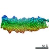
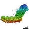
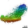
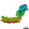
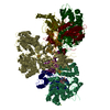
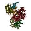
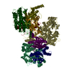
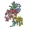
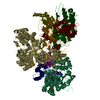
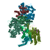
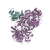
| Title | Respiratory complex I from Escherichia coli - focused refinement of cytoplasmic arm | ||||||||||||||||||||||||
 Components Components | (NADH-quinone oxidoreductase subunit ...) x 6 | ||||||||||||||||||||||||
 Keywords Keywords | ELECTRON TRANSPORT / NADH:ubiquinone reductase (H+-translocating) / oxidative phosphorylation | ||||||||||||||||||||||||
| Function / homology |  Function and homology information Function and homology informationNADH dehydrogenase (quinone) (non-electrogenic) activity / Translocases; Catalysing the translocation of protons; Linked to oxidoreductase reactions / NADH dehydrogenase complex / molybdopterin cofactor binding / cellular respiration / oxidoreductase activity, acting on NAD(P)H / respiratory chain complex I / NADH dehydrogenase (ubiquinone) activity / quinone binding / ATP synthesis coupled electron transport ...NADH dehydrogenase (quinone) (non-electrogenic) activity / Translocases; Catalysing the translocation of protons; Linked to oxidoreductase reactions / NADH dehydrogenase complex / molybdopterin cofactor binding / cellular respiration / oxidoreductase activity, acting on NAD(P)H / respiratory chain complex I / NADH dehydrogenase (ubiquinone) activity / quinone binding / ATP synthesis coupled electron transport / proton transmembrane transport / aerobic respiration / respiratory electron transport chain / 2 iron, 2 sulfur cluster binding / NAD binding / FMN binding / 4 iron, 4 sulfur cluster binding / oxidoreductase activity / iron ion binding / membrane / metal ion binding / plasma membrane / cytoplasm Similarity search - Function | ||||||||||||||||||||||||
| Biological species |  | ||||||||||||||||||||||||
| Method | ELECTRON MICROSCOPY / single particle reconstruction / cryo EM / Resolution: 2.1 Å | ||||||||||||||||||||||||
 Authors Authors | Kolata, P. / Efremov, R.G. | ||||||||||||||||||||||||
| Funding support |  Belgium, 1items Belgium, 1items
| ||||||||||||||||||||||||
 Citation Citation | Journal: Elife / Year: 2021 Title: Structure of respiratory complex I reconstituted into lipid nanodiscs reveals an uncoupled conformation. Authors: Piotr Kolata / Rouslan G Efremov / Abstract: Respiratory complex I is a multi-subunit membrane protein complex that reversibly couples NADH oxidation and ubiquinone reduction with proton translocation against transmembrane potential. Complex I ...Respiratory complex I is a multi-subunit membrane protein complex that reversibly couples NADH oxidation and ubiquinone reduction with proton translocation against transmembrane potential. Complex I from is among the best functionally characterized complexes, but its structure remains unknown, hindering further studies to understand the enzyme coupling mechanism. Here, we describe the single particle cryo-electron microscopy (cryo-EM) structure of the entire catalytically active complex I reconstituted into lipid nanodiscs. The structure of this mesophilic bacterial complex I displays highly dynamic connection between the peripheral and membrane domains. The peripheral domain assembly is stabilized by unique terminal extensions and an insertion loop. The membrane domain structure reveals novel dynamic features. Unusual conformation of the conserved interface between the peripheral and membrane domains suggests an uncoupled conformation of the complex. Considering constraints imposed by the structural data, we suggest a new simple hypothetical coupling mechanism for the molecular machine. | ||||||||||||||||||||||||
| History |
|
- Structure visualization
| Movie |
Movie viewer |
|---|---|
| Structure viewer | Molecule: MolmilJmol/JSmol |
- Downloads & links
-Download
| PDBx/mmCIF format | 7nz1.cif.gz | 469.5 KB | Display | PDBx/mmCIF format |
|---|---|---|---|---|
| PDB format | pdb7nz1.ent.gz | 358.9 KB | Display | PDB format |
| PDBx/mmJSON format | 7nz1.json.gz | Tree view | PDBx/mmJSON format | |
| Others |  Other downloads Other downloads |
-Validation report
| Arichive directory | https://data.pdbj.org/pub/pdb/validation_reports/nz/7nz1ftp://data.pdbj.org/pub/pdb/validation_reports/nz/7nz1 | HTTPS FTP |
|---|
-Related structure data
| Related structure data |  12661MC  7nyhC  7nyrC 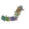 7nyuC 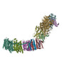 7nyvC C: citing same article ( M: map data used to model this data |
|---|---|
| Similar structure data |


-Links
 PDBj
PDBj


- Assembly
| Deposited unit | 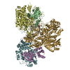
|
|---|---|
| 1 |
|
-Components
-NADH-quinone oxidoreductase subunit ... , 6 types, 6 molecules BDEFGI
| #1: Protein | Mass: 25097.809 Da / Num. of mol.: 1 / Source method: isolated from a natural source / Source: (natural) |
|---|---|
| #2: Protein | Mass: 68321.945 Da / Num. of mol.: 1 / Source method: isolated from a natural source / Source: (natural) References: UniProt: P33599, NADH:ubiquinone reductase (H+-translocating) |
| #3: Protein | Mass: 18630.049 Da / Num. of mol.: 1 / Source method: isolated from a natural source / Source: (natural) References: UniProt: P0AFD1, NADH:ubiquinone reductase (H+-translocating) |
| #4: Protein | Mass: 49368.332 Da / Num. of mol.: 1 / Source method: isolated from a natural source / Source: (natural) References: UniProt: P31979, NADH:ubiquinone reductase (H+-translocating) |
| #5: Protein | Mass: 100419.211 Da / Num. of mol.: 1 / Source method: isolated from a natural source / Source: (natural) References: UniProt: P33602, NADH:ubiquinone reductase (H+-translocating) |
| #6: Protein | Mass: 20562.771 Da / Num. of mol.: 1 / Source method: isolated from a natural source / Source: (natural) References: UniProt: P0AFD6, NADH:ubiquinone reductase (H+-translocating) |
-Non-polymers , 5 types, 1181 molecules 


| #7: Chemical | ChemComp-SF4 /  Mass: 351.640 Da / Num. of mol.: 7 / Source method: obtained synthetically / Formula: Fe4S4 Mass: 351.640 Da / Num. of mol.: 7 / Source method: obtained synthetically / Formula: Fe4S4#8: Chemical |  Mass: 175.820 Da / Num. of mol.: 2 / Source method: obtained synthetically / Formula: Fe2S2 Mass: 175.820 Da / Num. of mol.: 2 / Source method: obtained synthetically / Formula: Fe2S2#9: Chemical | ChemComp-FMN / |  Mass: 456.344 Da / Num. of mol.: 1 / Source method: obtained synthetically / Formula: C17H21N4O9P Mass: 456.344 Da / Num. of mol.: 1 / Source method: obtained synthetically / Formula: C17H21N4O9P#10: Chemical | ChemComp-CA / |  Mass: 40.078 Da / Num. of mol.: 1 / Source method: obtained synthetically / Formula: Ca Mass: 40.078 Da / Num. of mol.: 1 / Source method: obtained synthetically / Formula: Ca#11: Water | ChemComp-HOH / | Mass: 18.015 Da / Num. of mol.: 1170 / Source method: isolated from a natural source / Formula: H2O |
|---|
-Details
| Has ligand of interest | N |
|---|---|
| Has protein modification | Y |
-Experimental details
-Experiment
| Experiment | Method: ELECTRON MICROSCOPY |
|---|---|
| EM experiment | Aggregation state: PARTICLE / 3D reconstruction method: single particle reconstruction |
- Sample preparation
| Component | Name: Respiratory complex I from Escherichia coli - focused refinement of cytoplasmic arm Type: COMPLEX / Entity ID: #1-#6 / Source: NATURAL | ||||||||||||||||||||
|---|---|---|---|---|---|---|---|---|---|---|---|---|---|---|---|---|---|---|---|---|---|
| Molecular weight | Value: 0.28 MDa / Experimental value: NO | ||||||||||||||||||||
| Source (natural) | Organism: | ||||||||||||||||||||
| Buffer solution | pH: 6.8 Details: The buffer was used for gel filtration of protein reconstituted in lipid nanodiscs | ||||||||||||||||||||
| Buffer component |
| ||||||||||||||||||||
| Specimen | Conc.: 0.1 mg/ml / Embedding applied: NO / Shadowing applied: NO / Staining applied: NO / Vitrification applied: YES | ||||||||||||||||||||
| Specimen support | Grid material: COPPER / Grid mesh size: 300 divisions/in. / Grid type: Quantifoil R0.6/1 | ||||||||||||||||||||
| Vitrification | Instrument: GATAN CRYOPLUNGE 3 / Cryogen name: ETHANE / Humidity: 97 % / Chamber temperature: 296 K |
- Electron microscopy imaging
| Microscopy | Model: JEOL CRYO ARM 300 |
|---|---|
| Electron gun | Electron source:  FIELD EMISSION GUN / Accelerating voltage: 300 kV / Illumination mode: FLOOD BEAM FIELD EMISSION GUN / Accelerating voltage: 300 kV / Illumination mode: FLOOD BEAM |
| Electron lens | Mode: BRIGHT FIELD / Nominal magnification: 60000 X / Nominal defocus max: 2200 nm / Nominal defocus min: 900 nm / Cs: 2.55 mm / Alignment procedure: COMA FREE |
| Specimen holder | Cryogen: NITROGEN / Specimen holder model: JEOL CRYOSPECPORTER |
| Image recording | Average exposure time: 3 sec. / Electron dose: 64.7 e/Å2 / Film or detector model: GATAN K3 (6k x 4k) / Num. of real images: 9122 |
| EM imaging optics | Energyfilter name: In-column Omega Filter / Energyfilter slit width: 20 eV |
- Processing
| Software |
| ||||||||||||||||||||||||||||||||||||||||
|---|---|---|---|---|---|---|---|---|---|---|---|---|---|---|---|---|---|---|---|---|---|---|---|---|---|---|---|---|---|---|---|---|---|---|---|---|---|---|---|---|---|
| EM software |
| ||||||||||||||||||||||||||||||||||||||||
| CTF correction | Type: PHASE FLIPPING AND AMPLITUDE CORRECTION | ||||||||||||||||||||||||||||||||||||||||
| Particle selection | Num. of particles selected: 1256734 | ||||||||||||||||||||||||||||||||||||||||
| Symmetry | Point symmetry: C1 (asymmetric) | ||||||||||||||||||||||||||||||||||||||||
| 3D reconstruction | Resolution: 2.1 Å / Resolution method: FSC 0.143 CUT-OFF / Num. of particles: 286333 / Symmetry type: POINT | ||||||||||||||||||||||||||||||||||||||||
| Atomic model building | B value: 30 / Protocol: AB INITIO MODEL / Space: REAL / Target criteria: Correlation coefficient | ||||||||||||||||||||||||||||||||||||||||
| Atomic model building | PDB-ID: 4HEA Accession code: 4HEA / Source name: PDB / Type: experimental model | ||||||||||||||||||||||||||||||||||||||||
| Refinement | Cross valid method: NONE Stereochemistry target values: GeoStd + Monomer Library + CDL v1.2 | ||||||||||||||||||||||||||||||||||||||||
| Displacement parameters | Biso mean: 30.33 Å2 | ||||||||||||||||||||||||||||||||||||||||
| Refine LS restraints |
|

