- EMDB-10224: Cryo-EM Structure of T. kodakarensis 70S ribosome in TkNat10 dele... -
+
Open data
ID or keywords:
Loading...
-
Basic information
Entry
Database: EMDB / ID: EMD-10224
Title
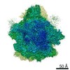
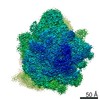
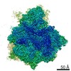
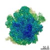
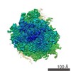
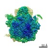
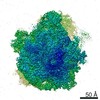
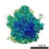
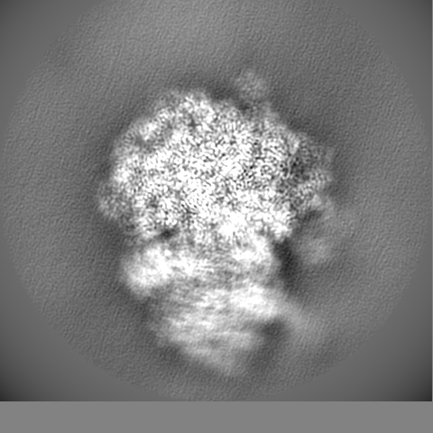
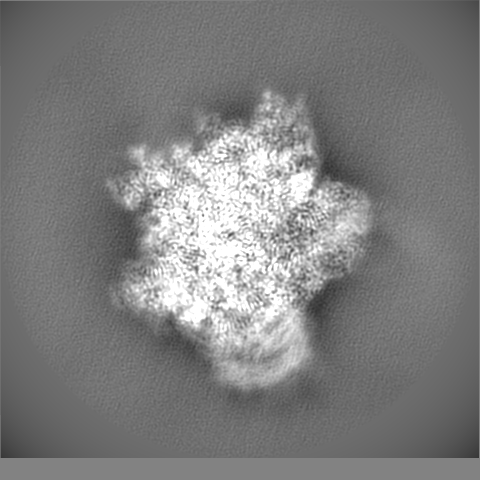
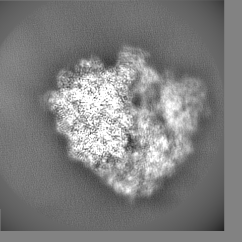
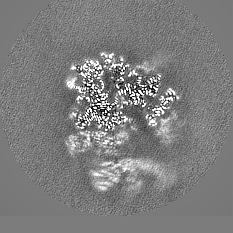
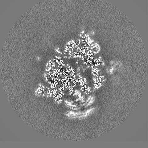
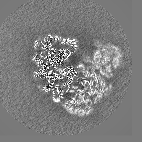
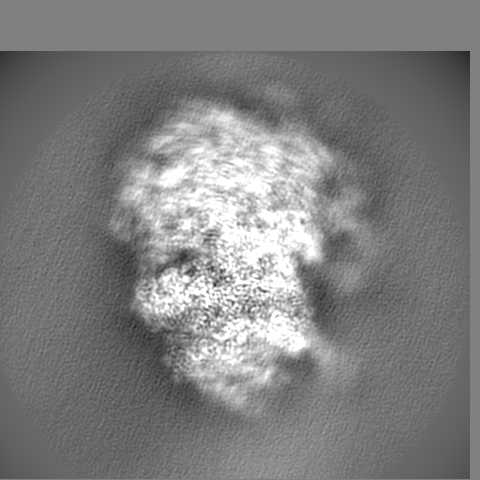
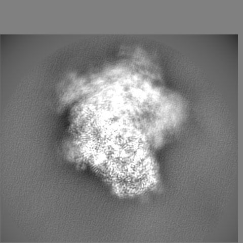
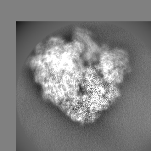
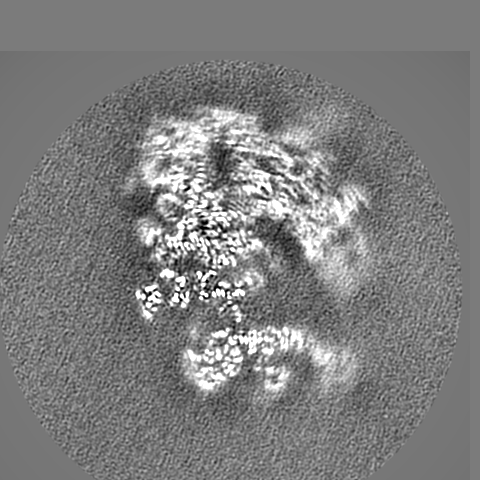
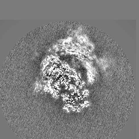
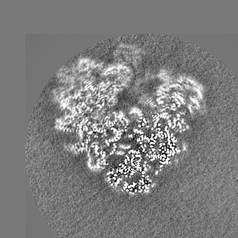
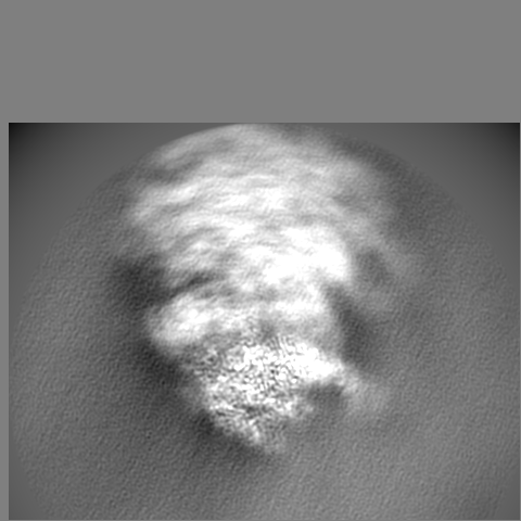
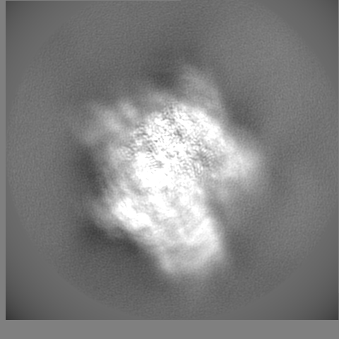
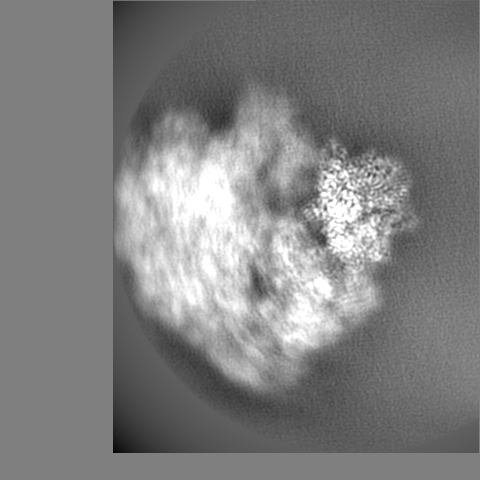
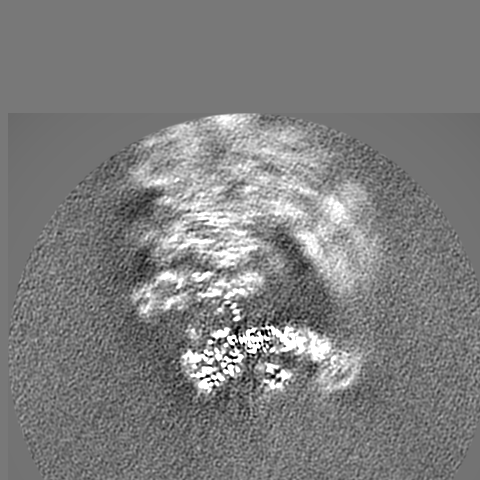
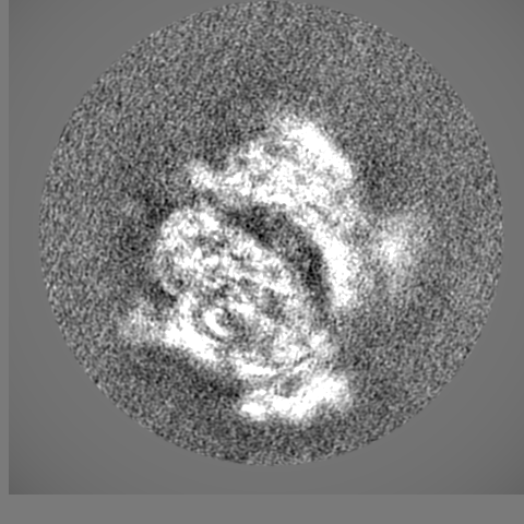
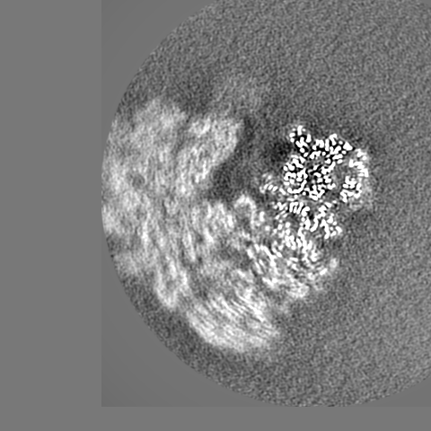
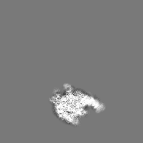
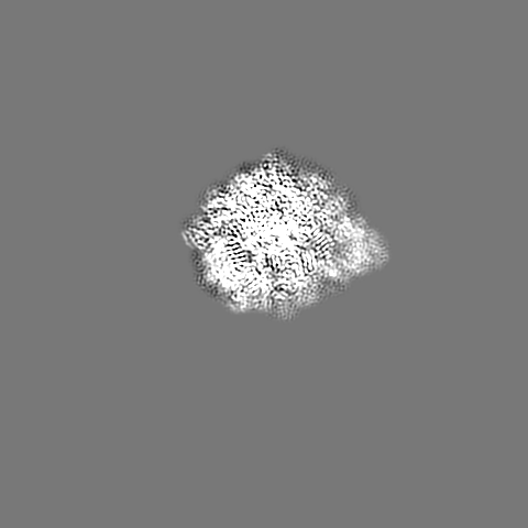
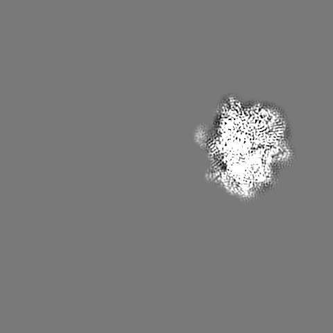
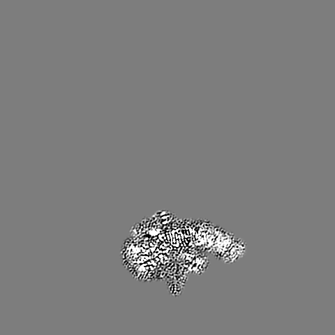
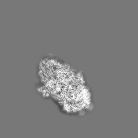
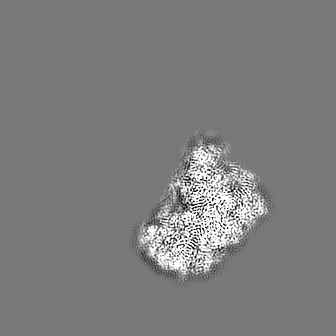
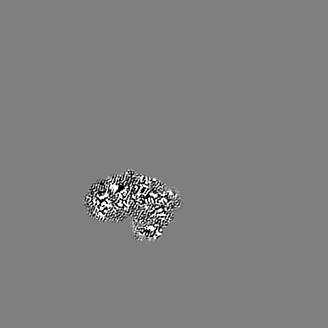
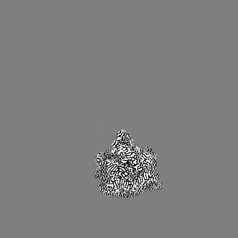
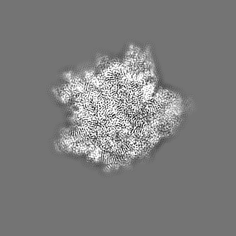
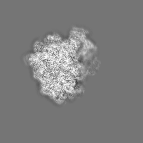
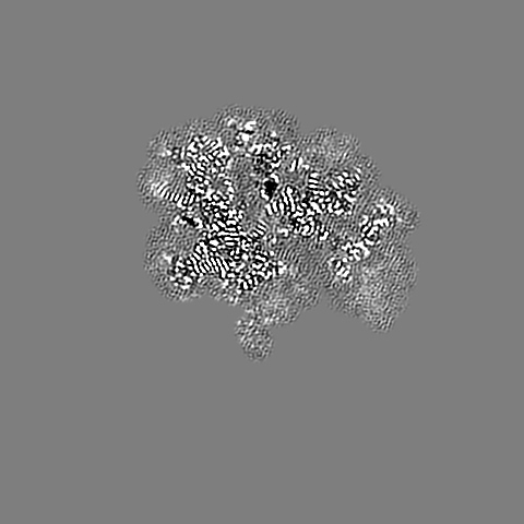
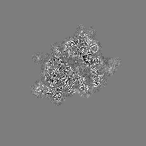
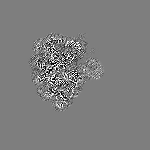
Cryo-EM Structure of T. kodakarensis 70S ribosome in TkNat10 deleted strain
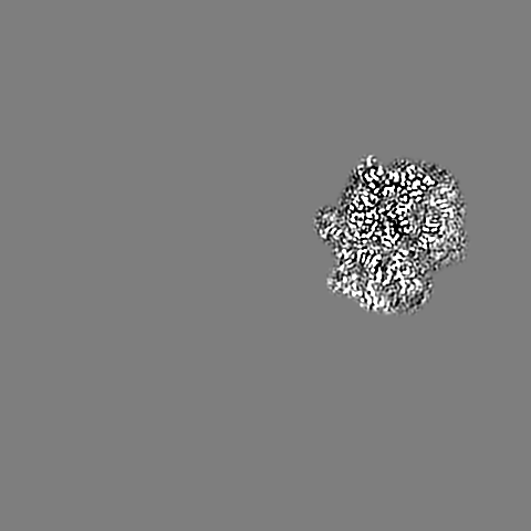
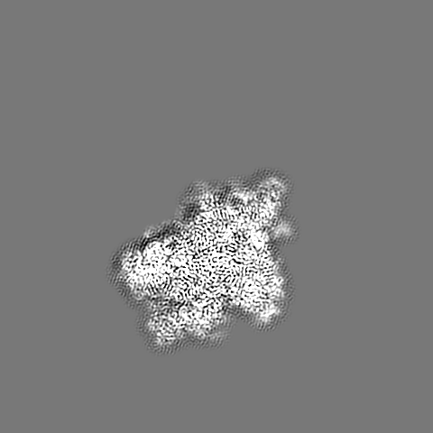
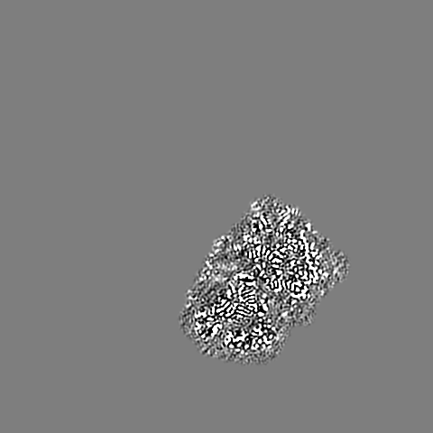
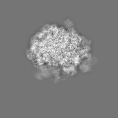
Map data
30S head body postprocess
Sample
Complex: 70S ribosome from Thermococcus kodakarensis
RNA: x 3 types
Protein or peptide: x 62 types
Ligand: x 2 types
Keywords
T. kodakarensis / ac4C deletion / Ribosome / cryo-EM
Function / homology
Function and homology information
ribonuclease P activity / tRNA 5'-leader removal / maturation of LSU-rRNA from tricistronic rRNA transcript (SSU-rRNA, 5.8S rRNA, LSU-rRNA) / ribosomal large subunit biogenesis / maturation of SSU-rRNA from tricistronic rRNA transcript (SSU-rRNA, 5.8S rRNA, LSU-rRNA) / maturation of SSU-rRNA / cytosolic ribosome assembly / ribosomal small subunit biogenesis / small ribosomal subunit rRNA binding / ribosome biogenesis ...ribonuclease P activity / tRNA 5'-leader removal / maturation of LSU-rRNA from tricistronic rRNA transcript (SSU-rRNA, 5.8S rRNA, LSU-rRNA) / ribosomal large subunit biogenesis / maturation of SSU-rRNA from tricistronic rRNA transcript (SSU-rRNA, 5.8S rRNA, LSU-rRNA) / maturation of SSU-rRNA / cytosolic ribosome assembly / ribosomal small subunit biogenesis / small ribosomal subunit rRNA binding / ribosome biogenesis / ribosomal small subunit assembly / ribosomal large subunit assembly / small ribosomal subunit / large ribosomal subunit rRNA binding / 5S rRNA binding / cytosolic small ribosomal subunit / cytoplasmic translation / cytosolic large ribosomal subunit / tRNA binding / negative regulation of translation / rRNA binding / ribosome / structural constituent of ribosome / ribonucleoprotein complex / translation / mRNA binding / RNA binding / zinc ion binding / cytosol / cytoplasm Similarity search - Function
Ribosome maturation protein Sdo1/SBDS / Ribosome maturation protein SBDS, conserved site / Ribosome maturation protein SDO1/SBDS, central domain / Ribosome maturation protein Sdo1/SBDS, central domain superfamily / Ribosome maturation protein Sdo1/SBDS-like / Ribosome maturation protein SDO1/SBDS, C-terminal domain / SBDS protein, domain II / SBDS protein, C-terminal domain / Uncharacterized protein family UPF0023 signature. / Ribosome maturation protein SDO1/SBDS, N-terminal ...Ribosome maturation protein Sdo1/SBDS / Ribosome maturation protein SBDS, conserved site / Ribosome maturation protein SDO1/SBDS, central domain / Ribosome maturation protein Sdo1/SBDS, central domain superfamily / Ribosome maturation protein Sdo1/SBDS-like / Ribosome maturation protein SDO1/SBDS, C-terminal domain / SBDS protein, domain II / SBDS protein, C-terminal domain / Uncharacterized protein family UPF0023 signature. / Ribosome maturation protein SDO1/SBDS, N-terminal / Ribosome maturation protein SBDS, N-terminal domain superfamily / Shwachman-Bodian-Diamond syndrome (SBDS) protein / Small zinc finger protein HVO_2753-like, zinc-binding pocket / Small zinc finger protein HVO_2753-like / Small zinc finger protein HVO_2753-like, Zn-binding pocket / Ribosomal protein L14e / : / Ribosomal protein L40e, archaeal / Ribosomal protein S9, archaeal / Ribosomal protein S13, archaeal / Ribosomal protein S17, archaeal / Ribosomal protein S6e, archaeal / Ribosomal protein S4, archaeal / Ribosomal protein S12, archaea / Ribosomal protein S3, archaeal / 30S ribosomal protein S3Ae / : / Ribosomal protein S7, archaeal / Ribosomal protein S19e, archaeal / Ribosomal protein S11, archaeal / Ribosomal protein S2, archaeal / Ribosomal protein S14, type Z, archaeal / Ribosomal protein S8e, archaeal / Ribosomal protein L15e, archaeal / Ribosomal protein L30, archaeal / Ribosomal protein L6P, archaea / Ribosomal protein L14P, archaeal / Ribosomal protein L21e, archaeal / Ribosomal protein L18e, archaea / Ribosomal protein L10e, archaea / Ribosomal protein L32e, archaeal / Ribosomal protein L3, archaeal / Ribosomal protein L4, archaea / Ribosomal protein L5, archaeal / Ribosomal protein L7Ae, archaea / Ribosomal protein L24e / Ribosomal protein L23 / Ribosomal protein L30e / Ribosomal protein L2, archaeal-type / Ribosomal L15/L27a, N-terminal / Ribosomal protein L18/L18-A/B/e, conserved site / Ribosomal protein L18e signature. / : / EF-G domain III/V-like / Ribosomal protein L18e / Zinc finger, C2H2 type / Ribosomal protein 60S L18 and 50S L18e / metallochaperone-like domain / TRASH domain / Ribosomal protein L14 / Ribosomal protein L14, KOW motif / Ribosomal protein S5, eukaryotic/archaeal / Ribosomal protein S19e, conserved site / Ribosomal protein S19e signature. / zinc finger / Ribosomal protein S10, eukaryotic/archaeal / Ribosomal protein L44e signature. / Ribosomal protein L10e, conserved site / Ribosomal protein L10e signature. / Ribosomal protein L10e / Ribosomal protein S2, eukaryotic/archaeal / Ribosomal protein S17e, conserved site / Ribosomal protein S17e signature. / : / 40S ribosomal protein S29/30S ribosomal protein S14 type Z / Ribosomal protein L24e, conserved site / Ribosomal protein L19/L19e conserved site / Ribosomal protein L19e signature. / Ribosomal protein L24e signature. / Ribosomal protein S3, eukaryotic/archaeal / Ribosomal protein L44e / Ribosomal protein L44 / Ribosomal protein L34e, conserved site / Ribosomal protein L34e signature. / Ribosomal protein S19e / Ribosomal protein S19e / Ribosomal_S19e / Ribosomal protein S3Ae, conserved site / Ribosomal protein S3Ae signature. / 50S ribosomal protein L18Ae/60S ribosomal protein L20 and L18a / Ribosomal protein S27e signature. / Ribosomal protein 50S-L18Ae/60S-L20/60S-L18A / Ribosomal proteins 50S-L18Ae/60S-L20/60S-L18A / Ribosomal protein S4e, N-terminal, conserved site / Ribosomal protein S4e signature. / Ribosomal_L40e / Ribosomal protein L40e / Ribosomal protein L40e superfamily / Ribosomal protein S19A/S15e / Ribosomal protein S8e, conserved site Similarity search - Domain/homology
Small ribosomal subunit protein eS17 / Large ribosomal subunit protein uL23 / Large ribosomal subunit protein uL2 / Small ribosomal subunit protein uS19 / Large ribosomal subunit protein uL22 / Small ribosomal subunit protein uS3 / Large ribosomal subunit protein uL29 / Small ribosomal subunit protein uS17 / Large ribosomal subunit protein uL4 / Large ribosomal subunit protein uL16 ...Small ribosomal subunit protein eS17 / Large ribosomal subunit protein uL23 / Large ribosomal subunit protein uL2 / Small ribosomal subunit protein uS19 / Large ribosomal subunit protein uL22 / Small ribosomal subunit protein uS3 / Large ribosomal subunit protein uL29 / Small ribosomal subunit protein uS17 / Large ribosomal subunit protein uL4 / Large ribosomal subunit protein uL16 / Large ribosomal subunit protein uL3 / Small ribosomal subunit protein eS6 / Large ribosomal subunit protein eL43 / Small ribosomal subunit protein uS7 / Small ribosomal subunit protein uS12 / Large ribosomal subunit protein eL30 / Small ribosomal subunit protein eS27 / Large ribosomal subunit protein eL42 / LSU ribosomal protein L41E / Predicted zinc-ribbon RNA-binding protein involved in translation / Nucleic acid-binding protein, containing C2H2 zinc-finger / Small ribosomal subunit protein uS10 / Small ribosomal subunit protein eS8 / Small ribosomal subunit protein uS15 / Small ribosomal subunit protein eS1 / Small ribosomal subunit protein eS19 / Large ribosomal subunit protein eL8 / Small ribosomal subunit protein eS28 / Large ribosomal subunit protein eL24 / Large ribosomal subunit protein eL20 / Large ribosomal subunit protein eL31 / Large ribosomal subunit protein eL39 / Large ribosomal subunit protein eL15 / Large ribosomal subunit protein eL21 / Large ribosomal subunit protein eL37 / Ribosome maturation protein SDO1 homolog / Small ribosomal subunit protein eS24 / Small ribosomal subunit protein uS2 / Large ribosomal subunit protein eL40 / Small ribosomal subunit protein uS9 / Large ribosomal subunit protein eL14 / Small ribosomal subunit protein uS13 / Small ribosomal subunit protein uS4 / Small ribosomal subunit protein uS11 / Large ribosomal subunit protein eL18 / Large ribosomal subunit protein uL13 / Large ribosomal subunit protein eL34 / Large ribosomal subunit protein uL14 / Large ribosomal subunit protein uL24 / Small ribosomal subunit protein eS4 / Large ribosomal subunit protein uL5 / Small ribosomal subunit protein uS14 / Small ribosomal subunit protein uS8 / Large ribosomal subunit protein uL6 / Large ribosomal subunit protein eL32 / Large ribosomal subunit protein eL19 / Large ribosomal subunit protein uL18 / Small ribosomal subunit protein uS5 / Large ribosomal subunit protein uL30 / Large ribosomal subunit protein uL15 Similarity search - Component
Biological species
Thermococcus kodakarensis (archaea)
Method
single particle reconstruction / cryo EM / Resolution: 2.65 Å
Journal: Nature / Year: 2020 Title: Dynamic RNA acetylation revealed by quantitative cross-evolutionary mapping. Authors: Aldema Sas-Chen / Justin M Thomas / Donna Matzov / Masato Taoka / Kellie D Nance / Ronit Nir / Keri M Bryson / Ran Shachar / Geraldy L S Liman / Brett W Burkhart / Supuni Thalalla Gamage / ...Authors: Aldema Sas-Chen / Justin M Thomas / Donna Matzov / Masato Taoka / Kellie D Nance / Ronit Nir / Keri M Bryson / Ran Shachar / Geraldy L S Liman / Brett W Burkhart / Supuni Thalalla Gamage / Yuko Nobe / Chloe A Briney / Michaella J Levy / Ryan T Fuchs / G Brett Robb / Jesse Hartmann / Sunny Sharma / Qishan Lin / Laurence Florens / Michael P Washburn / Toshiaki Isobe / Thomas J Santangelo / Moran Shalev-Benami / Jordan L Meier / Schraga Schwartz / Abstract: N-acetylcytidine (acC) is an ancient and highly conserved RNA modification that is present on tRNA and rRNA and has recently been investigated in eukaryotic mRNA. However, the distribution, dynamics ...N-acetylcytidine (acC) is an ancient and highly conserved RNA modification that is present on tRNA and rRNA and has recently been investigated in eukaryotic mRNA. However, the distribution, dynamics and functions of cytidine acetylation have yet to be fully elucidated. Here we report acC-seq, a chemical genomic method for the transcriptome-wide quantitative mapping of acC at single-nucleotide resolution. In human and yeast mRNAs, acC sites are not detected but can be induced-at a conserved sequence motif-via the ectopic overexpression of eukaryotic acetyltransferase complexes. By contrast, cross-evolutionary profiling revealed unprecedented levels of acC across hundreds of residues in rRNA, tRNA, non-coding RNA and mRNA from hyperthermophilic archaea. AcC is markedly induced in response to increases in temperature, and acetyltransferase-deficient archaeal strains exhibit temperature-dependent growth defects. Visualization of wild-type and acetyltransferase-deficient archaeal ribosomes by cryo-electron microscopy provided structural insights into the temperature-dependent distribution of acC and its potential thermoadaptive role. Our studies quantitatively define the acC landscape, providing a technical and conceptual foundation for elucidating the role of this modification in biology and disease.
History
Deposition
Aug 15, 2019
-
Header (metadata) release
Jul 29, 2020
-
Map release
Jul 29, 2020
-
Update
May 22, 2024
-
Current status
May 22, 2024
Processing site: PDBe / Status: Released
-
Structure visualization
Movie
Surface view with section colored by density value
Model: Quantifoil R2/2 / Material: COPPER / Mesh: 200 / Support film - Material: CARBON / Support film - topology: CONTINUOUS / Support film - Film thickness: 2
Vitrification
Cryogen name: ETHANE / Chamber humidity: 100 % / Chamber temperature: 277.15 K / Instrument: FEI VITROBOT MARK IV
-
Electron microscopy
Microscope
FEI TITAN KRIOS
Image recording
Film or detector model: FEI FALCON III (4k x 4k) / Detector mode: COUNTING / Average electron dose: 34.0 e/Å2
Electron beam
Acceleration voltage: 300 kV / Electron source: FIELD EMISSION GUN
In the structure databanks used in Yorodumi, some data are registered as the other names, "COVID-19 virus" and "2019-nCoV". Here are the details of the virus and the list of structure data.
Jan 31, 2019. EMDB accession codes are about to change! (news from PDBe EMDB page)
EMDB accession codes are about to change! (news from PDBe EMDB page)
The allocation of 4 digits for EMDB accession codes will soon come to an end. Whilst these codes will remain in use, new EMDB accession codes will include an additional digit and will expand incrementally as the available range of codes is exhausted. The current 4-digit format prefixed with “EMD-” (i.e. EMD-XXXX) will advance to a 5-digit format (i.e. EMD-XXXXX), and so on. It is currently estimated that the 4-digit codes will be depleted around Spring 2019, at which point the 5-digit format will come into force.
The EM Navigator/Yorodumi systems omit the EMD- prefix.
Related info.:Q: What is EMD? / ID/Accession-code notation in Yorodumi/EM Navigator
Yorodumi is a browser for structure data from EMDB, PDB, SASBDB, etc.
This page is also the successor to EM Navigator detail page, and also detail information page/front-end page for Omokage search.
The word "yorodu" (or yorozu) is an old Japanese word meaning "ten thousand". "mi" (miru) is to see.
Related info.:EMDB / PDB / SASBDB / Comparison of 3 databanks / Yorodumi Search / Aug 31, 2016. New EM Navigator & Yorodumi / Yorodumi Papers / Jmol/JSmol / Function and homology information / Changes in new EM Navigator and Yorodumi
 Movie
Movie Controller

 Yorodumi
 Open data
 Basic information
 Map data
 Sample
 Keywords
 Function and homology information
 Thermococcus kodakarensis (archaea)
 Authors
 Citation


 Structure visualization
 Downloads & links
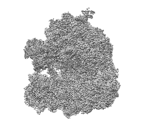 emd_10224.png
 http://ftp.pdbj.org/pub/emdb/structures/EMD-10224


 Z (Sec.)
 Y (Row.)
 X (Col.)


 Sample components
 Processing
 Electron microscopy
 FIELD EMISSION GUN
