National Institutes of Health/National Institute of General Medical Sciences (NIH/NIGMS)
R01 GM114213
United States
Citation
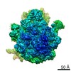
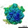
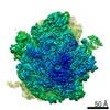
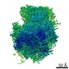
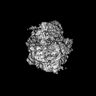
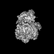
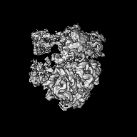
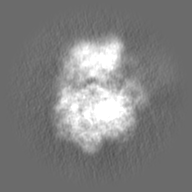
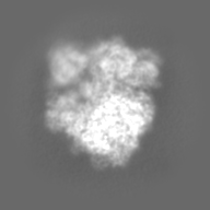
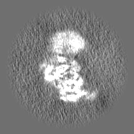
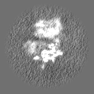
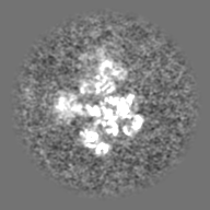
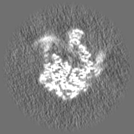
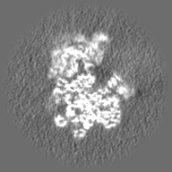
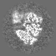
Journal: J Bacteriol / Year: 2020 Title: Ribosome Dimerization Protects the Small Subunit. Authors: Heather A Feaga / Mykhailo Kopylov / Jenny Kim Kim / Marko Jovanovic / Jonathan Dworkin / Abstract: When nutrients become scarce, bacteria can enter an extended state of quiescence. A major challenge of this state is how to preserve ribosomes for the return to favorable conditions. Here, we show ...When nutrients become scarce, bacteria can enter an extended state of quiescence. A major challenge of this state is how to preserve ribosomes for the return to favorable conditions. Here, we show that the ribosome dimerization protein hibernation-promoting factor (HPF) functions to protect essential ribosomal proteins. Ribosomes isolated from strains lacking HPF (Δ) or encoding a mutant allele of HPF that binds the ribosome but does not mediate dimerization were substantially depleted of the small subunit proteins S2 and S3. Strikingly, these proteins are located directly at the ribosome dimer interface. We used single-particle cryo-electron microscopy (cryo-EM) to further characterize these ribosomes and observed that a high percentage of ribosomes were missing S2, S3, or both. These data support a model in which the ribosome dimerization activity of HPF evolved to protect labile proteins that are essential for ribosome function. HPF is almost universally conserved in bacteria, and HPF deletions in diverse species exhibit decreased viability during starvation. Our data provide mechanistic insight into this phenotype and establish a mechanism for how HPF protects ribosomes during quiescence. The formation of ribosome dimers during periods of dormancy is widespread among bacteria. Dimerization is typically mediated by a single protein, hibernation-promoting factor (HPF). Bacteria lacking HPF exhibit strong defects in viability and pathogenesis and, in some species, extreme loss of rRNA. The mechanistic basis of these phenotypes has not been determined. Here, we report that HPF from the Gram-positive bacterium preserves ribosomes by preventing the loss of essential ribosomal proteins at the dimer interface. This protection may explain phenotypes associated with the loss of HPF, since ribosome protection would aid survival during nutrient limitation and impart a strong selective advantage when the bacterial cell rapidly reinitiates growth in the presence of sufficient nutrients.
History
Deposition
Feb 27, 2020
-
Header (metadata) release
Mar 25, 2020
-
Map release
Mar 25, 2020
-
Update
May 13, 2020
-
Current status
May 13, 2020
Processing site: RCSB / Status: Released
-
Structure visualization
Movie
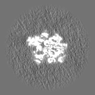
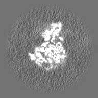
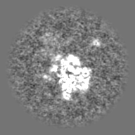
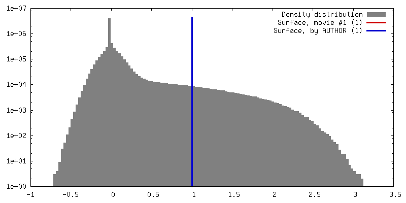
Surface view with section colored by density value
In the structure databanks used in Yorodumi, some data are registered as the other names, "COVID-19 virus" and "2019-nCoV". Here are the details of the virus and the list of structure data.
Jan 31, 2019. EMDB accession codes are about to change! (news from PDBe EMDB page)
EMDB accession codes are about to change! (news from PDBe EMDB page)
The allocation of 4 digits for EMDB accession codes will soon come to an end. Whilst these codes will remain in use, new EMDB accession codes will include an additional digit and will expand incrementally as the available range of codes is exhausted. The current 4-digit format prefixed with “EMD-” (i.e. EMD-XXXX) will advance to a 5-digit format (i.e. EMD-XXXXX), and so on. It is currently estimated that the 4-digit codes will be depleted around Spring 2019, at which point the 5-digit format will come into force.
The EM Navigator/Yorodumi systems omit the EMD- prefix.
Related info.:Q: What is EMD? / ID/Accession-code notation in Yorodumi/EM Navigator
Yorodumi is a browser for structure data from EMDB, PDB, SASBDB, etc.
This page is also the successor to EM Navigator detail page, and also detail information page/front-end page for Omokage search.
The word "yorodu" (or yorozu) is an old Japanese word meaning "ten thousand". "mi" (miru) is to see.
Related info.:EMDB / PDB / SASBDB / Comparison of 3 databanks / Yorodumi Search / Aug 31, 2016. New EM Navigator & Yorodumi / Yorodumi Papers / Jmol/JSmol / Function and homology information / Changes in new EM Navigator and Yorodumi
 Movie
Movie Controller

 Yorodumi
 Open data
 Basic information
 Map data
 Sample
 Authors
 United States, 1 items
  Citation
 Structure visualization
 Movie viewer
 Downloads & links
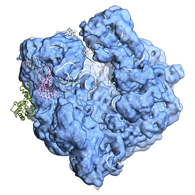 emd_21480.png
 http://ftp.pdbj.org/pub/emdb/structures/EMD-21480


 Z (Sec.)
 Y (Row.)
 X (Col.)


 Sample components
 Processing
 Electron microscopy
 FIELD EMISSION GUN
