 Movie
Movie Controller


+ Open data

- Basic information
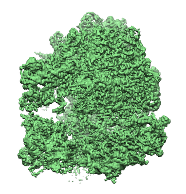
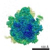
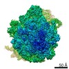
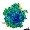
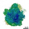
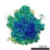
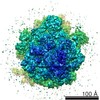
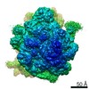
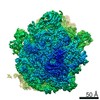
| Entry | Database: EMDB / ID: EMD-3806 | |||||||||
|---|---|---|---|---|---|---|---|---|---|---|
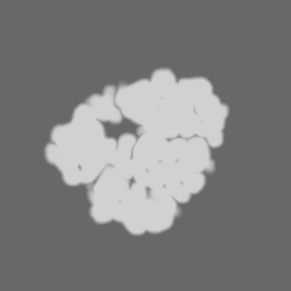
| Title | Chloroplast Ribosome, collected with pixel size 1.06 | |||||||||
 Map data Map data | ||||||||||
 Sample Sample |
| |||||||||
| Biological species |  Spinacia oleracea (spinach) Spinacia oleracea (spinach) | |||||||||
| Method | single particle reconstruction / cryo EM / Resolution: 3.2 Å | |||||||||
 Authors Authors | Forsberg BO / Aibara S / Kimanius D / Paul B / Lindahl E / Amunts A | |||||||||
 Citation Citation | Journal: IUCrJ / Year: 2017 Title: Cryo-EM reconstruction of the chlororibosome to 3.2 Å resolution within 24 h. Authors: Björn O Forsberg / Shintaro Aibara / Dari Kimanius / Bijoya Paul / Erik Lindahl / Alexey Amunts /  Abstract: The introduction of direct detectors and the automation of data collection in cryo-EM have led to a surge in data, creating new opportunities for advancing computational processing. In particular, on- ...The introduction of direct detectors and the automation of data collection in cryo-EM have led to a surge in data, creating new opportunities for advancing computational processing. In particular, on-the-fly workflows that connect data collection with three-dimensional reconstruction would be valuable for more efficient use of cryo-EM and its application as a sample-screening tool. Here, accelerated on-the-fly analysis is reported with optimized organization of the data-processing tools, image acquisition and particle alignment that make it possible to reconstruct the three-dimensional density of the 70S chlororibosome to 3.2 Å resolution within 24 h of tissue harvesting. It is also shown that it is possible to achieve even faster processing at comparable quality by imposing some limits to data use, as illustrated by a 3.7 Å resolution map that was obtained in only 80 min on a desktop computer. These on-the-fly methods can be employed as an assessment of data quality from small samples and extended to high-throughput approaches. | |||||||||
| History |
|
- Structure visualization
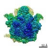
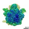
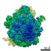
| Movie |
 Movie viewer Movie viewer |
|---|---|
| Structure viewer | EM map: SurfViewMolmilJmol/JSmol |
| Supplemental images |
- Downloads & links
-EMDB archive
| Map data | emd_3806.map.gz | 27.8 MB | EMDB map data format | |
|---|---|---|---|---|
| Header (meta data) | emd-3806-v30.xmlemd-3806.xml | 11.4 KB 11.4 KB | Display Display | EMDB header |
| Images |  emd_3806.png emd_3806.png | 197.5 KB | ||
| Masks | emd_3806_msk_1.map | 282.6 MB | Mask map | |
| Others | emd_3806_half_map_1.map.gzemd_3806_half_map_2.map.gz | 224.5 MB 224.8 MB | ||
| Archive directory |  http://ftp.pdbj.org/pub/emdb/structures/EMD-3806ftp://ftp.pdbj.org/pub/emdb/structures/EMD-3806 http://ftp.pdbj.org/pub/emdb/structures/EMD-3806ftp://ftp.pdbj.org/pub/emdb/structures/EMD-3806 | HTTPS FTP |
-Related structure data
-Links
| EMDB pages | EMDB (EBI/PDBe) / EMDataResource |
|---|---|
| Related items in Molecule of the Month |

-Map
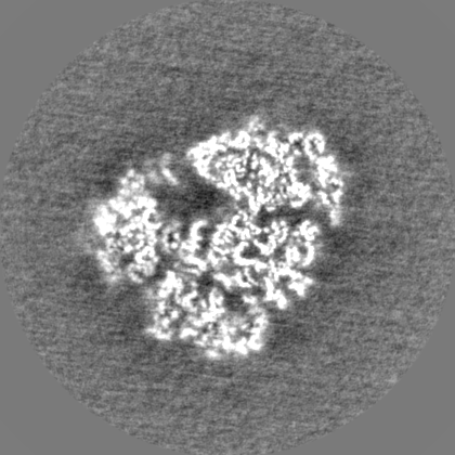
| File | Download / File: emd_3806.map.gz / Format: CCP4 / Size: 282.6 MB / Type: IMAGE STORED AS FLOATING POINT NUMBER (4 BYTES) | ||||||||||||||||||||||||||||||||||||||||||||||||||||||||||||
|---|---|---|---|---|---|---|---|---|---|---|---|---|---|---|---|---|---|---|---|---|---|---|---|---|---|---|---|---|---|---|---|---|---|---|---|---|---|---|---|---|---|---|---|---|---|---|---|---|---|---|---|---|---|---|---|---|---|---|---|---|---|
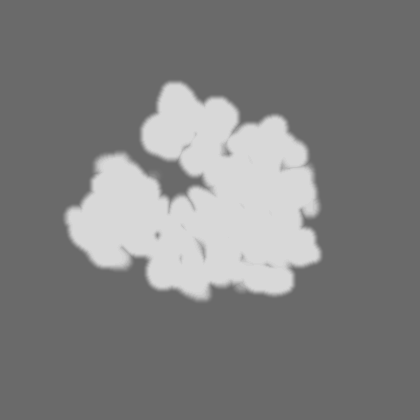
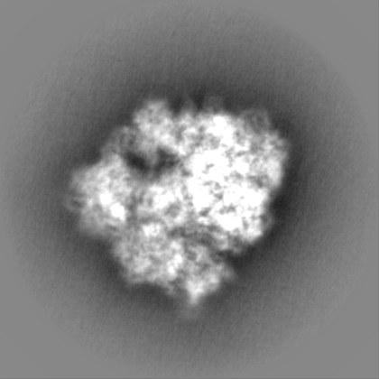
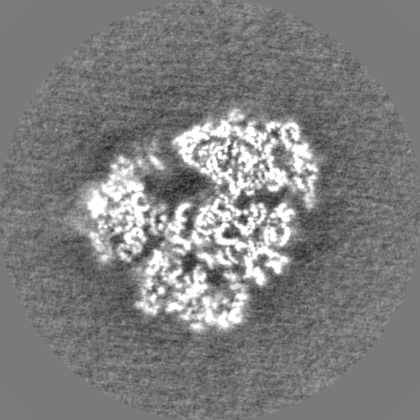
| Projections & slices | Image control
Images are generated by Spider. | ||||||||||||||||||||||||||||||||||||||||||||||||||||||||||||
| Voxel size | X=Y=Z: 1.06 Å | ||||||||||||||||||||||||||||||||||||||||||||||||||||||||||||
| Density |
| ||||||||||||||||||||||||||||||||||||||||||||||||||||||||||||
| Symmetry | Space group: 1 | ||||||||||||||||||||||||||||||||||||||||||||||||||||||||||||
| Details | EMDB XML:
CCP4 map header:
| ||||||||||||||||||||||||||||||||||||||||||||||||||||||||||||
 Z (Sec.)
 Y (Row.)
 X (Col.)


-Supplemental data
-Mask #1
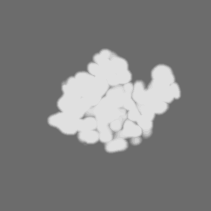
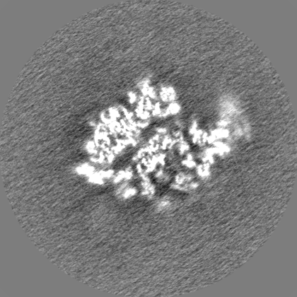
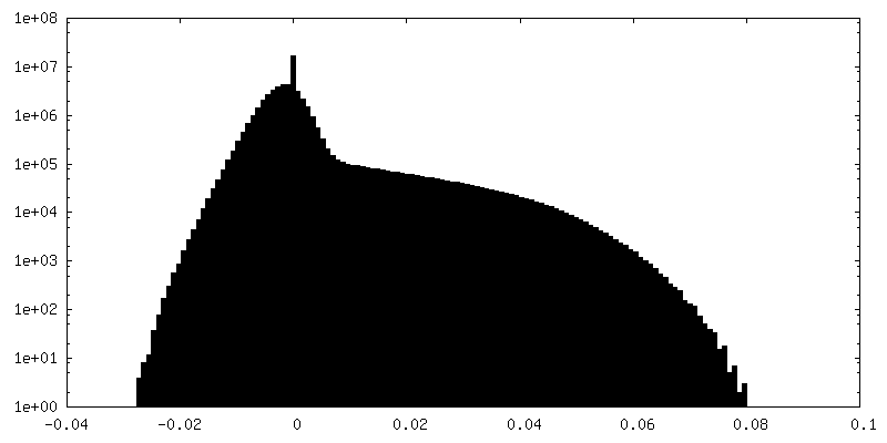
| File | emd_3806_msk_1.map | ||||||||||||
|---|---|---|---|---|---|---|---|---|---|---|---|---|---|
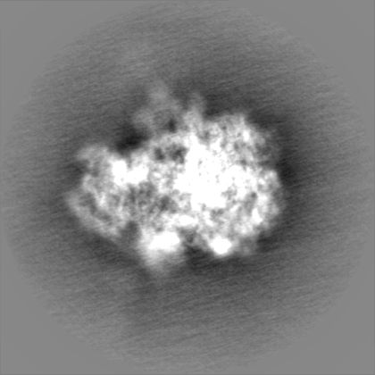
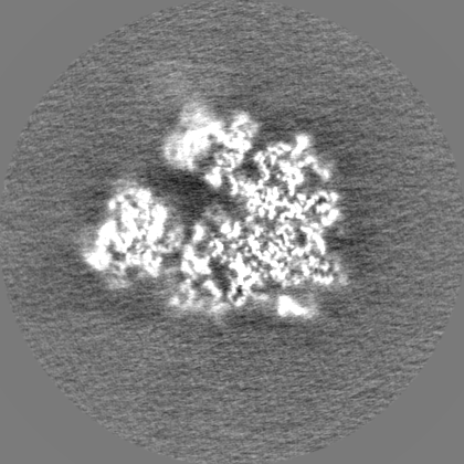
| Projections & Slices |
| ||||||||||||
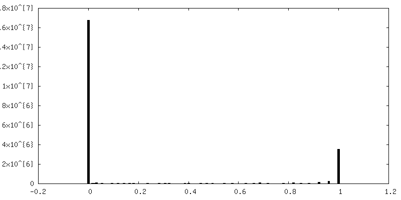
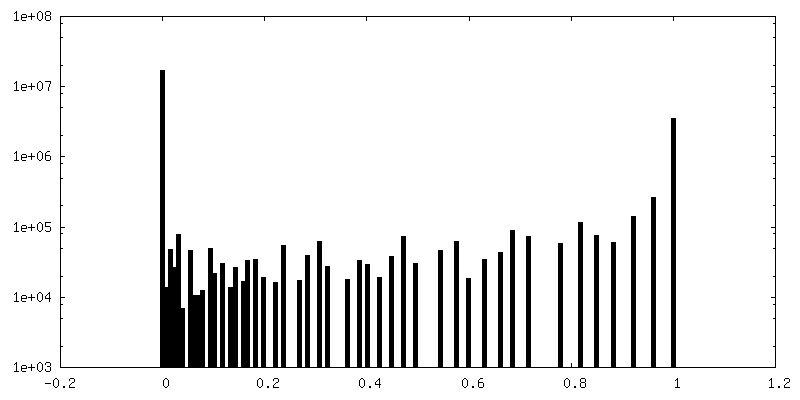
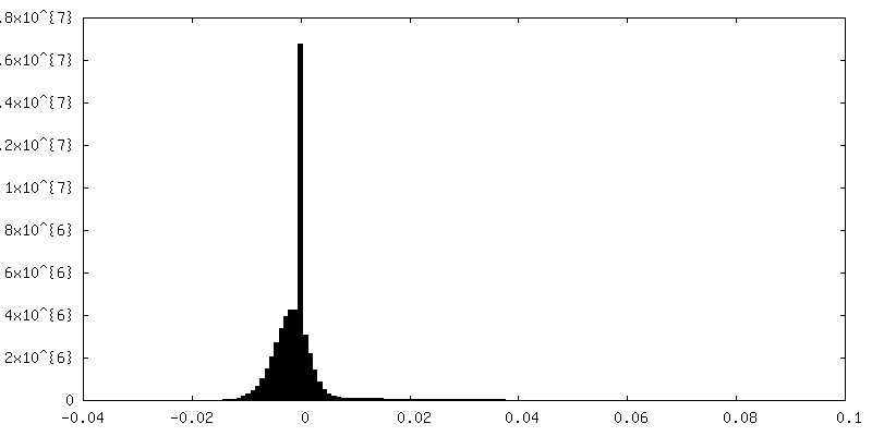
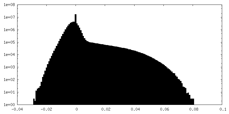
| Density Histograms |
-Half map: #1
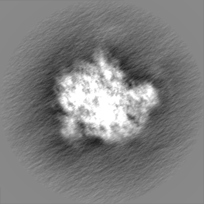
| File | emd_3806_half_map_1.map | ||||||||||||
|---|---|---|---|---|---|---|---|---|---|---|---|---|---|
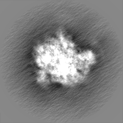
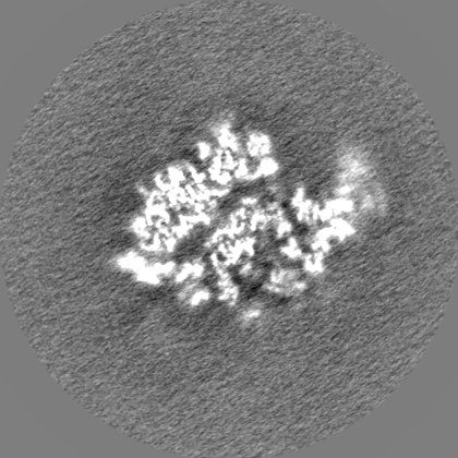
| Projections & Slices |
| ||||||||||||
| Density Histograms |


-Half map: #2
| File | emd_3806_half_map_2.map | ||||||||||||
|---|---|---|---|---|---|---|---|---|---|---|---|---|---|
| Projections & Slices |
| ||||||||||||
| Density Histograms |


- Sample components
-Entire : Chloroplast ribosome from spinach
| Entire | Name: Chloroplast ribosome from spinach |
|---|---|
| Components |
|
-Supramolecule #1: Chloroplast ribosome from spinach
| Supramolecule | Name: Chloroplast ribosome from spinach / type: complex / ID: 1 / Parent: 0 |
|---|---|
| Source (natural) | Organism: Spinacia oleracea (spinach) |
-Experimental details
-Structure determination
| Method | cryo EM |
|---|---|
 Processing Processing | single particle reconstruction |
| Aggregation state | particle |
-Sample preparation
| Buffer | pH: 7.5 |
|---|---|
| Vitrification | Cryogen name: ETHANE |
- Electron microscopy
| Microscope | FEI TITAN KRIOS |
|---|---|
| Image recording | Film or detector model: FEI FALCON II (4k x 4k) / Average electron dose: 19.0 e/Å2 |
| Electron beam | Acceleration voltage: 300 kV / Electron source:  FIELD EMISSION GUN FIELD EMISSION GUN |
| Electron optics | Illumination mode: FLOOD BEAM / Imaging mode: BRIGHT FIELD |
| Experimental equipment |  Model: Titan Krios / Image courtesy: FEI Company |
-Image processing
| Final reconstruction | Applied symmetry - Point group: C1 (asymmetric) / Resolution.type: BY AUTHOR / Resolution: 3.2 Å / Resolution method: FSC 0.143 CUT-OFF / Number images used: 100475 |
|---|---|
| Initial angle assignment | Type: NOT APPLICABLE |
| Final angle assignment | Type: OTHER |
