 Movie
Movie Controller


+ Open data

- Basic information
| Entry | Database: EMDB / ID: EMD-10188 | |||||||||||||||||||||
|---|---|---|---|---|---|---|---|---|---|---|---|---|---|---|---|---|---|---|---|---|---|---|
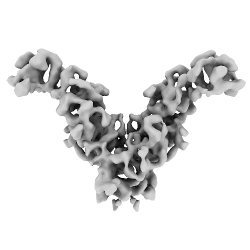
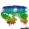
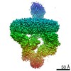
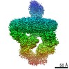
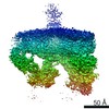
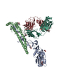
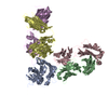
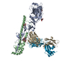
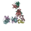
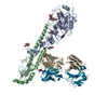
| Title | Structure of EccB3 dimer from the ESX-3 core complex | |||||||||||||||||||||
 Map data Map data | EccB3 dimer. | |||||||||||||||||||||
 Sample Sample |
| |||||||||||||||||||||
 Keywords Keywords | Type VII Secretion System ESX-3 secretion system T7SS ESX-3 Mycobacterium smegmatis / MEMBRANE PROTEIN | |||||||||||||||||||||
| Function / homology | Type VII secretion system EccB / Type VII secretion system EccB, repeat 3 domain / Type VII secretion system EccB, repeat 1 domain / Type VII secretion system ESX-1, transport TM domain B / Hydrolases; Acting on acid anhydrides / hydrolase activity / ATP binding / plasma membrane / ESX-3 secretion system ATPase EccB3 Function and homology information Function and homology information | |||||||||||||||||||||
| Biological species |  Mycolicibacterium smegmatis MC2 155 (bacteria) / Mycobacterium smegmatis (strain ATCC 700084 / mc(2)155) (bacteria) Mycolicibacterium smegmatis MC2 155 (bacteria) / Mycobacterium smegmatis (strain ATCC 700084 / mc(2)155) (bacteria) | |||||||||||||||||||||
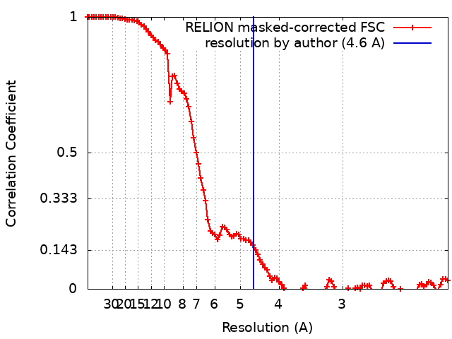
| Method | single particle reconstruction / cryo EM / Resolution: 4.6 Å | |||||||||||||||||||||
 Authors Authors | Rivera-Calzada A / Famelis N / Geibel S / Llorca O | |||||||||||||||||||||
| Funding support |  Germany, Germany,  Spain, 6 items Spain, 6 items
| |||||||||||||||||||||
 Citation Citation | Journal: Nature / Year: 2019 Title: Architecture of the mycobacterial type VII secretion system. Authors: Nikolaos Famelis / Angel Rivera-Calzada / Gianluca Degliesposti / Maria Wingender / Nicole Mietrach / J Mark Skehel / Rafael Fernandez-Leiro / Bettina Böttcher / Andreas Schlosser / Oscar ...Authors: Nikolaos Famelis / Angel Rivera-Calzada / Gianluca Degliesposti / Maria Wingender / Nicole Mietrach / J Mark Skehel / Rafael Fernandez-Leiro / Bettina Böttcher / Andreas Schlosser / Oscar Llorca / Sebastian Geibel /  Abstract: Host infection by pathogenic mycobacteria, such as Mycobacterium tuberculosis, is facilitated by virulence factors that are secreted by type VII secretion systems. A molecular understanding of the ...Host infection by pathogenic mycobacteria, such as Mycobacterium tuberculosis, is facilitated by virulence factors that are secreted by type VII secretion systems. A molecular understanding of the type VII secretion mechanism has been hampered owing to a lack of three-dimensional structures of the fully assembled secretion apparatus. Here we report the cryo-electron microscopy structure of a membrane-embedded core complex of the ESX-3/type VII secretion system from Mycobacterium smegmatis. The core of the ESX-3 secretion machine consists of four protein components-EccB3, EccC3, EccD3 and EccE3, in a 1:1:2:1 stoichiometry-which form two identical protomers. The EccC3 coupling protein comprises a flexible array of four ATPase domains, which are linked to the membrane through a stalk domain. The domain of unknown function (DUF) adjacent to the stalk is identified as an ATPase domain that is essential for secretion. EccB3 is predominantly periplasmatic, but a small segment crosses the membrane and contacts the stalk domain. This suggests that conformational changes in the stalk domain-triggered by substrate binding at the distal end of EccC3 and subsequent ATP hydrolysis in the DUF-could be coupled to substrate secretion to the periplasm. Our results reveal that the architecture of type VII secretion systems differs markedly from that of other known secretion machines, and provide a structural understanding of these systems that will be useful for the design of antimicrobial strategies that target bacterial virulence. | |||||||||||||||||||||
| History |
|
- Structure visualization
| Movie |
Movie viewer |
|---|---|
| Structure viewer | EM map: SurfViewMolmilJmol/JSmol |
| Supplemental images |
- Downloads & links
-EMDB archive
| Map data | emd_10188.map.gz | 5.9 MB | EMDB map data format | |
|---|---|---|---|---|
| Header (meta data) | emd-10188-v30.xmlemd-10188.xml | 14.7 KB 14.7 KB | Display Display | EMDB header |
| FSC (resolution estimation) | emd_10188_fsc.xml | 10.9 KB | Display | FSC data file |
| Images |  emd_10188.png emd_10188.png | 69.6 KB | ||
| Filedesc metadata | emd-10188.cif.gz | 6.3 KB | ||
| Archive directory |  http://ftp.pdbj.org/pub/emdb/structures/EMD-10188ftp://ftp.pdbj.org/pub/emdb/structures/EMD-10188 http://ftp.pdbj.org/pub/emdb/structures/EMD-10188ftp://ftp.pdbj.org/pub/emdb/structures/EMD-10188 | HTTPS FTP |
-Validation report
| Summary document | emd_10188_validation.pdf.gz | 350.5 KB | Display | EMDB validaton report |
|---|---|---|---|---|
| Full document | emd_10188_full_validation.pdf.gz | 350.1 KB | Display | |
| Data in XML | emd_10188_validation.xml.gz | 11.9 KB | Display | |
| Data in CIF | emd_10188_validation.cif.gz | 16 KB | Display | |
| Arichive directory | https://ftp.pdbj.org/pub/emdb/validation_reports/EMD-10188ftp://ftp.pdbj.org/pub/emdb/validation_reports/EMD-10188 | HTTPS FTP |
-Related structure data
| Related structure data |  6sgyMC  6sgwC  6sgxC 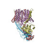 6sgzC M: atomic model generated by this map C: citing same article ( |
|---|---|
| Similar structure data |


-Links
| EMDB pages | EMDB (EBI/PDBe) / EMDataResource |
|---|
-Map
| File | Download / File: emd_10188.map.gz / Format: CCP4 / Size: 107.2 MB / Type: IMAGE STORED AS FLOATING POINT NUMBER (4 BYTES) | ||||||||||||||||||||||||||||||||||||||||||||||||||||||||||||
|---|---|---|---|---|---|---|---|---|---|---|---|---|---|---|---|---|---|---|---|---|---|---|---|---|---|---|---|---|---|---|---|---|---|---|---|---|---|---|---|---|---|---|---|---|---|---|---|---|---|---|---|---|---|---|---|---|---|---|---|---|---|
| Annotation | EccB3 dimer. | ||||||||||||||||||||||||||||||||||||||||||||||||||||||||||||
| Projections & slices | Image control
Images are generated by Spider. | ||||||||||||||||||||||||||||||||||||||||||||||||||||||||||||
| Voxel size | X=Y=Z: 1.0635 Å | ||||||||||||||||||||||||||||||||||||||||||||||||||||||||||||
| Density |
| ||||||||||||||||||||||||||||||||||||||||||||||||||||||||||||
| Symmetry | Space group: 1 | ||||||||||||||||||||||||||||||||||||||||||||||||||||||||||||
| Details | EMDB XML:
CCP4 map header:
| ||||||||||||||||||||||||||||||||||||||||||||||||||||||||||||
 Z (Sec.)
 Y (Row.)
 X (Col.)


-Supplemental data
- Sample components
-Entire : EccB3 dimeric structure from ESX-3/Type VII secretion system
| Entire | Name: EccB3 dimeric structure from ESX-3/Type VII secretion system |
|---|---|
| Components |
|
-Supramolecule #1: EccB3 dimeric structure from ESX-3/Type VII secretion system
| Supramolecule | Name: EccB3 dimeric structure from ESX-3/Type VII secretion system type: complex / ID: 1 / Parent: 0 / Macromolecule list: all Details: The sample consists of four protein components, EccB3:EccC3:EccD3:EccE3 in a 1:1:2:1 stoichiometry Molecular weight of the complex without the amphipol micelle: 0.65 MDa |
|---|---|
| Source (natural) | Organism: Mycolicibacterium smegmatis MC2 155 (bacteria) |
| Molecular weight | Theoretical: 83 KDa |
-Macromolecule #1: ESX-3 secretion system protein EccB3
| Macromolecule | Name: ESX-3 secretion system protein EccB3 / type: protein_or_peptide / ID: 1 / Number of copies: 2 / Enantiomer: LEVO |
|---|---|
| Source (natural) | Organism: Mycobacterium smegmatis (strain ATCC 700084 / mc(2)155) (bacteria) |
| Molecular weight | Theoretical: 42.746754 KDa |
| Recombinant expression | Organism: Mycolicibacterium smegmatis MC2 155 (bacteria) |
| Sequence | String: NAILADRSTS ALYVRVGEQL HPVLNLTSAR LISGSPDNPT MVKTSEIDKF PRGNLLGIPG APERMVQNAA TDAEWTVCDA VGGANPGVT VIAGPLGADG ERAAPLPPDH AVLVHSDAEP NPGDWLLWDG KRSPIDLADR AVTDALGLGG QALAPRPIAA G LFNAVPAA ...String: NAILADRSTS ALYVRVGEQL HPVLNLTSAR LISGSPDNPT MVKTSEIDKF PRGNLLGIPG APERMVQNAA TDAEWTVCDA VGGANPGVT VIAGPLGADG ERAAPLPPDH AVLVHSDAEP NPGDWLLWDG KRSPIDLADR AVTDALGLGG QALAPRPIAA G LFNAVPAA PALTAPVIPD AGAAPQFELS LPVPVGAVVV AYDADNTARY YAVLSDGLQP ISPVLAAILR NTDSHGFAQP PR LGPDEVA RTPMSRGLDT SAYPDNPVTL VEASAHPVTC AHWTKPSDAA ESSLSVLSGA VLPLAEGLHT VDLVGAGAGG AAN RVALTP GTGYFVQTVG AEPGSPTAGS MFWVSDTGVR YGIDTAEDDK VVAALGLSTS PLPVPWSVLS QFAAGPALSR GDAL VAHDA VSTNPNSARM EASR UniProtKB: ESX-3 secretion system ATPase EccB3 |
-Experimental details
-Structure determination
| Method | cryo EM |
|---|---|
 Processing Processing | single particle reconstruction |
| Aggregation state | particle |
-Sample preparation
| Concentration | 0.3 mg/mL |
|---|---|
| Buffer | pH: 8 / Details: 30 mM Hepes pH 8.0, 150 mM NaCl |
| Vitrification | Cryogen name: ETHANE / Chamber humidity: 100 % / Chamber temperature: 278 K / Instrument: FEI VITROBOT MARK IV / Details: Blotting time: 3s Blotting force: -10. |
| Details | bound to Amphipol A8-35 |
- Electron microscopy
| Microscope | FEI TITAN KRIOS |
|---|---|
| Image recording | Film or detector model: FEI FALCON III (4k x 4k) / Detector mode: COUNTING / Number real images: 11903 / Average electron dose: 50.0 e/Å2 Details: Images acquired as 55 frames movies at a calibrated magnification of 1.0635 Angs/px |
| Electron beam | Acceleration voltage: 300 kV / Electron source:  FIELD EMISSION GUN FIELD EMISSION GUN |
| Electron optics | Illumination mode: SPOT SCAN / Imaging mode: BRIGHT FIELD / Cs: 2.7 mm / Nominal defocus max: 2.6 µm / Nominal defocus min: 1.6 µm / Nominal magnification: 75000 |
| Sample stage | Specimen holder model: FEI TITAN KRIOS AUTOGRID HOLDER / Cooling holder cryogen: NITROGEN |
| Experimental equipment |  Model: Titan Krios / Image courtesy: FEI Company |
+Image processing
-Atomic model buiding 1
| Initial model | PDB ID: Chain - Chain ID: A / Chain - Residue range: 100-518 / Chain - Source name: PDB / Chain - Initial model type: experimental model |
|---|---|
| Details | Two copies of the periplasmic domain of a EccB3 homology model (based on pdb 3X3M) were fitted into the density and then morphing was performed using PHENIX Real-Space Refinement. |
| Refinement | Space: REAL / Protocol: AB INITIO MODEL / Target criteria: Map correlation coefficient |
| Output model | PDB-6sgy: |

