 Movie
Movie Controller

[English] 日本語
 Yorodumi
- PDB-5y2l: Crystal structure of a group 2 HA binding antibody AF4H1K1 Fab in... -
+ Open data

- Basic information
| Entry | Database: PDB / ID: 5y2l | |||||||||
|---|---|---|---|---|---|---|---|---|---|---|
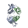
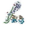
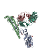
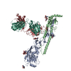
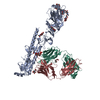
| Title | Crystal structure of a group 2 HA binding antibody AF4H1K1 Fab in complex with the 1968 H3N2 pandemic (H3-AC/68) hemagglutinin | |||||||||
 Components Components |
| |||||||||
 Keywords Keywords | ANTIMICROBIAL PROTEIN / neutralizing antibody / influenza virus / HA / H3-clade | |||||||||
| Function / homology |  Function and homology information Function and homology informationviral budding from plasma membrane / clathrin-dependent endocytosis of virus by host cell / host cell surface receptor binding / fusion of virus membrane with host plasma membrane / fusion of virus membrane with host endosome membrane / viral envelope / virion attachment to host cell / host cell plasma membrane / virion membrane / membrane Similarity search - Function | |||||||||
| Biological species |   Influenza A virus Influenza A virus Homo sapiens (human) Homo sapiens (human) | |||||||||
| Method |  X-RAY DIFFRACTION / SYNCHROTRON / Resolution: 2.902 Å X-RAY DIFFRACTION / SYNCHROTRON / Resolution: 2.902 Å | |||||||||
 Authors Authors | Xiao, H. / Qi, J. / Gao, F.G. | |||||||||
 Citation Citation | Journal: To Be Published Title: An H3-clade neutralizing antibody screened from an H7N9 patient that binds group 2 influenza A hemagglutinins Authors: Xiao, H. / Qi, J. / Gao, F.G. | |||||||||
| History |
|
- Structure visualization
| Structure viewer | Molecule: MolmilJmol/JSmol |
|---|
- Downloads & links
-Download
| PDBx/mmCIF format | 5y2l.cif.gz | 366.6 KB | Display | PDBx/mmCIF format |
|---|---|---|---|---|
| PDB format | pdb5y2l.ent.gz | 299.5 KB | Display | PDB format |
| PDBx/mmJSON format | 5y2l.json.gz | Tree view | PDBx/mmJSON format | |
| Others |  Other downloads Other downloads |
-Validation report
| Arichive directory | https://data.pdbj.org/pub/pdb/validation_reports/y2/5y2lftp://data.pdbj.org/pub/pdb/validation_reports/y2/5y2l | HTTPS FTP |
|---|
-Related structure data


-Links
 PDBj
PDBj


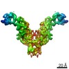
- Assembly
| Deposited unit | 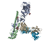
| ||||||||
|---|---|---|---|---|---|---|---|---|---|
| 1 | 
| ||||||||
| Unit cell |
|
-Components
-Protein , 2 types, 2 molecules AB
| #1: Protein | Mass: 36222.652 Da / Num. of mol.: 1 Source method: isolated from a genetically manipulated source Source: (gene. exp.) Influenza A virus (strain A/Aichi/2/1968 H3N2)Strain: A/Aichi/2/1968 H3N2 / Gene: HA / Production host:  Trichoplusia ni (cabbage looper) / References: UniProt: P03437 Trichoplusia ni (cabbage looper) / References: UniProt: P03437 |
|---|---|
| #2: Protein | Mass: 20311.480 Da / Num. of mol.: 1 / Fragment: UNP residues 346-521 Source method: isolated from a genetically manipulated source Source: (gene. exp.) Influenza A virus (strain A/Aichi/2/1968 H3N2)Strain: A/Aichi/2/1968 H3N2 / Gene: HA / Production host: Trichoplusia ni (cabbage looper) / References: UniProt: P03437 |
-Antibody , 2 types, 2 molecules IJ
| #3: Antibody | Mass: 24977.014 Da / Num. of mol.: 1 Source method: isolated from a genetically manipulated source Source: (gene. exp.) Homo sapiens (human) / Production host: Homo sapiens (human) |
|---|---|
| #4: Antibody | Mass: 24243.838 Da / Num. of mol.: 1 Source method: isolated from a genetically manipulated source Source: (gene. exp.) Homo sapiens (human) / Production host: Homo sapiens (human) |
-Sugars , 2 types, 4 molecules 
| #5: Polysaccharide | Source method: isolated from a genetically manipulated source #6: Sugar |  Type: D-saccharide, beta linking / Mass: 221.208 Da / Num. of mol.: 2 Type: D-saccharide, beta linking / Mass: 221.208 Da / Num. of mol.: 2Source method: isolated from a genetically manipulated source Formula: C8H15NO6 |
|---|
-Details
| Has protein modification | Y |
|---|
-Experimental details
-Experiment
| Experiment | Method: X-RAY DIFFRACTION / Number of used crystals: 1 |
|---|
- Sample preparation
| Crystal | Density Matthews: 4.01 Å3/Da / Density % sol: 69.32 % |
|---|---|
| Crystal grow | Temperature: 291 K / Method: vapor diffusion, sitting drop / pH: 7.5 Details: 0.19 mM CYMAL7, 0.1M HEPES pH 7.5, 40% ployethylene glycol 400 |
-Data collection
| Diffraction | Mean temperature: 100 K |
|---|---|
| Diffraction source | Source: SYNCHROTRON / Site: SSRF  / Beamline: BL17U1 / Wavelength: 1 Å / Beamline: BL17U1 / Wavelength: 1 Å |
| Detector | Type: ADSC QUANTUM 315 / Detector: CCD / Date: Jul 8, 2014 |
| Radiation | Protocol: SINGLE WAVELENGTH / Monochromatic (M) / Laue (L): M / Scattering type: x-ray |
| Radiation wavelength | Wavelength: 1 Å / Relative weight: 1 |
| Reflection | Resolution: 2.9→50 Å / Num. obs: 37831 / % possible obs: 99.9 % / Redundancy: 10.7 % / Rmerge(I) obs: 0.097 / Net I/σ(I): 23 |
| Reflection shell | Resolution: 2.9→3 Å / Redundancy: 10.7 % / Mean I/σ(I) obs: 2.5 / Num. unique obs: 3739 / % possible all: 100 |
- Processing
| Software |
| ||||||||||||||||||||||||||||||||||||||||||||||||||||||||||||||||||||||||||||||||||||||||||||||||||
|---|---|---|---|---|---|---|---|---|---|---|---|---|---|---|---|---|---|---|---|---|---|---|---|---|---|---|---|---|---|---|---|---|---|---|---|---|---|---|---|---|---|---|---|---|---|---|---|---|---|---|---|---|---|---|---|---|---|---|---|---|---|---|---|---|---|---|---|---|---|---|---|---|---|---|---|---|---|---|---|---|---|---|---|---|---|---|---|---|---|---|---|---|---|---|---|---|---|---|---|
| Refinement | Resolution: 2.902→47.331 Å / SU ML: 0.41 / Cross valid method: FREE R-VALUE / σ(F): 1.34 / Phase error: 31.53
| ||||||||||||||||||||||||||||||||||||||||||||||||||||||||||||||||||||||||||||||||||||||||||||||||||
| Solvent computation | Shrinkage radii: 0.9 Å / VDW probe radii: 1.11 Å | ||||||||||||||||||||||||||||||||||||||||||||||||||||||||||||||||||||||||||||||||||||||||||||||||||
| Refinement step | Cycle: LAST / Resolution: 2.902→47.331 Å
| ||||||||||||||||||||||||||||||||||||||||||||||||||||||||||||||||||||||||||||||||||||||||||||||||||
| Refine LS restraints |
| ||||||||||||||||||||||||||||||||||||||||||||||||||||||||||||||||||||||||||||||||||||||||||||||||||
| LS refinement shell |
| ||||||||||||||||||||||||||||||||||||||||||||||||||||||||||||||||||||||||||||||||||||||||||||||||||
| Refinement TLS params. | Method: refined / Origin x: 45.6564 Å / Origin y: -47.1799 Å / Origin z: -27.6507 Å
| ||||||||||||||||||||||||||||||||||||||||||||||||||||||||||||||||||||||||||||||||||||||||||||||||||
| Refinement TLS group | Selection details: all |
