 Movie
Movie Controller

[English] 日本語
 Yorodumi
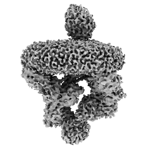
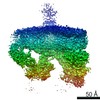
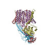
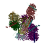
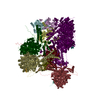
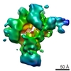
- EMDB-10189: ESX-3 core complex centred at the cytoplasmic region, conformation 1. -
+ Open data

- Basic information
| Entry | Database: EMDB / ID: EMD-10189 | |||||||||||||||||||||
|---|---|---|---|---|---|---|---|---|---|---|---|---|---|---|---|---|---|---|---|---|---|---|
| Title | ESX-3 core complex centred at the cytoplasmic region, conformation 1. | |||||||||||||||||||||
 Map data Map data | ESX-3 core dimer centred at the cytoplasmic region, conformation 1. | |||||||||||||||||||||
 Sample Sample |
| |||||||||||||||||||||
| Biological species |  Mycolicibacterium smegmatis MC2 155 (bacteria) Mycolicibacterium smegmatis MC2 155 (bacteria) | |||||||||||||||||||||
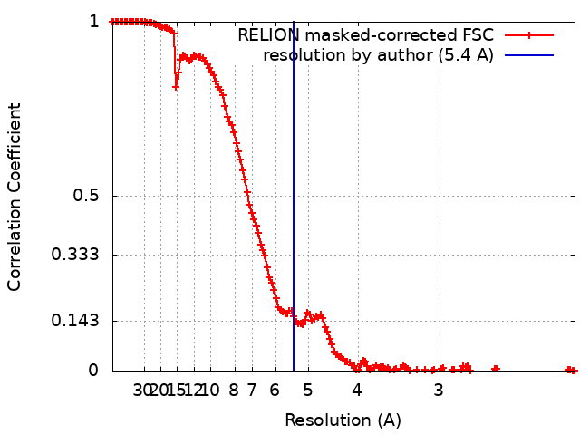
| Method | single particle reconstruction / cryo EM / Resolution: 5.4 Å | |||||||||||||||||||||
 Authors Authors | Rivera-Calzada A / Famelis N / Geibel S / Llorca O | |||||||||||||||||||||
| Funding support |  Germany, Germany,  Spain, 6 items Spain, 6 items
| |||||||||||||||||||||
 Citation Citation | Journal: Nature / Year: 2019 Title: Architecture of the mycobacterial type VII secretion system. Authors: Nikolaos Famelis / Angel Rivera-Calzada / Gianluca Degliesposti / Maria Wingender / Nicole Mietrach / J Mark Skehel / Rafael Fernandez-Leiro / Bettina Böttcher / Andreas Schlosser / Oscar ...Authors: Nikolaos Famelis / Angel Rivera-Calzada / Gianluca Degliesposti / Maria Wingender / Nicole Mietrach / J Mark Skehel / Rafael Fernandez-Leiro / Bettina Böttcher / Andreas Schlosser / Oscar Llorca / Sebastian Geibel /  Abstract: Host infection by pathogenic mycobacteria, such as Mycobacterium tuberculosis, is facilitated by virulence factors that are secreted by type VII secretion systems. A molecular understanding of the ...Host infection by pathogenic mycobacteria, such as Mycobacterium tuberculosis, is facilitated by virulence factors that are secreted by type VII secretion systems. A molecular understanding of the type VII secretion mechanism has been hampered owing to a lack of three-dimensional structures of the fully assembled secretion apparatus. Here we report the cryo-electron microscopy structure of a membrane-embedded core complex of the ESX-3/type VII secretion system from Mycobacterium smegmatis. The core of the ESX-3 secretion machine consists of four protein components-EccB3, EccC3, EccD3 and EccE3, in a 1:1:2:1 stoichiometry-which form two identical protomers. The EccC3 coupling protein comprises a flexible array of four ATPase domains, which are linked to the membrane through a stalk domain. The domain of unknown function (DUF) adjacent to the stalk is identified as an ATPase domain that is essential for secretion. EccB3 is predominantly periplasmatic, but a small segment crosses the membrane and contacts the stalk domain. This suggests that conformational changes in the stalk domain-triggered by substrate binding at the distal end of EccC3 and subsequent ATP hydrolysis in the DUF-could be coupled to substrate secretion to the periplasm. Our results reveal that the architecture of type VII secretion systems differs markedly from that of other known secretion machines, and provide a structural understanding of these systems that will be useful for the design of antimicrobial strategies that target bacterial virulence. | |||||||||||||||||||||
| History |
|
- Structure visualization
| Movie |
 Movie viewer Movie viewer |
|---|---|
| Structure viewer | EM map: SurfViewMolmilJmol/JSmol |
| Supplemental images |
- Downloads & links
-EMDB archive
| Map data | emd_10189.map.gz | 253.9 MB | EMDB map data format | |
|---|---|---|---|---|
| Header (meta data) | emd-10189-v30.xmlemd-10189.xml | 18.8 KB 18.8 KB | Display Display | EMDB header |
| FSC (resolution estimation) | emd_10189_fsc.xml | 14.9 KB | Display | FSC data file |
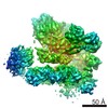
| Images |  emd_10189.png emd_10189.png | 142.4 KB | ||
| Archive directory |  http://ftp.pdbj.org/pub/emdb/structures/EMD-10189ftp://ftp.pdbj.org/pub/emdb/structures/EMD-10189 http://ftp.pdbj.org/pub/emdb/structures/EMD-10189ftp://ftp.pdbj.org/pub/emdb/structures/EMD-10189 | HTTPS FTP |
-Related structure data


-Links
| EMDB pages | EMDB (EBI/PDBe) / EMDataResource |
|---|
-Map
| File | Download / File: emd_10189.map.gz / Format: CCP4 / Size: 274.6 MB / Type: IMAGE STORED AS FLOATING POINT NUMBER (4 BYTES) | ||||||||||||||||||||||||||||||||||||||||||||||||||||||||||||||||||||
|---|---|---|---|---|---|---|---|---|---|---|---|---|---|---|---|---|---|---|---|---|---|---|---|---|---|---|---|---|---|---|---|---|---|---|---|---|---|---|---|---|---|---|---|---|---|---|---|---|---|---|---|---|---|---|---|---|---|---|---|---|---|---|---|---|---|---|---|---|---|
| Annotation | ESX-3 core dimer centred at the cytoplasmic region, conformation 1. | ||||||||||||||||||||||||||||||||||||||||||||||||||||||||||||||||||||
| Projections & slices | Image control
Images are generated by Spider. | ||||||||||||||||||||||||||||||||||||||||||||||||||||||||||||||||||||
| Voxel size | X=Y=Z: 1.0635 Å | ||||||||||||||||||||||||||||||||||||||||||||||||||||||||||||||||||||
| Density |
| ||||||||||||||||||||||||||||||||||||||||||||||||||||||||||||||||||||
| Symmetry | Space group: 1 | ||||||||||||||||||||||||||||||||||||||||||||||||||||||||||||||||||||
| Details | EMDB XML:
CCP4 map header:
| ||||||||||||||||||||||||||||||||||||||||||||||||||||||||||||||||||||
 Z (Sec.)
 Y (Row.)
 X (Col.)


-Supplemental data
- Sample components
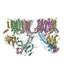
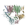
-Entire : ESX-3 core complex, conformation 1
| Entire | Name: ESX-3 core complex, conformation 1 |
|---|---|
| Components |
|
-Supramolecule #1: ESX-3 core complex, conformation 1
| Supramolecule | Name: ESX-3 core complex, conformation 1 / type: complex / ID: 1 / Parent: 0 / Macromolecule list: all Details: The sample consists of four protein components, EccB3:EccC3:EccD3:EccE3 in a 1:1:2:1 stoichiometry Molecular weight of the complex without the amphipol micelle: 0.65 MDa |
|---|---|
| Source (natural) | Organism: Mycolicibacterium smegmatis MC2 155 (bacteria) |
| Recombinant expression | Organism: Mycolicibacterium smegmatis MC2 155 (bacteria) |
| Molecular weight | Experimental: 650 KDa |
-Macromolecule #1: EccB3
| Macromolecule | Name: EccB3 / type: protein_or_peptide / ID: 1 / Enantiomer: LEVO |
|---|---|
| Source (natural) | Organism: Mycolicibacterium smegmatis MC2 155 (bacteria) |
| Recombinant expression | Organism: Mycolicibacterium smegmatis MC2 155 (bacteria) |
| Sequence | String: MTGPVNPDDR RSFSSRTPVN ENPDGVQYRR GFVTRHQVSG WRFVMRRIAS GVALHDTRML VDPLRTQSR AVLTGALILV TGLVGCFIFS LFRPGGVPGN NAILADRSTS ALYVRVGEQL H PVLNLTSA RLISGSPDNP TMVKTSEIDK FPRGNLLGIP GAPERMVQNA ...String: MTGPVNPDDR RSFSSRTPVN ENPDGVQYRR GFVTRHQVSG WRFVMRRIAS GVALHDTRML VDPLRTQSR AVLTGALILV TGLVGCFIFS LFRPGGVPGN NAILADRSTS ALYVRVGEQL H PVLNLTSA RLISGSPDNP TMVKTSEIDK FPRGNLLGIP GAPERMVQNA ATDAEWTVCD AV GGANPGV TVIAGPLGAD GERAAPLPPD HAVLVHSDAE PNPGDWLLWD GKRSPIDLAD RAV TDALGL GGQALAPRPI AAGLFNAVPA APALTAPVIP DAGAAPQFEL SLPVPVGAVV VAYD ADNTA RYYAVLSDGL QPISPVLAAI LRNTDSHGFA QPPRLGPDEV ARTPMSRGLD TSAYP DNPV TLVEASAHPV TCAHWTKPSD AAESSLSVLS GAVLPLAEGL HTVDLVGAGA GGAANR VAL TPGTGYFVQT VGAEPGSPTA GSMFWVSDTG VRYGIDTAED DKVVAALGLS TSPLPVP WS VLSQFAAGPA LSRGDALVAH DAVSTNPNSA RMEASR |
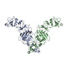
-Macromolecule #2: EccD3
| Macromolecule | Name: EccD3 / type: protein_or_peptide / ID: 2 / Enantiomer: LEVO |
|---|---|
| Source (natural) | Organism: Mycolicibacterium smegmatis MC2 155 (bacteria) |
| Recombinant expression | Organism: Mycolicibacterium smegmatis MC2 155 (bacteria) |
| Sequence | String: MASWSHPQFE KGSMSENTVM PIVRVAVLAA GDDGGRLTEM ALPSELPLRE ILPAVQRIVQ PARENDGAAD PAAAPNPVRL SLAPIGGAPF SLDATLDTVG VVDGDLLALQ AVPSGPPAPR IVEDIADAAV IFSEARRRQW GPTHIARGAA LALIGLILVG TGLSVAHRVI ...String: MASWSHPQFE KGSMSENTVM PIVRVAVLAA GDDGGRLTEM ALPSELPLRE ILPAVQRIVQ PARENDGAAD PAAAPNPVRL SLAPIGGAPF SLDATLDTVG VVDGDLLALQ AVPSGPPAPR IVEDIADAAV IFSEARRRQW GPTHIARGAA LALIGLILVG TGLSVAHRVI TGDLLGQFIV SGIALATVIA ALAVRNRSAV LATSLAVTAL VPVAAAFALG VPGDFGAPNV LLAAAGVAAW SLISMAGSPD DRGIAVFTAT AVTGVGVLLV AGAASLWVIS SDVIGCALVL LGLIVTVQAA QLSAMWARFP LPVIPAPGDP TPAARPLSVL ADLPRRVRVS QAHQTGVIAA GVLLGVAGSV ALVSSANASP WAWYIVVAAA AGAALRARVW DSAACKAWLL GHSYLLAVAL LVAFVIGDRY QAALWALAAL AVLVLVWIVA ALNPKIASPD TYSLPMRRMV GFLATGLDAS LIPVMALLVG LFSLVLDR |
-Macromolecule #3: EccC3
| Macromolecule | Name: EccC3 / type: protein_or_peptide / ID: 3 / Enantiomer: LEVO |
|---|---|
| Source (natural) | Organism: Mycolicibacterium smegmatis MC2 155 (bacteria) |
| Recombinant expression | Organism: Mycolicibacterium smegmatis MC2 155 (bacteria) |
| Sequence | String: MSRLIFEHQR RLTPPTTRKG TITIEPPPQL PRVVPPSLLR RVLPFLIVIL IVGMIVALFA TGMRLISPT MLFFPFVLLL AATALYRGGD NKMRTEEVDA ERADYLRYLS VVRDNVRAHA A EQRAALEW SHPEPEVLAT IPGTRRQWER DPRDRDFLVL RAGRHDVPLD ...String: MSRLIFEHQR RLTPPTTRKG TITIEPPPQL PRVVPPSLLR RVLPFLIVIL IVGMIVALFA TGMRLISPT MLFFPFVLLL AATALYRGGD NKMRTEEVDA ERADYLRYLS VVRDNVRAHA A EQRAALEW SHPEPEVLAT IPGTRRQWER DPRDRDFLVL RAGRHDVPLD AALKVKDTAD EI DLEPVAH SALRGLLDVQ RTVRDAPTGL DVAKLARITV IGEADEARAA IRAWIAQAVT WHD PTMLGV ALAAPDLESG DWSWLKWLPH VDVPNEADGV GPARYLTTST AELRERLAPA LADR PLFPA ESGAALKHLL VVLDDPDADP DDIARKPGLT GVTVIHRTTE LPNREQYPDP ERPIL RVAD GRIERWQVGG WQPCVDVADA MSAAEAAHIA RRLSRWDSNP GYIRSTSTGS ATFTTL LGI PDASALDVAS LWAPRPRDEE LRVPIGVTST GEPLYFDLKD EAEGGMGPHG LMIGMTG SG KSQTLMSILL SLLTTHPADR LIVIYADFKG EAGADIFRHF PQVVAVISNM AEKRSLAD R FADTLRGEVA RREQILKEAG RRVQGSAFNS VAEYESAIAA GHDLPPMPTL FVVADEFTL MLAEHPEYAD LFDYVARKGR SFRIHLLFAS QTLDVGRIKD IDKNTSYRIG LKVASPSISR QIIGVEDAY HIESGREHKG EGFLVPAPGA VPIKFRSTYV DGIYDPPRAE KSIVVHALPQ P QVFTAGRV EPEPDTVIAT GDVEVHTAPP RKLIATIGDQ LAAYGPKAPQ LWLPPLDEPI AL ADVLAGA DVEPGQLRWP LGEIDKPFEM RRDVLVYDAH TAAANVLIHG GPRSGKSTAL QAF VLSAAA LHSPRAITFY CLDYGGGKLA DLADLAHVGS VATPLEPERI RRTFGELEQL LRAR QRQGA VNRTGSYTDG YGEVFLVIDN LYAFSRDNTD TFNTRNPLLA KVTELANSGL AYGIH VVIT TPNWLEVPLA MRDGLGLRLE LKLHDSHDSI VRVAGALRRP ADSVPADQPG RGLTMA AEH FLFAEPALSD IAVINARYPG VSAPPVRLLP TDLSPDALAP LYPAPETVVI GQREEDL AP VAVDFANHPL LMVFGDSKSG KTTLLRHIIR TVRENSTPDQ VAFTVIDRRL HLVDEPLF P DNEYTANIDR VLPAMLGLSA LIEKRRPPAG LSAQELSRWT YTGHTHYLIV DDVDQIPDT PAVSGPFVGQ RPWTNIVGLL AEAADLGLRV IVTARATGSA HAVMTAPLLR RLNDLQATTL MLSGNPTDS GKIRGHRFAR FPAGRGLLLT DTDTPDHIQL VNPLGDAALS GNIGNNGNHN R GGEYRGSM GGSHHHHHH |
-Macromolecule #4: EccE3
| Macromolecule | Name: EccE3 / type: protein_or_peptide / ID: 4 / Enantiomer: LEVO |
|---|---|
| Source (natural) | Organism: Mycolicibacterium smegmatis MC2 155 (bacteria) |
| Recombinant expression | Organism: Mycolicibacterium smegmatis MC2 155 (bacteria) |
| Sequence | String: MTARIALASL FVVAAVLAQP WQTTTQRWVL GVSIAAVIVL LAWWKGMFLT TRIGRALAMV RRNRAEDTV ETDAHRATVV LRVDPAAPAQ LPVVVGYLDR YGITCDKVRI THRDAGGTRR S WISLTVDA VDNLAALQAR SARIPLQDTT EVVGRRLADH LREQGWTVTV ...String: MTARIALASL FVVAAVLAQP WQTTTQRWVL GVSIAAVIVL LAWWKGMFLT TRIGRALAMV RRNRAEDTV ETDAHRATVV LRVDPAAPAQ LPVVVGYLDR YGITCDKVRI THRDAGGTRR S WISLTVDA VDNLAALQAR SARIPLQDTT EVVGRRLADH LREQGWTVTV VEGVDTPLPV SG KETWRGV ADDAGVVAAY RVKVDDRLDE VLAEIGHLPA EETWTALEFT GSPAEPLLTV CAA VRTSDR PAAKAPLAGL TPARGRHRPA LAALNPLSTE RLDGTAVPLP AVVRTSVKGS VEHE AAQEA GHPA |
-Experimental details
-Structure determination
| Method | cryo EM |
|---|---|
 Processing Processing | single particle reconstruction |
| Aggregation state | particle |
-Sample preparation
| Concentration | 0.3 mg/mL |
|---|---|
| Buffer | pH: 8 / Details: 30 mM Hepes pH 8.0, 150 mM NaCl |
| Vitrification | Cryogen name: ETHANE / Chamber humidity: 100 % / Chamber temperature: 278 K / Instrument: FEI VITROBOT MARK IV / Details: Blotting time: 3s Blotting force: -10. |
| Details | bound to Amphipol A8-35 |
- Electron microscopy
| Microscope | FEI TITAN KRIOS |
|---|---|
| Image recording | Film or detector model: FEI FALCON III (4k x 4k) / Detector mode: COUNTING / Number real images: 11903 / Average electron dose: 50.0 e/Å2 Details: Images acquired as 55 frames movies at a calibrated magnification of 1.0635 Angs/px |
| Electron beam | Acceleration voltage: 300 kV / Electron source:  FIELD EMISSION GUN FIELD EMISSION GUN |
| Electron optics | Illumination mode: SPOT SCAN / Imaging mode: BRIGHT FIELD / Cs: 2.7 mm / Nominal defocus max: 2.6 µm / Nominal defocus min: 1.6 µm / Nominal magnification: 75000 |
| Sample stage | Specimen holder model: FEI TITAN KRIOS AUTOGRID HOLDER / Cooling holder cryogen: NITROGEN |
| Experimental equipment |  Model: Titan Krios / Image courtesy: FEI Company |
+Image processing
-Atomic model buiding 1
| Initial model | PDB ID: Chain - Chain ID: B / Chain - Residue range: 401-772 |
|---|---|
| Details | A homology model based on pdb 4NH0 was created using Phyre2 and fitted into the density using Chimera. |
| Refinement | Protocol: RIGID BODY FIT / Target criteria: Map correlation coefficient |

