[English] 日本語
 Yorodumi
- PDB-6emk: Cryo-EM Structure of Saccharomyces cerevisiae Target of Rapamycin... -
+ Open data
- Basic information
| Entry | Database: PDB / ID: 6emk | |||||||||||||||
|---|---|---|---|---|---|---|---|---|---|---|---|---|---|---|---|---|
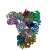
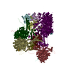
| Title | Cryo-EM Structure of Saccharomyces cerevisiae Target of Rapamycin Complex 2 | |||||||||||||||
 Components Components |
| |||||||||||||||
 Keywords Keywords | SIGNALING PROTEIN / target of rapamycin / torc2 / FRB domain / Tor2-Lst8 / kinases | |||||||||||||||
| Function / homology |  Function and homology information Function and homology informationPIP3 activates AKT signaling / CD28 dependent PI3K/Akt signaling / TOR complex / regulation of snRNA pseudouridine synthesis / High laminar flow shear stress activates signaling by PIEZO1 and PECAM1:CDH5:KDR in endothelial cells / Regulation of TP53 Degradation / mitochondria-nucleus signaling pathway / 1-phosphatidylinositol 4-kinase / 1-phosphatidylinositol 4-kinase activity / establishment or maintenance of actin cytoskeleton polarity ...PIP3 activates AKT signaling / CD28 dependent PI3K/Akt signaling / TOR complex / regulation of snRNA pseudouridine synthesis / High laminar flow shear stress activates signaling by PIEZO1 and PECAM1:CDH5:KDR in endothelial cells / Regulation of TP53 Degradation / mitochondria-nucleus signaling pathway / 1-phosphatidylinositol 4-kinase / 1-phosphatidylinositol 4-kinase activity / establishment or maintenance of actin cytoskeleton polarity / VEGFR2 mediated vascular permeability / HSF1-dependent transactivation / fungal-type cell wall organization / Amino acids regulate mTORC1 / TORC2 signaling / TORC2 complex / TORC1 complex / TORC1 signaling / fungal-type vacuole membrane / cellular response to nitrogen starvation / vacuolar membrane / negative regulation of macroautophagy / positive regulation of Rho protein signal transduction / TOR signaling / positive regulation of endocytosis / phosphatidylinositol-4,5-bisphosphate binding / cytoskeleton organization / response to nutrient / nuclear periphery / negative regulation of autophagy / protein serine/threonine kinase activator activity / regulation of actin cytoskeleton organization / regulation of cell growth / ribosome biogenesis / molecular adaptor activity / non-specific serine/threonine protein kinase / endosome membrane / regulation of cell cycle / Golgi membrane / protein serine kinase activity / protein serine/threonine kinase activity / protein-containing complex binding / signal transduction / mitochondrion / ATP binding / nucleus / plasma membrane / cytosol / cytoplasm Similarity search - Function | |||||||||||||||
| Biological species |  | |||||||||||||||
| Method | ELECTRON MICROSCOPY / single particle reconstruction / cryo EM / Resolution: 7.9 Å | |||||||||||||||
 Authors Authors | Karuppasamy, M. / Kusmider, B. / Oliveira, T.M. / Gaubitz, C. / Prouteau, M. / Loewith, R. / Schaffitzel, C. | |||||||||||||||
| Funding support | 4items
| |||||||||||||||
 Citation Citation |  Journal: Nat Commun / Year: 2017 Journal: Nat Commun / Year: 2017Title: Cryo-EM structure of Saccharomyces cerevisiae target of rapamycin complex 2. Authors: Manikandan Karuppasamy / Beata Kusmider / Taiana M Oliveira / Christl Gaubitz / Manoel Prouteau / Robbie Loewith / Christiane Schaffitzel /    Abstract: The target of rapamycin (TOR) kinase assembles into two distinct multiprotein complexes, conserved across eukaryote evolution. In contrast to TOR complex 1 (TORC1), TORC2 kinase activity is not ...The target of rapamycin (TOR) kinase assembles into two distinct multiprotein complexes, conserved across eukaryote evolution. In contrast to TOR complex 1 (TORC1), TORC2 kinase activity is not inhibited by the macrolide rapamycin. Here, we present the structure of Saccharomyces cerevisiae TORC2 determined by electron cryo-microscopy. TORC2 contains six subunits assembling into a 1.4 MDa rhombohedron. Tor2 and Lst8 form the common core of both TOR complexes. Avo3/Rictor is unique to TORC2, but interacts with the same HEAT repeats of Tor2 that are engaged by Kog1/Raptor in mammalian TORC1, explaining the mutual exclusivity of these two proteins. Density, which we conclude is Avo3, occludes the FKBP12-rapamycin-binding site of Tor2's FRB domain rendering TORC2 rapamycin insensitive and recessing the kinase active site. Although mobile, Avo1/hSin1 further restricts access to the active site as its conserved-region-in-the-middle (CRIM) domain is positioned along an edge of the TORC2 active-site-cleft, consistent with a role for CRIM in substrate recruitment. #1:  Journal: Mol Cell / Year: 2015 Journal: Mol Cell / Year: 2015Title: Molecular Basis of the Rapamycin Insensitivity of Target Of Rapamycin Complex 2. Authors: Christl Gaubitz / Taiana M Oliveira / Manoel Prouteau / Alexander Leitner / Manikandan Karuppasamy / Georgia Konstantinidou / Delphine Rispal / Sandra Eltschinger / Graham C Robinson / ...Authors: Christl Gaubitz / Taiana M Oliveira / Manoel Prouteau / Alexander Leitner / Manikandan Karuppasamy / Georgia Konstantinidou / Delphine Rispal / Sandra Eltschinger / Graham C Robinson / Stéphane Thore / Ruedi Aebersold / Christiane Schaffitzel / Robbie Loewith /    Abstract: Target of Rapamycin (TOR) plays central roles in the regulation of eukaryote growth as the hub of two essential multiprotein complexes: TORC1, which is rapamycin-sensitive, and the lesser ...Target of Rapamycin (TOR) plays central roles in the regulation of eukaryote growth as the hub of two essential multiprotein complexes: TORC1, which is rapamycin-sensitive, and the lesser characterized TORC2, which is not. TORC2 is a key regulator of lipid biosynthesis and Akt-mediated survival signaling. In spite of its importance, its structure and the molecular basis of its rapamycin insensitivity are unknown. Using crosslinking-mass spectrometry and electron microscopy, we determined the architecture of TORC2. TORC2 displays a rhomboid shape with pseudo-2-fold symmetry and a prominent central cavity. Our data indicate that the C-terminal part of Avo3, a subunit unique to TORC2, is close to the FKBP12-rapamycin-binding domain of Tor2. Removal of this sequence generated a FKBP12-rapamycin-sensitive TORC2 variant, which provides a powerful tool for deciphering TORC2 function in vivo. Using this variant, we demonstrate a role for TORC2 in G2/M cell-cycle progression. | |||||||||||||||
| History |
|
- Structure visualization
| Movie |
 Movie viewer Movie viewer |
|---|---|
| Structure viewer | Molecule:  Molmil Molmil Jmol/JSmol Jmol/JSmol |
- Downloads & links
- Download
| PDBx/mmCIF format |  6emk.cif.gz 6emk.cif.gz | 1.2 MB | Display |  PDBx/mmCIF format PDBx/mmCIF format |
|---|---|---|---|---|
| PDB format |  pdb6emk.ent.gz pdb6emk.ent.gz | 983 KB | Display |  PDB format PDB format |
| PDBx/mmJSON format |  6emk.json.gz 6emk.json.gz | Tree view |  PDBx/mmJSON format PDBx/mmJSON format | |
| Others |  Other downloads Other downloads |
-Validation report
| Summary document |  6emk_validation.pdf.gz 6emk_validation.pdf.gz | 517.2 KB | Display |  wwPDB validaton report wwPDB validaton report |
|---|---|---|---|---|
| Full document |  6emk_full_validation.pdf.gz 6emk_full_validation.pdf.gz | 798.2 KB | Display | |
| Data in XML |  6emk_validation.xml.gz 6emk_validation.xml.gz | 131 KB | Display | |
| Data in CIF |  6emk_validation.cif.gz 6emk_validation.cif.gz | 205.9 KB | Display | |
| Arichive directory |  https://data.pdbj.org/pub/pdb/validation_reports/em/6emk https://data.pdbj.org/pub/pdb/validation_reports/em/6emk ftp://data.pdbj.org/pub/pdb/validation_reports/em/6emk ftp://data.pdbj.org/pub/pdb/validation_reports/em/6emk | HTTPS FTP |
-Related structure data
| Related structure data |  3896MC M: map data used to model this data C: citing same article ( |
|---|---|
| Similar structure data |
- Links
Links
- Assembly
| Deposited unit | 
|
|---|---|
| 1 |
|
- Components
| #1: Protein | Mass: 281915.438 Da / Num. of mol.: 2 Source method: isolated from a genetically manipulated source Source: (gene. exp.)  Gene: TOR2, DRR2, TSC14, YKL203C / Production host:  References: UniProt: P32600, 1-phosphatidylinositol 4-kinase, non-specific serine/threonine protein kinase #2: Protein | Mass: 34077.879 Da / Num. of mol.: 2 Source method: isolated from a genetically manipulated source Source: (gene. exp.)  Gene: LST8, YNL006W, N2005 / Production host:  #3: Protein | Mass: 25804.662 Da / Num. of mol.: 2 Source method: isolated from a genetically manipulated source Source: (gene. exp.)  Production host:  #4: Protein | Mass: 47206.457 Da / Num. of mol.: 2 Source method: isolated from a genetically manipulated source Source: (gene. exp.)  Gene: AVO2, YMR068W, YM9916.07 / Production host:  #5: Protein | Mass: 131565.453 Da / Num. of mol.: 2 Source method: isolated from a genetically manipulated source Source: (gene. exp.)  Gene: AVO1, YOL078W, O1110 / Production host:  |
|---|
-Experimental details
-Experiment
| Experiment | Method: ELECTRON MICROSCOPY |
|---|---|
| EM experiment | Aggregation state: PARTICLE / 3D reconstruction method: single particle reconstruction |
- Sample preparation
| Component | Name: Target of rapamycin protein complex 2 / Type: COMPLEX / Entity ID: all / Source: RECOMBINANT |
|---|---|
| Molecular weight | Value: 1.4 MDa / Experimental value: NO |
| Source (natural) | Organism:  |
| Source (recombinant) | Organism:  |
| Buffer solution | pH: 7.5 |
| Specimen | Embedding applied: NO / Shadowing applied: NO / Staining applied: NO / Vitrification applied: YES |
| Specimen support | Grid material: COPPER / Grid mesh size: 300 divisions/in. / Grid type: Quantifoil R2/2 |
| Vitrification | Instrument: FEI VITROBOT MARK IV / Cryogen name: ETHANE / Humidity: 100 % / Chamber temperature: 4 K / Details: 2 - 3 sec blotting |
- Electron microscopy imaging
| Experimental equipment |  Model: Titan Krios / Image courtesy: FEI Company | |||||||||||||||||||||
|---|---|---|---|---|---|---|---|---|---|---|---|---|---|---|---|---|---|---|---|---|---|---|
| Microscopy | Model: FEI TITAN KRIOS | |||||||||||||||||||||
| Electron gun | Electron source:  FIELD EMISSION GUN / Accelerating voltage: 300 kV / Illumination mode: FLOOD BEAM FIELD EMISSION GUN / Accelerating voltage: 300 kV / Illumination mode: FLOOD BEAM | |||||||||||||||||||||
| Electron lens | Mode: BRIGHT FIELD / Nominal magnification: 105000 X / Nominal defocus max: 3500 nm / Nominal defocus min: 1500 nm / Cs: 2.7 mm / C2 aperture diameter: 50 µm / Alignment procedure: COMA FREE | |||||||||||||||||||||
| Specimen holder | Cryogen: NITROGEN / Specimen holder model: FEI TITAN KRIOS AUTOGRID HOLDER / Temperature (max): 70 K / Temperature (min): 70 K | |||||||||||||||||||||
| Image recording | Imaging-ID: 1
| |||||||||||||||||||||
| Image scans | Width: 3710 / Height: 3838 / Movie frames/image: 40 / Used frames/image: 1-40 |
- Processing
| Software | Name: REFMAC / Version: 5.8.0158 / Classification: refinement | ||||||||||||||||||||||||||||||||||||||||||||||||||||||||||||||||||||||||||||||||||||||||||||||||||||||||||
|---|---|---|---|---|---|---|---|---|---|---|---|---|---|---|---|---|---|---|---|---|---|---|---|---|---|---|---|---|---|---|---|---|---|---|---|---|---|---|---|---|---|---|---|---|---|---|---|---|---|---|---|---|---|---|---|---|---|---|---|---|---|---|---|---|---|---|---|---|---|---|---|---|---|---|---|---|---|---|---|---|---|---|---|---|---|---|---|---|---|---|---|---|---|---|---|---|---|---|---|---|---|---|---|---|---|---|---|
| EM software |
| ||||||||||||||||||||||||||||||||||||||||||||||||||||||||||||||||||||||||||||||||||||||||||||||||||||||||||
| Image processing |
| ||||||||||||||||||||||||||||||||||||||||||||||||||||||||||||||||||||||||||||||||||||||||||||||||||||||||||
| CTF correction |
| ||||||||||||||||||||||||||||||||||||||||||||||||||||||||||||||||||||||||||||||||||||||||||||||||||||||||||
| Particle selection |
| ||||||||||||||||||||||||||||||||||||||||||||||||||||||||||||||||||||||||||||||||||||||||||||||||||||||||||
| Symmetry |
| ||||||||||||||||||||||||||||||||||||||||||||||||||||||||||||||||||||||||||||||||||||||||||||||||||||||||||
| 3D reconstruction |
| ||||||||||||||||||||||||||||||||||||||||||||||||||||||||||||||||||||||||||||||||||||||||||||||||||||||||||
| Atomic model building | Protocol: FLEXIBLE FIT / Space: REAL | ||||||||||||||||||||||||||||||||||||||||||||||||||||||||||||||||||||||||||||||||||||||||||||||||||||||||||
| Atomic model building | PDB-ID: 5FVM Accession code: 5FVM / Pdb chain residue range: 81-2474 / Source name: PDB / Type: experimental model | ||||||||||||||||||||||||||||||||||||||||||||||||||||||||||||||||||||||||||||||||||||||||||||||||||||||||||
| Refinement | Resolution: 7.9→7.9 Å / Cor.coef. Fo:Fc: 0.987 / SU B: 111.293 / SU ML: 0.794 Stereochemistry target values: MAXIMUM LIKELIHOOD WITH PHASES
| ||||||||||||||||||||||||||||||||||||||||||||||||||||||||||||||||||||||||||||||||||||||||||||||||||||||||||
| Solvent computation | Solvent model: PARAMETERS FOR MASK CACLULATION | ||||||||||||||||||||||||||||||||||||||||||||||||||||||||||||||||||||||||||||||||||||||||||||||||||||||||||
| Displacement parameters | Biso mean: 295.421 Å2
| ||||||||||||||||||||||||||||||||||||||||||||||||||||||||||||||||||||||||||||||||||||||||||||||||||||||||||
| Refinement step | Cycle: 1 / Total: 48016 | ||||||||||||||||||||||||||||||||||||||||||||||||||||||||||||||||||||||||||||||||||||||||||||||||||||||||||
| Refine LS restraints |
|
 Movie
Movie Controller


 PDBj
PDBj


