Movie
Movie Controller


+ Open data

- Basic information
| Entry | Database: PDB / ID: 5fvm | ||||||
|---|---|---|---|---|---|---|---|
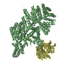
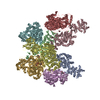
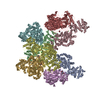
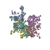
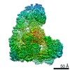
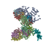
| Title | Cryo electron microscopy of a complex of Tor and Lst8 | ||||||
 Components Components |
| ||||||
 Keywords Keywords | TRANSFERASE / CRYO-EM / TOR / LST8 / MTOR / KINASE / PIKK / S/T PROTEIN KINASE / TORC1 / MTORC1 | ||||||
| Function / homology | : / :  Function and homology information Function and homology information | ||||||
| Biological species |  KLUYVEROMYCES MARXIANUS (yeast) KLUYVEROMYCES MARXIANUS (yeast) | ||||||
| Method | ELECTRON MICROSCOPY / single particle reconstruction / cryo EM / Resolution: 6.7 Å | ||||||
 Authors Authors | Baretic, D. / Berndt, A. / Ohashi, Y. / Johnson, C.M. / Williams, R.L. | ||||||
 Citation Citation | Journal: Nat Commun / Year: 2016 Title: Tor forms a dimer through an N-terminal helical solenoid with a complex topology. Authors: Domagoj Baretić / Alex Berndt / Yohei Ohashi / Christopher M Johnson / Roger L Williams /  Abstract: The target of rapamycin (Tor) is a Ser/Thr protein kinase that regulates a range of anabolic and catabolic processes. Tor is present in two complexes, TORC1 and TORC2, in which the Tor-Lst8 ...The target of rapamycin (Tor) is a Ser/Thr protein kinase that regulates a range of anabolic and catabolic processes. Tor is present in two complexes, TORC1 and TORC2, in which the Tor-Lst8 heterodimer forms a common sub-complex. We have determined the cryo-electron microscopy (EM) structure of Tor bound to Lst8. Two Tor-Lst8 heterodimers assemble further into a dyad-symmetry dimer mediated by Tor-Tor interactions. The first 1,300 residues of Tor form a HEAT repeat-containing α-solenoid with four distinct segments: a highly curved 800-residue N-terminal 'spiral', followed by a 400-residue low-curvature 'bridge' and an extended 'railing' running along the bridge leading to the 'cap' that links to FAT region. This complex topology was verified by domain insertions and offers a new interpretation of the mTORC1 structure. The spiral of one TOR interacts with the bridge of another, which together form a joint platform for the Regulatory Associated Protein of TOR (RAPTOR) regulatory subunit. | ||||||
| History |
|
- Structure visualization
| Movie |
Movie viewer |
|---|---|
| Structure viewer | Molecule: MolmilJmol/JSmol |
- Downloads & links
-Download
| PDBx/mmCIF format | 5fvm.cif.gz | 728.3 KB | Display | PDBx/mmCIF format |
|---|---|---|---|---|
| PDB format | pdb5fvm.ent.gz | 491.8 KB | Display | PDB format |
| PDBx/mmJSON format | 5fvm.json.gz | Tree view | PDBx/mmJSON format | |
| Others |  Other downloads Other downloads |
-Validation report
| Arichive directory | https://data.pdbj.org/pub/pdb/validation_reports/fv/5fvmftp://data.pdbj.org/pub/pdb/validation_reports/fv/5fvm | HTTPS FTP |
|---|
-Related structure data
| Related structure data | 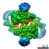 3329MC 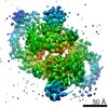 3334C 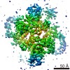 3335C 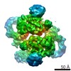 3336C M: map data used to model this data C: citing same article ( |
|---|---|
| Similar structure data |
-Links
 PDBj
PDBj- Assembly
| Deposited unit | 
|
|---|---|
| 1 |
|
| Noncrystallographic symmetry (NCS) | NCS oper: (Code: given / Matrix: (-1), |
-Components
| #1: Protein | Mass: 279407.938 Da / Num. of mol.: 2 Source method: isolated from a genetically manipulated source Source: (gene. exp.) KLUYVEROMYCES MARXIANUS (yeast) / Gene: KMAR_30040 / Production host: KLUYVEROMYCES MARXIANUS (yeast) / References: UniProt: A0A090BKR7#2: Protein | Mass: 33952.672 Da / Num. of mol.: 2 Source method: isolated from a genetically manipulated source Source: (gene. exp.) KLUYVEROMYCES MARXIANUS (yeast) / Gene: KMAR_20458 / Production host: KLUYVEROMYCES MARXIANUS (yeast) / References: UniProt: A0A090BJK6Sequence details | THE POLYPEPTIDE HAS AN N-TERMINAL 22 RESIDUE TAG AFTER TEV CLEAVGE. IT WAS CLONED FROM GENOMIC DNA. ...THE POLYPEPTID | |
|---|
-Experimental details
-Experiment
| Experiment | Method: ELECTRON MICROSCOPY |
|---|---|
| EM experiment | Aggregation state: PARTICLE / 3D reconstruction method: single particle reconstruction |
- Sample preparation
| Component | Name: COMPLEX OF TOR1 WITH LST8 / Type: COMPLEX |
|---|---|
| Buffer solution | Name: 50 MM HEPES PH 7.4 (23 DEG C), 75 MM KCL, 250 MM NACL, 0.3 % (V/V) CHAPS, 1 MM TCEP pH: 7.4 Details: 50 MM HEPES PH 7.4 (23 DEG C), 75 MM KCL, 250 MM NACL, 0.3 % (V/V) CHAPS, 1 MM TCEP |
| Specimen | Conc.: 2.5 mg/ml / Embedding applied: NO / Shadowing applied: NO / Staining applied: NO / Vitrification applied: YES |
| Specimen support | Details: HOLEY CARBON |
| Vitrification | Cryogen name: ETHANE Details: VITRIFICATION 1 -- CRYOGEN- ETHANE, INSTRUMENT- OTHER, METHOD- 11-13 S AT 4 DEG C, |
- Electron microscopy imaging
| Experimental equipment |  Model: Titan Krios / Image courtesy: FEI Company |
|---|---|
| Microscopy | Model: FEI TITAN KRIOS / Date: May 16, 2015 |
| Electron gun | Electron source:  FIELD EMISSION GUN / Accelerating voltage: 300 kV / Illumination mode: FLOOD BEAM FIELD EMISSION GUN / Accelerating voltage: 300 kV / Illumination mode: FLOOD BEAM |
| Electron lens | Mode: BRIGHT FIELD / Nominal magnification: 47000 X / Calibrated magnification: 105263 X / Nominal defocus max: 3500 nm / Nominal defocus min: 2500 nm / Cs: 2.7 mm |
| Image recording | Electron dose: 40 e/Å2 / Film or detector model: FEI FALCON II (4k x 4k) |
- Processing
| EM software |
| ||||||||||||
|---|---|---|---|---|---|---|---|---|---|---|---|---|---|
| CTF correction | Details: EACH PARTICLE (GCTF) | ||||||||||||
| Symmetry | Point symmetry: C2 (2 fold cyclic) | ||||||||||||
| 3D reconstruction | Resolution: 6.7 Å / Num. of particles: 28877 / Nominal pixel size: 1.33 Å / Actual pixel size: 1.33 Å Details: THE FAT AND KINASE DOMAINS (FATKIN) WERE FIT USING 4JSV FATKIN AS AN INITIAL MODEL. THE REMAINDER OF THE STRUCTURE WAS FIT WITH A POLY-ALANINE, AND AN APPROXIMATE SEQUENCE REGISTER WAS ...Details: THE FAT AND KINASE DOMAINS (FATKIN) WERE FIT USING 4JSV FATKIN AS AN INITIAL MODEL. THE REMAINDER OF THE STRUCTURE WAS FIT WITH A POLY-ALANINE, AND AN APPROXIMATE SEQUENCE REGISTER WAS ESTABLISHED BASED ON THE PREDICED HELICAL ELEMENTS IN THE N-TERMINAL REGION AND THE HELICES VISIBLE IN THE EM DENSITY. THE ENTIRE MODEL IS POLY-ALANINE. THE TOPOLOGY OF THE MODEL WAS VERIFIED BY DETERMINING THE STRUCTURES OF VARINTS IN WHICH TENDEM RFPS WERE INSERTED INTO THE STRUCTURE. SUBMISSION BASED ON EXPERIMENTAL DATA FROM EMDB EMD-3329. (DEPOSITION ID: 14248). Symmetry type: POINT | ||||||||||||
| Atomic model building | Protocol: FLEXIBLE FIT / Space: REAL / Target criteria: Cross-correlation coefficient / Details: METHOD--FLEXIBLE | ||||||||||||
| Atomic model building | PDB-ID: 4JSV Accession code: 4JSV / Source name: PDB / Type: experimental model | ||||||||||||
| Refinement | Highest resolution: 6.7 Å | ||||||||||||
| Refinement step | Cycle: LAST / Highest resolution: 6.7 Å
|