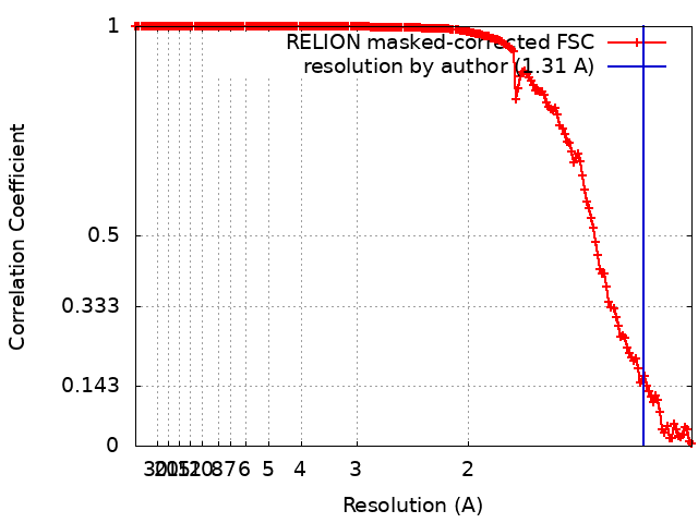
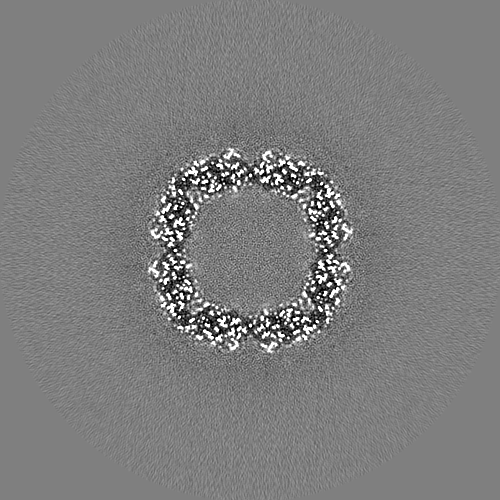
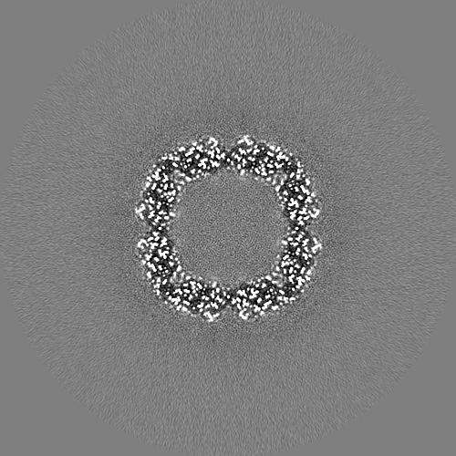
Journal: Microscopy (Oxf) / Year: 2021 Title: Cryo-EM performance testing of hardware and data acquisition strategies. Authors: Radostin Danev / Haruaki Yanagisawa / Masahide Kikkawa / Abstract: The increasing popularity and adoption rate of cryo-electron microscopy (cryo-EM) is evidenced by a growing number of new microscope installations around the world. The quality and reliability of the ...The increasing popularity and adoption rate of cryo-electron microscopy (cryo-EM) is evidenced by a growing number of new microscope installations around the world. The quality and reliability of the instruments improved dramatically in recent years, but site-specific issues or unnoticed problems during installation could undermine productivity. Newcomers to the field may also have limited experience and/or low confidence in the capabilities of the equipment or their own skills. Therefore, it is recommended to perform an initial test of the complete cryo-EM workflow with an 'easy' test sample, such as apoferritin, before starting work with real and challenging samples. Analogous test experiments are also recommended for the quantification of new data acquisition approaches or imaging hardware. Here, we present the results from our initial tests of a recently installed Krios G4 electron microscope equipped with two latest generation direct electron detector cameras-Gatan K3 and Falcon 4. Three beam-image shift-based data acquisition strategies were also tested. We detail the methodology and discuss the critical parameters and steps for performance testing. The two cameras performed equally, and the single- and multi-shot per-hole acquisition schemes produced comparable results. We also evaluated the effects of environmental factors and optical flaws on data quality. Our results reaffirmed the exceptional performance of the software aberration correction in Relion in dealing with severe coma aberration. We hope that this work will help cryo-EM teams in their testing and troubleshooting of hardware and data collection approaches.
History
Deposition
Nov 17, 2020
-
Header (metadata) release
Dec 2, 2020
-
Map release
Dec 2, 2020
-
Update
Mar 16, 2022
-
Current status
Mar 16, 2022
Processing site: PDBj / Status: Released
-
Structure visualization
Movie
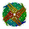
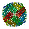
Surface view with section colored by density value
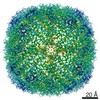
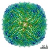
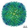
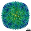
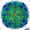
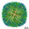
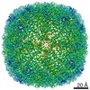
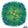
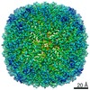
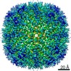
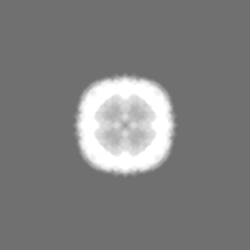
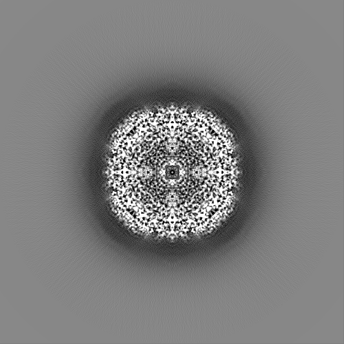
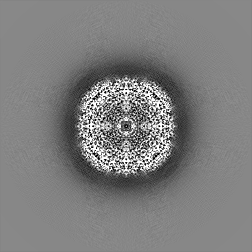
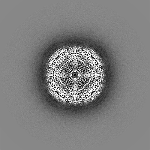
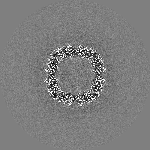
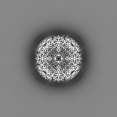
EMPIAR-10559 (Title: Krios G4 apoferritin test with K3/BioQuantum SerialEM BIS 3x3x1 Data size: 659.0 Data #1: Unaligned multi-frame non-gain-normalized movies in LZW compressed TIFF format [micrographs - multiframe])
In the structure databanks used in Yorodumi, some data are registered as the other names, "COVID-19 virus" and "2019-nCoV". Here are the details of the virus and the list of structure data.
Jan 31, 2019. EMDB accession codes are about to change! (news from PDBe EMDB page)
EMDB accession codes are about to change! (news from PDBe EMDB page)
The allocation of 4 digits for EMDB accession codes will soon come to an end. Whilst these codes will remain in use, new EMDB accession codes will include an additional digit and will expand incrementally as the available range of codes is exhausted. The current 4-digit format prefixed with “EMD-” (i.e. EMD-XXXX) will advance to a 5-digit format (i.e. EMD-XXXXX), and so on. It is currently estimated that the 4-digit codes will be depleted around Spring 2019, at which point the 5-digit format will come into force.
The EM Navigator/Yorodumi systems omit the EMD- prefix.
Related info.:Q: What is EMD? / ID/Accession-code notation in Yorodumi/EM Navigator
Yorodumi is a browser for structure data from EMDB, PDB, SASBDB, etc.
This page is also the successor to EM Navigator detail page, and also detail information page/front-end page for Omokage search.
The word "yorodu" (or yorozu) is an old Japanese word meaning "ten thousand". "mi" (miru) is to see.
Related info.:EMDB / PDB / SASBDB / Comparison of 3 databanks / Yorodumi Search / Aug 31, 2016. New EM Navigator & Yorodumi / Yorodumi Papers / Jmol/JSmol / Function and homology information / Changes in new EM Navigator and Yorodumi
 Movie
Movie Controller

 Yorodumi
 Open data
 Basic information
 Map data
 Sample
 Function and homology information
 Authors
 Japan, 1 items
  Citation
 Structure visualization
 Downloads & links
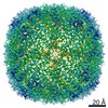
 emd_30683.png
 http://ftp.pdbj.org/pub/emdb/structures/EMD-30683
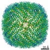


 Z (Sec.)
 Y (Row.)
 X (Col.)


 Sample components
 Processing
 Electron microscopy
 FIELD EMISSION GUN