Movie
Movie Controller

[English] 日本語
 Yorodumi
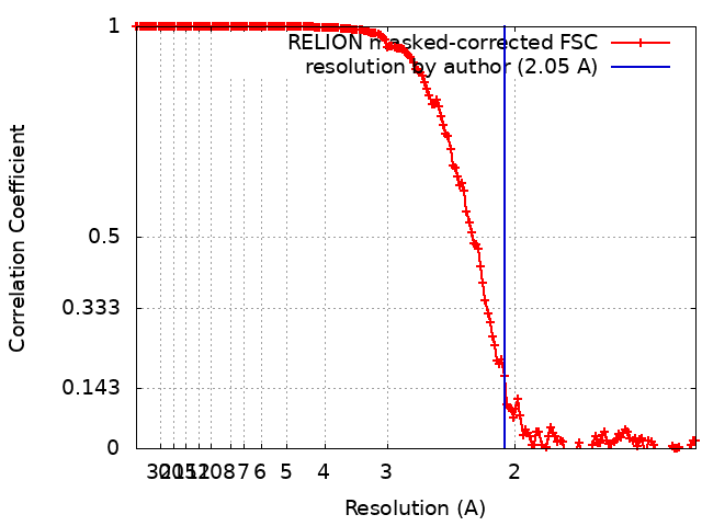
- EMDB-30362: 2.05 angstrom resolution structure determination of sulfur oxygen... -
+ Open data

- Basic information
| Entry | Database: EMDB / ID: EMD-30362 | |||||||||
|---|---|---|---|---|---|---|---|---|---|---|
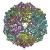
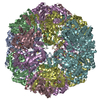
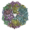
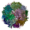
| Title | 2.05 angstrom resolution structure determination of sulfur oxygenase reductase using 200kV cryo-EM | |||||||||
 Map data Map data | ||||||||||
 Sample Sample |
| |||||||||
| Biological species |   Sulfurisphaera tokodaii str. 7 (archaea) Sulfurisphaera tokodaii str. 7 (archaea) | |||||||||
| Method | single particle reconstruction / cryo EM / Resolution: 2.05 Å | |||||||||
 Authors Authors | Moriya T / Naruhiko A / Sato Y / Arakawa T / Kawasaki M / Yamada C / Fushinobu S / Senda T | |||||||||
| Funding support |  Japan, 2 items Japan, 2 items
| |||||||||
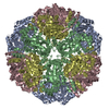
 Citation Citation | Journal: J Struct Biol X / Year: 2020 Title: Crystallographic and cryogenic electron microscopic structures and enzymatic characterization of sulfur oxygenase reductase from . Authors: Yuta Sato / Takashi Yabuki / Naruhiko Adachi / Toshio Moriya / Takatoshi Arakawa / Masato Kawasaki / Chihaya Yamada / Toshiya Senda / Shinya Fushinobu / Takayoshi Wakagi / Abstract: Sulfur oxygenase reductases (SORs) are present in thermophilic and mesophilic archaea and bacteria, and catalyze oxygen-dependent oxygenation and disproportionation of elemental sulfur. SOR has a ...Sulfur oxygenase reductases (SORs) are present in thermophilic and mesophilic archaea and bacteria, and catalyze oxygen-dependent oxygenation and disproportionation of elemental sulfur. SOR has a hollow, spherical homo-24-mer structure and reactions take place at active sites inside the chamber. The crystal structures of SORs from species have been reported. However, the states of the active site components (mononuclear iron and cysteines) and the entry and exit paths of the substrate and products are still in dispute. Here, we report the biochemical and structural characterizations of SORs from the thermoacidophilic archaeon (StSOR) and present high-resolution structures determined by X-ray crystallography and cryogenic electron microscopy (cryo-EM). The crystal structure of StSOR was determined at 1.73 Å resolution. At the catalytic center, iron is ligated to His86, His90, Glu114, and two water molecules. Three conserved cysteines in the cavity are located 9.5-13 Å from the iron and were observed as free thiol forms. A mutational analysis indicated that the iron and one of the cysteines (Cys31) were essential for both activities. The cryo-EM structure was determined at 2.24 Å resolution using an instrument operating at 200 kV. The two structures determined by different methodologies showed similar main chain traces, but the maps exhibited different features at catalytically important components. A possible role of StSOR in the sulfur metabolism of (an obligate aerobe) is discussed based on this study. Given the high resolution achieved in this study, StSOR was shown to be a good benchmark sample for cryo-EM. #1: Journal: J Struct Biol X / Year: 2020Title: Crystallographic and cryogenic electron microscopic structures and enzymatic characterization of sulfur oxygenase reductase from Sulfurisphaera tokodaii Authors: Sato Y / Yabuki T / Adachi N / Moriya T / Arakawa T / Kawasaki M / Yamada C / Senda T / Fushinobu S / Wakagi T | |||||||||
| History |
|
- Structure visualization
| Movie |
 Movie viewer Movie viewer |
|---|---|
| Structure viewer | EM map: SurfViewMolmilJmol/JSmol |
| Supplemental images |
- Downloads & links
-EMDB archive
| Map data | emd_30362.map.gz | 380.9 MB | EMDB map data format | |
|---|---|---|---|---|
| Header (meta data) | emd-30362-v30.xmlemd-30362.xml | 19.7 KB 19.7 KB | Display Display | EMDB header |
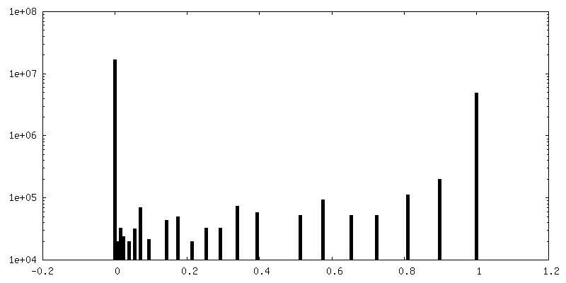
| FSC (resolution estimation) | emd_30362_fsc.xml | 16.9 KB | Display | FSC data file |
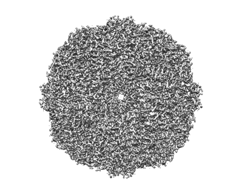
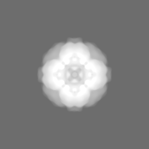
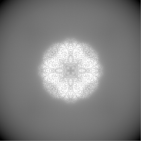
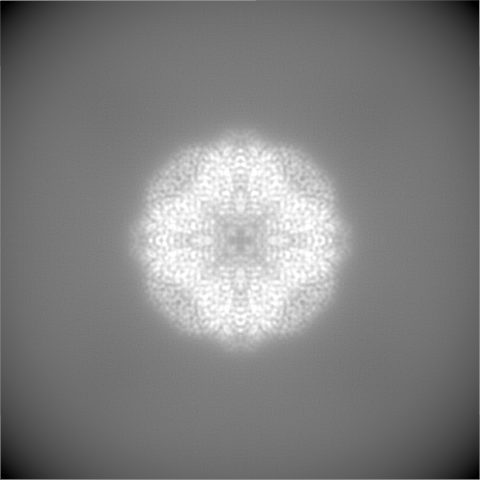
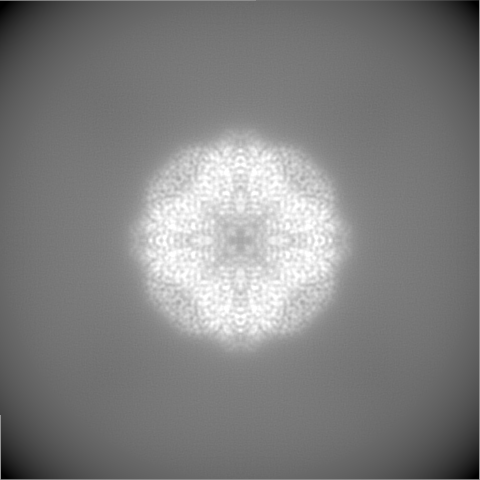
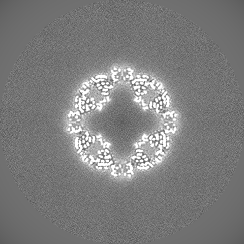
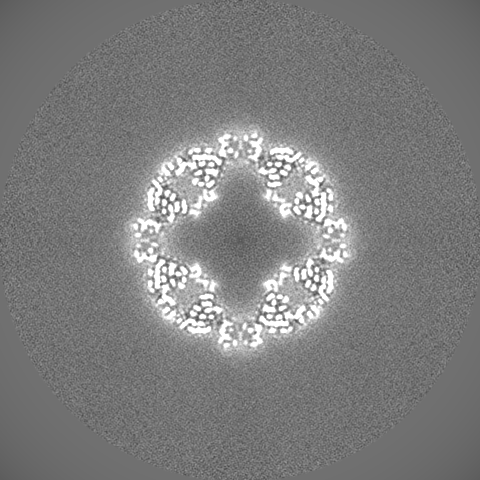
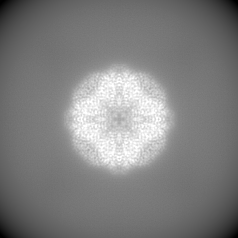
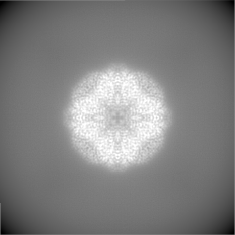
| Images |  emd_30362.png emd_30362.png | 108.6 KB | ||
| Masks | emd_30362_msk_1.map | 421.9 MB | Mask map | |
| Others | emd_30362_half_map_1.map.gzemd_30362_half_map_2.map.gz | 331.3 MB 331.3 MB | ||
| Archive directory |  http://ftp.pdbj.org/pub/emdb/structures/EMD-30362ftp://ftp.pdbj.org/pub/emdb/structures/EMD-30362 http://ftp.pdbj.org/pub/emdb/structures/EMD-30362ftp://ftp.pdbj.org/pub/emdb/structures/EMD-30362 | HTTPS FTP |
-Related structure data
| Related structure data | 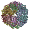 6m35C  6m3xC C: citing same article ( |
|---|---|
| Similar structure data | |
| EM raw data | EMPIAR-10546 (Title: 2.05 angstrom resolution structure determination of sulfur oxygenase reductase using 200kV cryo-EM Data size: 3.9 TB Data #1: 2.05 angstrom resolution structure determination of sulfur oxygenase reductase using 200kV cryo-EM [micrographs - multiframe]) |


-Links
| EMDB pages | EMDB (EBI/PDBe) / EMDataResource |
|---|
-Map
| File | Download / File: emd_30362.map.gz / Format: CCP4 / Size: 421.9 MB / Type: IMAGE STORED AS FLOATING POINT NUMBER (4 BYTES) | ||||||||||||||||||||||||||||||||||||||||||||||||||||||||||||||||||||
|---|---|---|---|---|---|---|---|---|---|---|---|---|---|---|---|---|---|---|---|---|---|---|---|---|---|---|---|---|---|---|---|---|---|---|---|---|---|---|---|---|---|---|---|---|---|---|---|---|---|---|---|---|---|---|---|---|---|---|---|---|---|---|---|---|---|---|---|---|---|
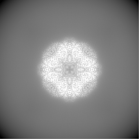
| Projections & slices | Image control
Images are generated by Spider. | ||||||||||||||||||||||||||||||||||||||||||||||||||||||||||||||||||||
| Voxel size | X=Y=Z: 0.676 Å | ||||||||||||||||||||||||||||||||||||||||||||||||||||||||||||||||||||
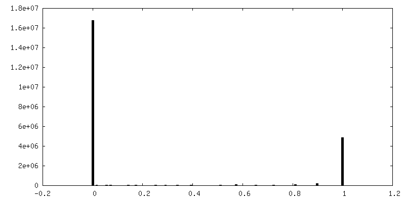
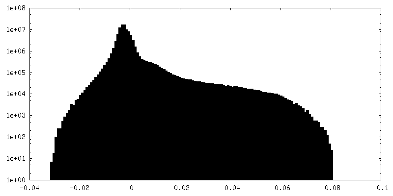
| Density |
| ||||||||||||||||||||||||||||||||||||||||||||||||||||||||||||||||||||
| Symmetry | Space group: 1 | ||||||||||||||||||||||||||||||||||||||||||||||||||||||||||||||||||||
| Details | EMDB XML:
CCP4 map header:
| ||||||||||||||||||||||||||||||||||||||||||||||||||||||||||||||||||||
 Z (Sec.)
 Y (Row.)
 X (Col.)


-Supplemental data
-Mask #1
| File | emd_30362_msk_1.map | ||||||||||||
|---|---|---|---|---|---|---|---|---|---|---|---|---|---|
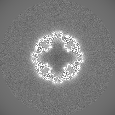
| Projections & Slices |
| ||||||||||||
| Density Histograms |


-Half map: #2
| File | emd_30362_half_map_1.map | ||||||||||||
|---|---|---|---|---|---|---|---|---|---|---|---|---|---|
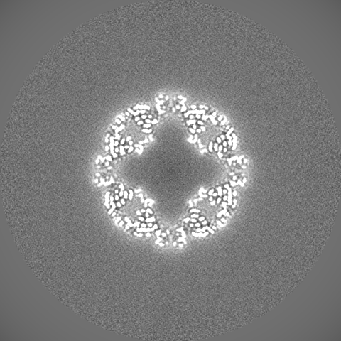
| Projections & Slices |
| ||||||||||||
| Density Histograms |


-Half map: #1
| File | emd_30362_half_map_2.map | ||||||||||||
|---|---|---|---|---|---|---|---|---|---|---|---|---|---|
| Projections & Slices |
| ||||||||||||
| Density Histograms |


- Sample components
-Entire : sulfur oxygenase reductase from Sulfurisphaera tokodaii
| Entire | Name: sulfur oxygenase reductase from Sulfurisphaera tokodaii |
|---|---|
| Components |
|
-Supramolecule #1: sulfur oxygenase reductase from Sulfurisphaera tokodaii
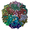
| Supramolecule | Name: sulfur oxygenase reductase from Sulfurisphaera tokodaii type: organelle_or_cellular_component / ID: 1 / Parent: 0 / Macromolecule list: all / Details: spherical homo 24-mer |
|---|---|
| Source (natural) | Organism: Sulfurisphaera tokodaii str. 7 (archaea) / Strain: strain 7 |
| Molecular weight | Theoretical: 857 KDa |
| Recombinant expression | Organism:  |
-Macromolecule #1: Sulfur oxygenase/reductase
| Macromolecule | Name: Sulfur oxygenase/reductase / type: protein_or_peptide / ID: 1 / Enantiomer: LEVO / EC number: sulfur oxygenase/reductase |
|---|---|
| Source (natural) | Organism: Sulfurisphaera tokodaii str. 7 (archaea) / Strain: DSM 16993 / JCM 10545 / NBRC 100140 / 7 |
| Recombinant expression | Organism: |
| Sequence | String: MPKPYVAINM VEVRNDPKTL ELFGKVGPKV CMVTARHPGF VGFQNHVQIG VVPLGTRWGG AKMEMSQEMH SLMLMQYTFW KNWKDHEEM HKQNWANLFR LCLQCADQMI WGPYEPLYEI VYANMPLNTE MTDFTVMVGK KFAAGEAVSI PPISQPYGKR V VAFGEHIV ...String: MPKPYVAINM VEVRNDPKTL ELFGKVGPKV CMVTARHPGF VGFQNHVQIG VVPLGTRWGG AKMEMSQEMH SLMLMQYTFW KNWKDHEEM HKQNWANLFR LCLQCADQMI WGPYEPLYEI VYANMPLNTE MTDFTVMVGK KFAAGEAVSI PPISQPYGKR V VAFGEHIV KEGLENQFEE YAIKTLEAFR SAPGFLGGMI LKEIGVSPLG SLQLNAKGFH QILETANGMD VPEPVTIYEA PE FRNRPQR YIVHTEWSDT NALMFGLGRV LIYPEVRQIH DKVLDTLVYG PYIRVLNPMM EGTYWREYLN EYHL |
-Experimental details
-Structure determination
| Method | cryo EM |
|---|---|
 Processing Processing | single particle reconstruction |
| Aggregation state | particle |
-Sample preparation
| Concentration | 10.0 mg/mL | |||||||||
|---|---|---|---|---|---|---|---|---|---|---|
| Buffer | pH: 8 Component:
| |||||||||
| Grid | Model: Quantifoil R1.2/1.3 / Material: COPPER / Mesh: 300 / Support film - Material: CARBON / Support film - topology: HOLEY / Pretreatment - Type: GLOW DISCHARGE / Pretreatment - Atmosphere: AIR / Details: The grid was washed by acetone prior to use. | |||||||||
| Vitrification | Cryogen name: ETHANE / Chamber humidity: 100 % / Chamber temperature: 291 K / Instrument: FEI VITROBOT MARK IV / Details: Blotting time was 5 seconds (blot force 20). | |||||||||
| Details | This sample was mono-disperse. |
- Electron microscopy
| Microscope | FEI TALOS ARCTICA |
|---|---|
| Image recording | Film or detector model: FEI FALCON III (4k x 4k) / Detector mode: COUNTING / Number grids imaged: 1 / Number real images: 2558 / Average exposure time: 50.89 sec. / Average electron dose: 50.0 e/Å2 |
| Electron beam | Acceleration voltage: 200 kV / Electron source:  FIELD EMISSION GUN FIELD EMISSION GUN |
| Electron optics | C2 aperture diameter: 50.0 µm / Illumination mode: FLOOD BEAM / Imaging mode: BRIGHT FIELD / Cs: 2.7 mm / Nominal defocus max: 1.5 µm / Nominal defocus min: 0.3 µm / Nominal magnification: 150000 |
| Sample stage | Cooling holder cryogen: NITROGEN |
| Experimental equipment |  Model: Talos Arctica / Image courtesy: FEI Company |