Movie
Movie Controller


+ Open data

- Basic information
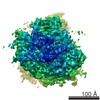
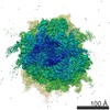
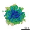
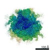
| Entry | Database: EMDB / ID: EMD-9237 | |||||||||
|---|---|---|---|---|---|---|---|---|---|---|
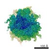
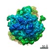
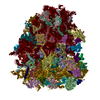
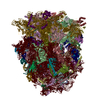
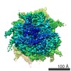
| Title | Rabbit 80S ribosome with P- and Z-site tRNAs (unrotated state) | |||||||||
 Map data Map data | Postprocessed map | |||||||||
 Sample Sample |
| |||||||||
 Keywords Keywords | Ribosome / translation | |||||||||
| Function / homology |  Function and homology information Function and homology informationubiquitin ligase inhibitor activity / 90S preribosome / positive regulation of signal transduction by p53 class mediator / phagocytic cup / gastrulation / translation regulator activity / rough endoplasmic reticulum / ribosomal small subunit export from nucleus / MDM2/MDM4 family protein binding / ribosomal large subunit biogenesis ...ubiquitin ligase inhibitor activity / 90S preribosome / positive regulation of signal transduction by p53 class mediator / phagocytic cup / gastrulation / translation regulator activity / rough endoplasmic reticulum / ribosomal small subunit export from nucleus / MDM2/MDM4 family protein binding / ribosomal large subunit biogenesis / maturation of LSU-rRNA from tricistronic rRNA transcript (SSU-rRNA, 5.8S rRNA, LSU-rRNA) / cytosolic ribosome / maturation of SSU-rRNA / small-subunit processome / rRNA processing / rhythmic process / antimicrobial humoral immune response mediated by antimicrobial peptide / large ribosomal subunit / ribosomal small subunit assembly / ribosome binding / ribosomal small subunit biogenesis / 5S rRNA binding / ribosomal large subunit assembly / small ribosomal subunit / small ribosomal subunit rRNA binding / cytosolic small ribosomal subunit / large ribosomal subunit rRNA binding / killing of cells of another organism / defense response to Gram-negative bacterium / cytosolic large ribosomal subunit / perikaryon / cytoplasmic translation / cell differentiation / tRNA binding / postsynaptic density / rRNA binding / structural constituent of ribosome / ribosome / translation / ribonucleoprotein complex / mRNA binding / centrosome / apoptotic process / synapse / dendrite / nucleolus / perinuclear region of cytoplasm / endoplasmic reticulum / RNA binding / zinc ion binding / metal ion binding / nucleus / cytosol / cytoplasm Similarity search - Function | |||||||||
| Biological species |  | |||||||||
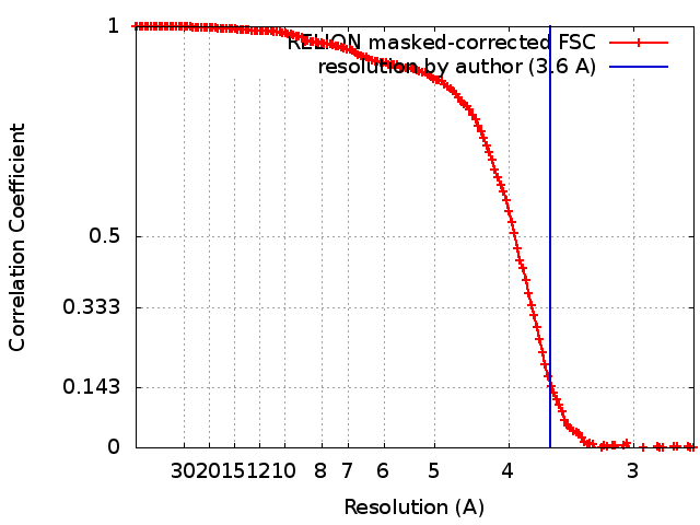
| Method | single particle reconstruction / cryo EM / Resolution: 3.6 Å | |||||||||
 Authors Authors | Brown A / Baird MR / Yip MCJ / Murray J / Shao S | |||||||||
 Citation Citation | Journal: Elife / Year: 2018 Title: Structures of translationally inactive mammalian ribosomes. Authors: Alan Brown / Matthew R Baird / Matthew Cj Yip / Jason Murray / Sichen Shao /   Abstract: The cellular levels and activities of ribosomes directly regulate gene expression during numerous physiological processes. The mechanisms that globally repress translation are incompletely understood. ...The cellular levels and activities of ribosomes directly regulate gene expression during numerous physiological processes. The mechanisms that globally repress translation are incompletely understood. Here, we use electron cryomicroscopy to analyze inactive ribosomes isolated from mammalian reticulocytes, the penultimate stage of red blood cell differentiation. We identify two types of ribosomes that are translationally repressed by protein interactions. The first comprises ribosomes sequestered with elongation factor 2 (eEF2) by SERPINE mRNA binding protein 1 (SERBP1) occupying the ribosomal mRNA entrance channel. The second type are translationally repressed by a novel ribosome-binding protein, interferon-related developmental regulator 2 (IFRD2), which spans the P and E sites and inserts a C-terminal helix into the mRNA exit channel to preclude translation. IFRD2 binds ribosomes with a tRNA occupying a noncanonical binding site, the 'Z site', on the ribosome. These structures provide functional insights into how ribosomal interactions may suppress translation to regulate gene expression. | |||||||||
| History |
|
- Structure visualization
| Movie |
Movie viewer |
|---|---|
| Structure viewer | EM map: SurfViewMolmilJmol/JSmol |
| Supplemental images |

- Downloads & links
-EMDB archive
| Map data | emd_9237.map.gz | 16.2 MB | EMDB map data format | |
|---|---|---|---|---|
| Header (meta data) | emd-9237-v30.xmlemd-9237.xml | 114.7 KB 114.7 KB | Display Display | EMDB header |
| FSC (resolution estimation) | emd_9237_fsc.xml | 14.2 KB | Display | FSC data file |
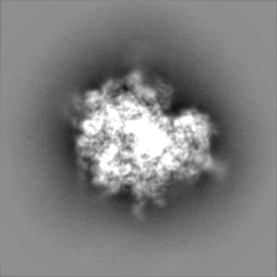
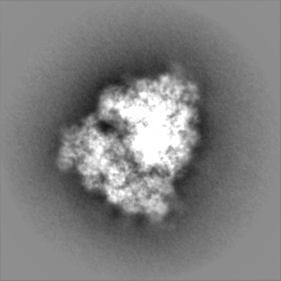
| Images | 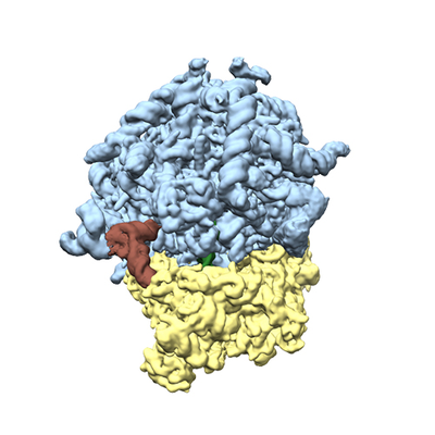 emd_9237.png emd_9237.png | 125.2 KB | ||
| Filedesc metadata | emd-9237.cif.gz | 22.3 KB | ||
| Others | emd_9237_additional.map.gzemd_9237_half_map_1.map.gzemd_9237_half_map_2.map.gz | 216.4 MB 214.7 MB 214.7 MB | ||
| Archive directory |  http://ftp.pdbj.org/pub/emdb/structures/EMD-9237ftp://ftp.pdbj.org/pub/emdb/structures/EMD-9237 http://ftp.pdbj.org/pub/emdb/structures/EMD-9237ftp://ftp.pdbj.org/pub/emdb/structures/EMD-9237 | HTTPS FTP |
-Related structure data
| Related structure data | 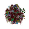 6mtbMC 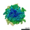 9234C 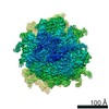 9235C 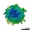 9236C 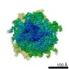 9239C 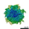 9240C 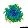 9241C 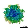 9242C 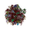 6mtcC  6mtdC 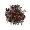 6mteC C: citing same article ( M: atomic model generated by this map |
|---|---|
| Similar structure data |
-Links
| EMDB pages | EMDB (EBI/PDBe) / EMDataResource |
|---|---|
| Related items in Molecule of the Month |


-Map
| File | Download / File: emd_9237.map.gz / Format: CCP4 / Size: 244.1 MB / Type: IMAGE STORED AS FLOATING POINT NUMBER (4 BYTES) | ||||||||||||||||||||||||||||||||||||||||||||||||||||||||||||||||||||
|---|---|---|---|---|---|---|---|---|---|---|---|---|---|---|---|---|---|---|---|---|---|---|---|---|---|---|---|---|---|---|---|---|---|---|---|---|---|---|---|---|---|---|---|---|---|---|---|---|---|---|---|---|---|---|---|---|---|---|---|---|---|---|---|---|---|---|---|---|---|
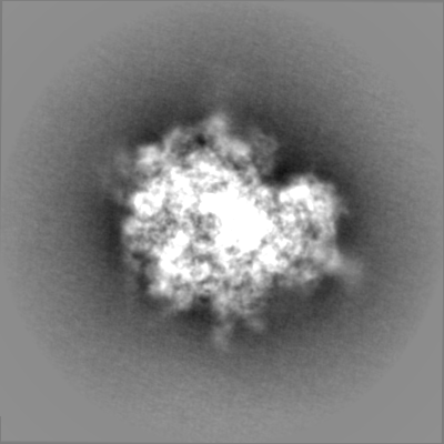
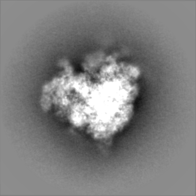
| Annotation | Postprocessed map | ||||||||||||||||||||||||||||||||||||||||||||||||||||||||||||||||||||
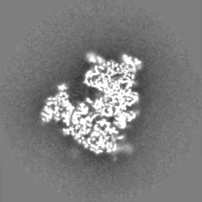
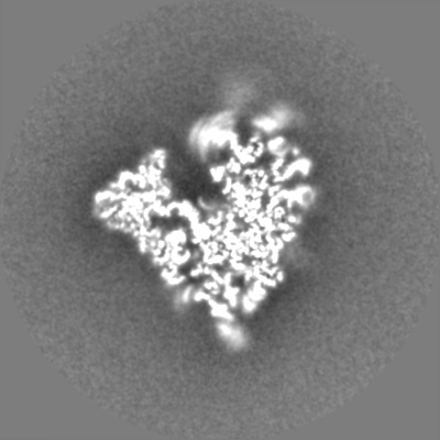
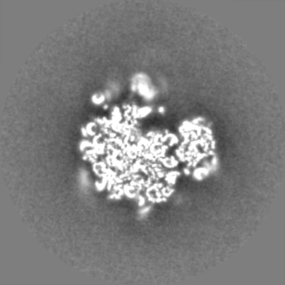
| Projections & slices | Image control
Images are generated by Spider. | ||||||||||||||||||||||||||||||||||||||||||||||||||||||||||||||||||||
| Voxel size | X=Y=Z: 1.34 Å | ||||||||||||||||||||||||||||||||||||||||||||||||||||||||||||||||||||
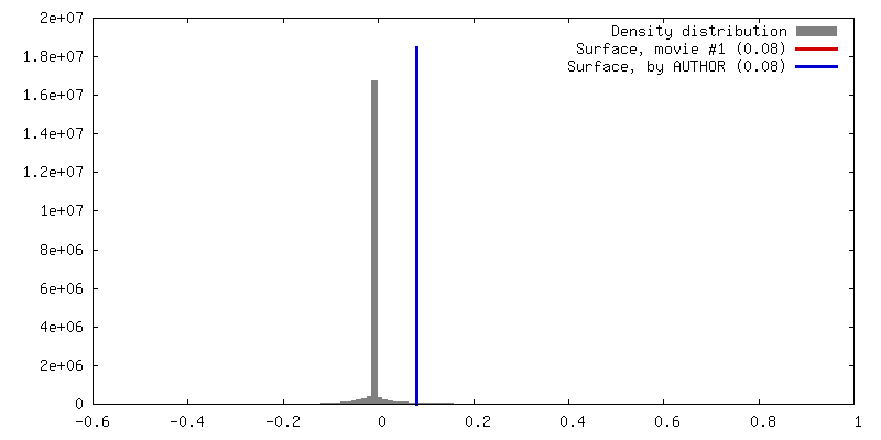
| Density |
| ||||||||||||||||||||||||||||||||||||||||||||||||||||||||||||||||||||
| Symmetry | Space group: 1 | ||||||||||||||||||||||||||||||||||||||||||||||||||||||||||||||||||||
| Details | EMDB XML:
CCP4 map header:
| ||||||||||||||||||||||||||||||||||||||||||||||||||||||||||||||||||||
 Z (Sec.)
 Y (Row.)
 X (Col.)


-Supplemental data
-Additional map: Pre-postprocessed map
| File | emd_9237_additional.map | ||||||||||||
|---|---|---|---|---|---|---|---|---|---|---|---|---|---|
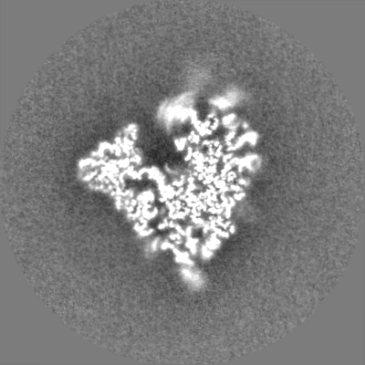
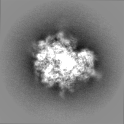
| Annotation | Pre-postprocessed map | ||||||||||||
| Projections & Slices |
| ||||||||||||
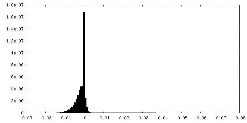
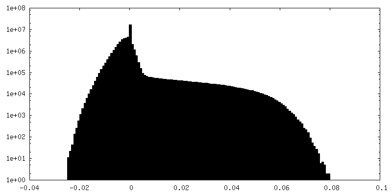
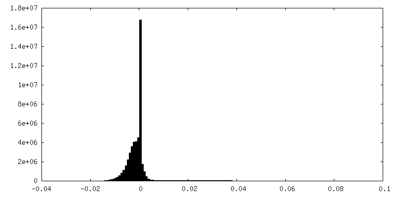
| Density Histograms |

-Half map: Half map 1
| File | emd_9237_half_map_1.map | ||||||||||||
|---|---|---|---|---|---|---|---|---|---|---|---|---|---|
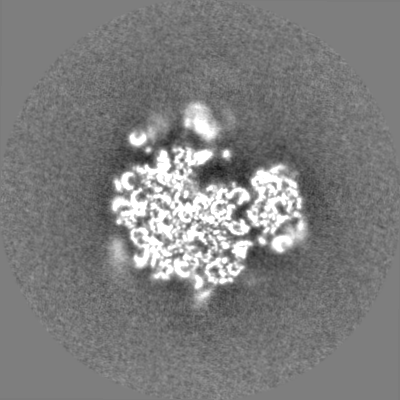
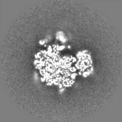
| Annotation | Half map 1 | ||||||||||||
| Projections & Slices |
| ||||||||||||
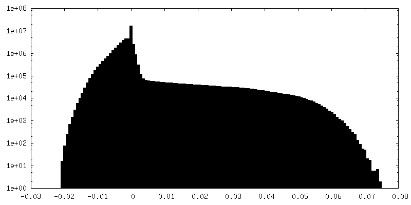
| Density Histograms |


-Half map: None
| File | emd_9237_half_map_2.map | ||||||||||||
|---|---|---|---|---|---|---|---|---|---|---|---|---|---|
| Annotation | None | ||||||||||||
| Projections & Slices |
| ||||||||||||
| Density Histograms |


- Sample components
+Entire : Rabbit 80S ribosome with a P- and Z-site tRNA (unrotated state)
+Supramolecule #1: Rabbit 80S ribosome with a P- and Z-site tRNA (unrotated state)
+Macromolecule #1: mRNA
+Macromolecule #2: P-site tRNA
+Macromolecule #3: Z-site tRNA
+Macromolecule #4: 28S rRNA
+Macromolecule #5: 5S rRNA
+Macromolecule #6: 5.8S rRNA
+Macromolecule #50: 18S rRNA
+Macromolecule #7: 60S ribosomal protein L8
+Macromolecule #8: 60S ribosomal protein L3
+Macromolecule #9: 60S ribosomal protein L4
+Macromolecule #10: 60S ribosomal protein L5
+Macromolecule #11: 60S ribosomal protein L6
+Macromolecule #12: 60S ribosomal protein L7
+Macromolecule #13: 60S ribosomal protein L7a
+Macromolecule #14: 60S ribosomal protein L9
+Macromolecule #15: 60S ribosomal protein L10
+Macromolecule #16: 60S ribosomal protein L11
+Macromolecule #17: 60S ribosomal protein L13
+Macromolecule #18: 60S ribosomal protein L14
+Macromolecule #19: 60S ribosomal protein L15
+Macromolecule #20: 60S ribosomal protein L13a
+Macromolecule #21: 60S ribosomal protein L17
+Macromolecule #22: 60S ribosomal protein L18
+Macromolecule #23: 60S ribosomal protein L19
+Macromolecule #24: 60S ribosomal protein L18a
+Macromolecule #25: 60S ribosomal protein L21
+Macromolecule #26: 60S ribosomal protein L22
+Macromolecule #27: 60S ribosomal protein L23
+Macromolecule #28: 60S ribosomal protein L24
+Macromolecule #29: 60S ribosomal protein L23a
+Macromolecule #30: 60S ribosomal protein L26
+Macromolecule #31: 60S ribosomal protein L27
+Macromolecule #32: 60S ribosomal protein L27a
+Macromolecule #33: 60S ribosomal protein L29
+Macromolecule #34: 60S ribosomal protein L30
+Macromolecule #35: 60S ribosomal protein L31
+Macromolecule #36: 60S ribosomal protein L32
+Macromolecule #37: 60S ribosomal protein L35a
+Macromolecule #38: 60S ribosomal protein L34
+Macromolecule #39: 60S ribosomal protein L35
+Macromolecule #40: 60S ribosomal protein L36
+Macromolecule #41: 60S ribosomal protein L37
+Macromolecule #42: 60S ribosomal protein L38
+Macromolecule #43: 60S ribosomal protein L39
+Macromolecule #44: 60S ribosomal protein L40
+Macromolecule #45: 60S ribosomal protein L41
+Macromolecule #46: 60S ribosomal protein L36a
+Macromolecule #47: 60S ribosomal protein L37a
+Macromolecule #48: 60S ribosomal protein L28
+Macromolecule #49: 60S ribosomal protein L10a
+Macromolecule #51: 40S ribosomal protein SA
+Macromolecule #52: 40S ribosomal protein S3a
+Macromolecule #53: 40S ribosomal protein S2
+Macromolecule #54: 40S ribosomal protein S3
+Macromolecule #55: 40S ribosomal protein S4, X isoform
+Macromolecule #56: 40S ribosomal protein S5
+Macromolecule #57: 40S ribosomal protein S6
+Macromolecule #58: 40S ribosomal protein S7
+Macromolecule #59: 40S ribosomal protein S8
+Macromolecule #60: 40S ribosomal protein S9
+Macromolecule #61: 40S ribosomal protein S10
+Macromolecule #62: 40S ribosomal protein S11
+Macromolecule #63: 40S ribosomal protein S12
+Macromolecule #64: 40S ribosomal protein S13
+Macromolecule #65: 40S ribosomal protein S14
+Macromolecule #66: 40S ribosomal protein S15
+Macromolecule #67: 40S ribosomal protein S16
+Macromolecule #68: 40S ribosomal protein S17
+Macromolecule #69: 40S ribosomal protein S18
+Macromolecule #70: 40S ribosomal protein S19
+Macromolecule #71: 40S ribosomal protein S20
+Macromolecule #72: 40S ribosomal protein S21
+Macromolecule #73: 40S ribosomal protein S15a
+Macromolecule #74: 40S ribosomal protein S23
+Macromolecule #75: 40S ribosomal protein S24
+Macromolecule #76: 40S ribosomal protein S25
+Macromolecule #77: 40S ribosomal protein S26
+Macromolecule #78: 40S ribosomal protein S27
+Macromolecule #79: 40S ribosomal protein S28
+Macromolecule #80: 40S ribosomal protein S29
+Macromolecule #81: 40S ribosomal protein S30
+Macromolecule #82: 40S ribosomal protein S27a
+Macromolecule #83: Receptor of activated protein C kinase 1
+Macromolecule #84: MAGNESIUM ION
+Macromolecule #85: ZINC ION
-Experimental details
-Structure determination
| Method | cryo EM |
|---|---|
 Processing Processing | single particle reconstruction |
| Aggregation state | particle |
-Sample preparation
| Buffer | pH: 7.4 |
|---|---|
| Grid | Support film - Material: CARBON / Support film - topology: CONTINUOUS / Support film - Film thickness: 5 / Details: unspecified |
| Vitrification | Cryogen name: ETHANE / Chamber humidity: 100 % / Chamber temperature: 277 K / Instrument: FEI VITROBOT MARK II |
- Electron microscopy
| Microscope | FEI TITAN KRIOS |
|---|---|
| Image recording | Film or detector model: FEI FALCON II (4k x 4k) / Detector mode: INTEGRATING / Digitization - Frames/image: 1-17 / Average exposure time: 1.1 sec. / Average electron dose: 40.0 e/Å2 |
| Electron beam | Acceleration voltage: 300 kV / Electron source:  FIELD EMISSION GUN FIELD EMISSION GUN |
| Electron optics | Illumination mode: FLOOD BEAM / Imaging mode: BRIGHT FIELD / Cs: 2.7 mm |
| Experimental equipment |  Model: Titan Krios / Image courtesy: FEI Company |