 Movie
Movie Controller


+ Open data

- Basic information
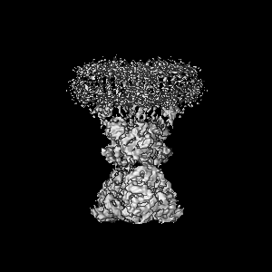
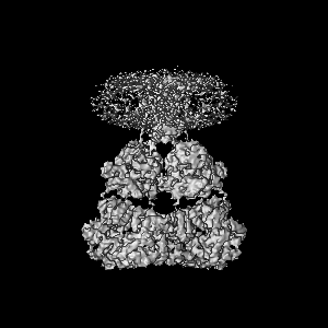
| Entry | Database: EMDB / ID: EMD-8819 | ||||||||||||||||||
|---|---|---|---|---|---|---|---|---|---|---|---|---|---|---|---|---|---|---|---|
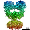
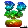
| Title | GluA2 bound to antagonist ZK and GSG1L in digitonin, state 1 | ||||||||||||||||||
 Map data Map data | GluA2 bound to antagonist ZK and GSG1L in digitonin, state 1 | ||||||||||||||||||
 Sample Sample |
| ||||||||||||||||||
 Keywords Keywords | Ion channel / TRANSPORT PROTEIN | ||||||||||||||||||
| Function / homology |  Function and homology information Function and homology informationregulation of short-term neuronal synaptic plasticity / regulation of AMPA receptor activity / spine synapse / dendritic spine neck / dendritic spine cytoplasm / dendritic spine head / cellular response to amine stimulus / regulation of postsynaptic neurotransmitter receptor internalization / Activation of AMPA receptors / ligand-gated monoatomic cation channel activity ...regulation of short-term neuronal synaptic plasticity / regulation of AMPA receptor activity / spine synapse / dendritic spine neck / dendritic spine cytoplasm / dendritic spine head / cellular response to amine stimulus / regulation of postsynaptic neurotransmitter receptor internalization / Activation of AMPA receptors / ligand-gated monoatomic cation channel activity / perisynaptic space / Trafficking of GluR2-containing AMPA receptors / response to lithium ion / AMPA glutamate receptor activity / AMPA glutamate receptor clustering / transmission of nerve impulse / regulation of receptor recycling / kainate selective glutamate receptor activity / immunoglobulin binding / AMPA glutamate receptor complex / extracellularly glutamate-gated ion channel activity / cellular response to glycine / ionotropic glutamate receptor complex / asymmetric synapse / Unblocking of NMDA receptors, glutamate binding and activation / glutamate receptor binding / positive regulation of synaptic transmission / conditioned place preference / regulation of synaptic transmission, glutamatergic / response to fungicide / extracellular ligand-gated monoatomic ion channel activity / cytoskeletal protein binding / glutamate-gated receptor activity / cellular response to brain-derived neurotrophic factor stimulus / regulation of long-term synaptic depression / glutamate-gated calcium ion channel activity / somatodendritic compartment / presynaptic active zone membrane / ionotropic glutamate receptor signaling pathway / ionotropic glutamate receptor binding / dendrite cytoplasm / dendrite membrane / excitatory synapse / ligand-gated monoatomic ion channel activity involved in regulation of presynaptic membrane potential / positive regulation of excitatory postsynaptic potential / dendritic shaft / SNARE binding / synaptic membrane / PDZ domain binding / establishment of protein localization / protein tetramerization / synaptic transmission, glutamatergic / transmitter-gated monoatomic ion channel activity involved in regulation of postsynaptic membrane potential / receptor internalization / cerebral cortex development / synapse organization / postsynaptic density membrane / modulation of chemical synaptic transmission / long-term synaptic potentiation / Schaffer collateral - CA1 synapse / terminal bouton / synaptic vesicle / synaptic vesicle membrane / amyloid-beta binding / presynapse / growth cone / signaling receptor activity / presynaptic membrane / scaffold protein binding / chemical synaptic transmission / dendritic spine / perikaryon / postsynaptic membrane / postsynaptic density / neuron projection / external side of plasma membrane / axon / neuronal cell body / synapse / dendrite / protein kinase binding / protein-containing complex binding / glutamatergic synapse / cell surface / endoplasmic reticulum / protein-containing complex / membrane / identical protein binding / plasma membrane Similarity search - Function | ||||||||||||||||||
| Biological species |  | ||||||||||||||||||
| Method | single particle reconstruction / cryo EM / Resolution: 4.6 Å | ||||||||||||||||||
 Authors Authors | Twomey EC / Yelshanskaya MV | ||||||||||||||||||
| Funding support |  United States, 5 items United States, 5 items
| ||||||||||||||||||
 Citation Citation | Journal: Nature / Year: 2017 Title: Channel opening and gating mechanism in AMPA-subtype glutamate receptors. Authors: Edward C Twomey / Maria V Yelshanskaya / Robert A Grassucci / Joachim Frank / Alexander I Sobolevsky / Abstract: AMPA (α-amino-3-hydroxy-5-methyl-4-isoxazole propionic acid)-subtype ionotropic glutamate receptors mediate fast excitatory neurotransmission throughout the central nervous system. Gated by the ...AMPA (α-amino-3-hydroxy-5-methyl-4-isoxazole propionic acid)-subtype ionotropic glutamate receptors mediate fast excitatory neurotransmission throughout the central nervous system. Gated by the neurotransmitter glutamate, AMPA receptors are critical for synaptic strength, and dysregulation of AMPA receptor-mediated signalling is linked to numerous neurological diseases. Here we use cryo-electron microscopy to solve the structures of AMPA receptor-auxiliary subunit complexes in the apo, antagonist- and agonist-bound states and determine the iris-like mechanism of ion channel opening. The ion channel selectivity filter is formed by the extended portions of the re-entrant M2 loops, while the helical portions of M2 contribute to extensive hydrophobic interfaces between AMPA receptor subunits in the ion channel. We show how the permeation pathway changes upon channel opening and identify conformational changes throughout the entire AMPA receptor that accompany activation and desensitization. Our findings provide a framework for understanding gating across the family of ionotropic glutamate receptors and the role of AMPA receptors in excitatory neurotransmission. | ||||||||||||||||||
| History |
|
- Structure visualization
| Movie |
Movie viewer |
|---|---|
| Structure viewer | EM map: SurfViewMolmilJmol/JSmol |
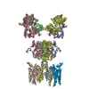
| Supplemental images |

- Downloads & links
-EMDB archive
| Map data | emd_8819.map.gz | 12 MB | EMDB map data format | |
|---|---|---|---|---|
| Header (meta data) | emd-8819-v30.xmlemd-8819.xml | 13.6 KB 13.6 KB | Display Display | EMDB header |
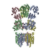
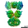
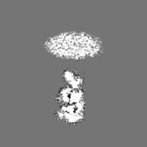
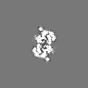
| Images | 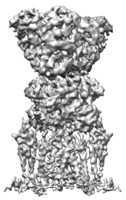 emd_8819.png emd_8819.png | 58.5 KB | ||
| Filedesc metadata | emd-8819.cif.gz | 6.5 KB | ||
| Archive directory |  http://ftp.pdbj.org/pub/emdb/structures/EMD-8819ftp://ftp.pdbj.org/pub/emdb/structures/EMD-8819 http://ftp.pdbj.org/pub/emdb/structures/EMD-8819ftp://ftp.pdbj.org/pub/emdb/structures/EMD-8819 | HTTPS FTP |
-Related structure data
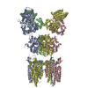
| Related structure data | 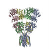 5wekMC 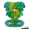 8820C 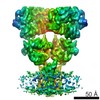 8821C  8822C 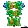 8823C  5welC  5wemC  5wenC 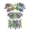 5weoC C: citing same article ( M: atomic model generated by this map |
|---|---|
| Similar structure data |
-Links
| EMDB pages | EMDB (EBI/PDBe) / EMDataResource |
|---|---|
| Related items in Molecule of the Month |


-Map
| File | Download / File: emd_8819.map.gz / Format: CCP4 / Size: 103 MB / Type: IMAGE STORED AS FLOATING POINT NUMBER (4 BYTES) | ||||||||||||||||||||||||||||||||||||||||||||||||||||||||||||
|---|---|---|---|---|---|---|---|---|---|---|---|---|---|---|---|---|---|---|---|---|---|---|---|---|---|---|---|---|---|---|---|---|---|---|---|---|---|---|---|---|---|---|---|---|---|---|---|---|---|---|---|---|---|---|---|---|---|---|---|---|---|
| Annotation | GluA2 bound to antagonist ZK and GSG1L in digitonin, state 1 | ||||||||||||||||||||||||||||||||||||||||||||||||||||||||||||
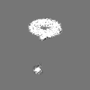
| Projections & slices | Image control
Images are generated by Spider. | ||||||||||||||||||||||||||||||||||||||||||||||||||||||||||||
| Voxel size | X=Y=Z: 0.98 Å | ||||||||||||||||||||||||||||||||||||||||||||||||||||||||||||
| Density |
| ||||||||||||||||||||||||||||||||||||||||||||||||||||||||||||
| Symmetry | Space group: 1 | ||||||||||||||||||||||||||||||||||||||||||||||||||||||||||||
| Details | EMDB XML:
CCP4 map header:
| ||||||||||||||||||||||||||||||||||||||||||||||||||||||||||||
 Z (Sec.)
 Y (Row.)
 X (Col.)


-Supplemental data
- Sample components
-Entire : GluA2 bound to antagonist ZK and GSG1L in digitonin, state 1
| Entire | Name: GluA2 bound to antagonist ZK and GSG1L in digitonin, state 1 |
|---|---|
| Components |
|
-Supramolecule #1: GluA2 bound to antagonist ZK and GSG1L in digitonin, state 1
| Supramolecule | Name: GluA2 bound to antagonist ZK and GSG1L in digitonin, state 1 type: complex / ID: 1 / Parent: 0 / Macromolecule list: #1 |
|---|---|
| Source (natural) | Organism: |
-Macromolecule #1: Chimera of Glutamate receptor 2,Germ cell-specific gene 1-like protein
| Macromolecule | Name: Chimera of Glutamate receptor 2,Germ cell-specific gene 1-like protein type: protein_or_peptide / ID: 1 / Number of copies: 4 / Enantiomer: LEVO |
|---|---|
| Source (natural) | Organism: |
| Molecular weight | Theoretical: 117.471211 KDa |
| Recombinant expression | Organism:  Homo sapiens (human) Homo sapiens (human) |
| Sequence | String: NSIQIGGLFP RGADQEYSAF RVGMVQFSTS EFRLTPHIDN LEVANSFAVT NAFCSQFSRG VYAIFGFYDK KSVNTITSFC GTLHVSFIT PSFPTDGTHP FVIQMRPDLK GALLSLIEYY QWDKFAYLYD SDRGLSTLQA VLDSAAEKKW QVTAINVGNI N NDKKDETY ...String: NSIQIGGLFP RGADQEYSAF RVGMVQFSTS EFRLTPHIDN LEVANSFAVT NAFCSQFSRG VYAIFGFYDK KSVNTITSFC GTLHVSFIT PSFPTDGTHP FVIQMRPDLK GALLSLIEYY QWDKFAYLYD SDRGLSTLQA VLDSAAEKKW QVTAINVGNI N NDKKDETY RSLFQDLELK KERRVILDCE RDKVNDIVDQ VITIGKHVKG YHYIIANLGF TDGDLLKIQF GGAEVSGFQI VD YDDSLVS KFIERWSTLE EKEYPGAHTA TIKYTSALTY DAVQVMTEAF RNLRKQRIEI SRRGNAGDCL ANPAVPWGQG VEI ERALKQ VQVEGLSGNI KFDQNGKRIN YTINIMELKT NGPRKIGYWS EVDKMVLTED DTSGLEQKTV VVTTILESPY VMMK KNHEM LEGNERYEGY CVDLAAEIAK HCGFKYKLTI VGDGKYGARD ADTKIWNGMV GELVYGKADI AIAPLTITLV REEVI DFSK PFMSLGISIM IKKPQKSKPG VFSFLDPLAY EIWMCIVFAY IGVSVVLFLV SRFSPYEWHT EEFEDGRETQ SSESTN EFG IFNSLWFSLG AFMQQGCDIS PRSLSGRIVG GVWWFFTLII ISSYTANLAA FLTVERMVSP IESAEDLSKQ TEIAYGT LD SGSTKEFFRR SKIAVFDKMW TYMRSAEPSV FVRTTAEGVA RVRKSKGKYA YLLESTMNEY IEQRKPCDTM KVGGNLDS K GYGIATPKGS SLGTPVNLAV LKLSEQGVLD KLKNKWWYDK GECGAKDSGS KEKTSALSLS NVAGVFYILV GGLGLAMLV ALIEFCYKSR AEAKRMKGTG KTSRRGRALL AVALNLLALL FATTAFLTTY WCQGTQRVPK PGCGQGGGAN CPNSGANATA NSTAAPVAA SPAGAPYSWE AGDERFQLRR FHTGIWYSCE EELGGPGEKC RSFIDLAPAS EKGVLWLSVV SEVLYILLLV V GFSLMCLE LLHSSSVIDG LKLNAFAAVF TVLSGLLGMV AHMMYTQVFQ VTVSLGPEDW RPHSWDYGWS FCLAWGSFTC CM AASVTTL NSYTKTVIEF UniProtKB: Glutamate receptor 2, Germ cell-specific gene 1-like protein |
-Macromolecule #2: {[7-morpholin-4-yl-2,3-dioxo-6-(trifluoromethyl)-3,4-dihydroquino...
| Macromolecule | Name: {[7-morpholin-4-yl-2,3-dioxo-6-(trifluoromethyl)-3,4-dihydroquinoxalin-1(2H)-yl]methyl}phosphonic acid type: ligand / ID: 2 / Number of copies: 4 / Formula: ZK1 |
|---|---|
| Molecular weight | Theoretical: 409.254 Da |
| Chemical component information | 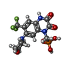 ChemComp-ZK1: |
-Experimental details
-Structure determination
| Method | cryo EM |
|---|---|
 Processing Processing | single particle reconstruction |
| Aggregation state | particle |
-Sample preparation
| Concentration | 5 mg/mL |
|---|---|
| Buffer | pH: 8 |
| Grid | Model: C-flat-1.2/1.3 / Material: GOLD / Mesh: 200 / Pretreatment - Type: GLOW DISCHARGE Details: Gold-gold grids, hydrogen and oxygen glow discharge (20s, 10 watts, 6.4 sccm H2, 27.5 sccm O2) |
| Vitrification | Cryogen name: ETHANE / Chamber humidity: 100 % / Chamber temperature: 295 K / Instrument: FEI VITROBOT MARK IV |
| Details | GluA2 bound to antagonist ZK and GSG1L in digitonin, state 1 |
- Electron microscopy
| Microscope | FEI POLARA 300 |
|---|---|
| Image recording | Film or detector model: GATAN K2 SUMMIT (4k x 4k) / Detector mode: COUNTING / Average electron dose: 67.0 e/Å2 |
| Electron beam | Acceleration voltage: 300 kV / Electron source:  FIELD EMISSION GUN FIELD EMISSION GUN |
| Electron optics | Illumination mode: SPOT SCAN / Imaging mode: BRIGHT FIELD |
| Experimental equipment |  Model: Tecnai Polara / Image courtesy: FEI Company |
-Image processing
| Startup model | Type of model: OTHER / Details: GluA2-2xGSG1L ZK DDM map |
|---|---|
| Final reconstruction | Applied symmetry - Point group: C2 (2 fold cyclic) / Resolution.type: BY AUTHOR / Resolution: 4.6 Å / Resolution method: FSC 0.143 CUT-OFF / Number images used: 26971 |
| Initial angle assignment | Type: RANDOM ASSIGNMENT |
| Final angle assignment | Type: ANGULAR RECONSTITUTION |
-Atomic model buiding 1
| Refinement | Space: REAL |
|---|---|
| Output model | PDB-5wek: |
