 Movie
Movie Controller

[English] 日本語
 Yorodumi
- EMDB-8596: 70S ribosome complex with dnaX mRNA stemloop and E-site tRNA ("in... -
+ Open data

- Basic information
| Entry | Database: EMDB / ID: EMD-8596 | |||||||||
|---|---|---|---|---|---|---|---|---|---|---|
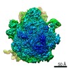
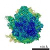
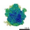
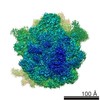
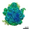
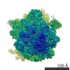
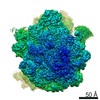
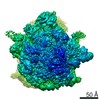
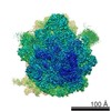
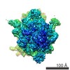
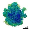
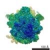
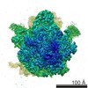
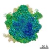
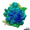
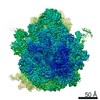
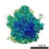
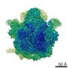
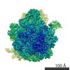
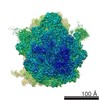
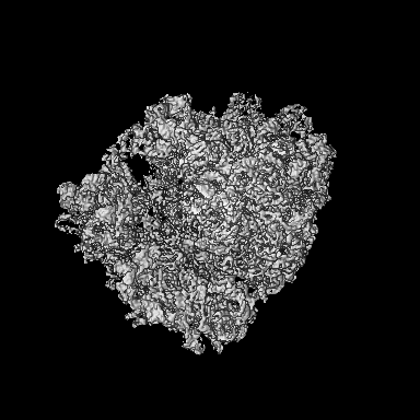
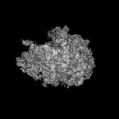
| Title | 70S ribosome complex with dnaX mRNA stemloop and E-site tRNA ("in" conformation) | |||||||||
 Map data Map data | 70S ribosome complex with dnaX mRNA stemloop and E-site tRNA ("in" conformation) | |||||||||
 Sample Sample |
| |||||||||
 Keywords Keywords | 70S ribosome / dnaX / stem-loop / structured mRNA / -1 programmed frameshifting / E site / RIBOSOME | |||||||||
| Function / homology |  Function and homology information Function and homology informationregulation of translation / large ribosomal subunit / transferase activity / ribosomal small subunit assembly / ribosomal small subunit biogenesis / 5S rRNA binding / ribosomal large subunit assembly / small ribosomal subunit / small ribosomal subunit rRNA binding / large ribosomal subunit rRNA binding ...regulation of translation / large ribosomal subunit / transferase activity / ribosomal small subunit assembly / ribosomal small subunit biogenesis / 5S rRNA binding / ribosomal large subunit assembly / small ribosomal subunit / small ribosomal subunit rRNA binding / large ribosomal subunit rRNA binding / cytosolic small ribosomal subunit / cytosolic large ribosomal subunit / cytoplasmic translation / tRNA binding / negative regulation of translation / rRNA binding / structural constituent of ribosome / ribosome / translation / ribonucleoprotein complex / mRNA binding / zinc ion binding / metal ion binding / cytoplasm / cytosol Similarity search - Function | |||||||||
| Biological species |   Thermus thermophilus (strain HB8 / ATCC 27634 / DSM 579) (bacteria) / Thermus thermophilus (strain HB8 / ATCC 27634 / DSM 579) (bacteria) / | |||||||||
| Method | single particle reconstruction / cryo EM / Resolution: 3.5 Å | |||||||||
 Authors Authors | Zhang Y / Hong S | |||||||||
 Citation Citation | Journal: Structure / Year: 2018 Title: Alternative Mode of E-Site tRNA Binding in the Presence of a Downstream mRNA Stem Loop at the Entrance Channel. Authors: Yan Zhang / Samuel Hong / Ajchareeya Ruangprasert / Georgios Skiniotis / Christine M Dunham /  Abstract: Structured mRNAs positioned downstream of the ribosomal decoding center alter gene expression by slowing protein synthesis. Here, we solved the cryo-EM structure of the bacterial ribosome bound to an ...Structured mRNAs positioned downstream of the ribosomal decoding center alter gene expression by slowing protein synthesis. Here, we solved the cryo-EM structure of the bacterial ribosome bound to an mRNA containing a 3' stem loop that regulates translation. Unexpectedly, the E-site tRNA adopts two distinct orientations. In the first structure, normal interactions with the 50S and 30S E site are observed. However, in the second structure, although the E-site tRNA makes normal interactions with the 50S E site, its anticodon stem loop moves ∼54 Å away from the 30S E site to interact with the 30S head domain and 50S uL5. This position of the E-site tRNA causes the uL1 stalk to adopt a more open conformation that likely represents an intermediate state during E-site tRNA dissociation. These results suggest that structured mRNAs at the entrance channel restrict 30S subunit movement required during translation to slow E-site tRNA dissociation. | |||||||||
| History |
|
- Structure visualization
| Movie |
Movie viewer |
|---|---|
| Structure viewer | EM map: SurfViewMolmilJmol/JSmol |
| Supplemental images |

- Downloads & links
-EMDB archive
| Map data | emd_8596.map.gz | 199.5 MB | EMDB map data format | |
|---|---|---|---|---|
| Header (meta data) | emd-8596-v30.xmlemd-8596.xml | 76.3 KB 76.3 KB | Display Display | EMDB header |
| Images | 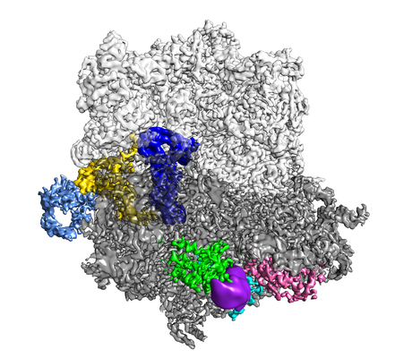 emd_8596.png emd_8596.png | 190.8 KB | ||
| Filedesc metadata | emd-8596.cif.gz | 14.2 KB | ||
| Archive directory |  http://ftp.pdbj.org/pub/emdb/structures/EMD-8596ftp://ftp.pdbj.org/pub/emdb/structures/EMD-8596 http://ftp.pdbj.org/pub/emdb/structures/EMD-8596ftp://ftp.pdbj.org/pub/emdb/structures/EMD-8596 | HTTPS FTP |
-Related structure data
| Related structure data |  5uq7MC  8597C  5uq8C C: citing same article ( M: atomic model generated by this map |
|---|---|
| Similar structure data |
-Links
| EMDB pages | EMDB (EBI/PDBe) / EMDataResource |
|---|---|
| Related items in Molecule of the Month |


-Map
| File | Download / File: emd_8596.map.gz / Format: CCP4 / Size: 216 MB / Type: IMAGE STORED AS FLOATING POINT NUMBER (4 BYTES) | ||||||||||||||||||||||||||||||||||||||||||||||||||||||||||||||||||||
|---|---|---|---|---|---|---|---|---|---|---|---|---|---|---|---|---|---|---|---|---|---|---|---|---|---|---|---|---|---|---|---|---|---|---|---|---|---|---|---|---|---|---|---|---|---|---|---|---|---|---|---|---|---|---|---|---|---|---|---|---|---|---|---|---|---|---|---|---|---|
| Annotation | 70S ribosome complex with dnaX mRNA stemloop and E-site tRNA ("in" conformation) | ||||||||||||||||||||||||||||||||||||||||||||||||||||||||||||||||||||
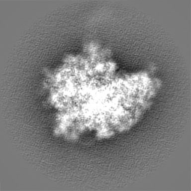
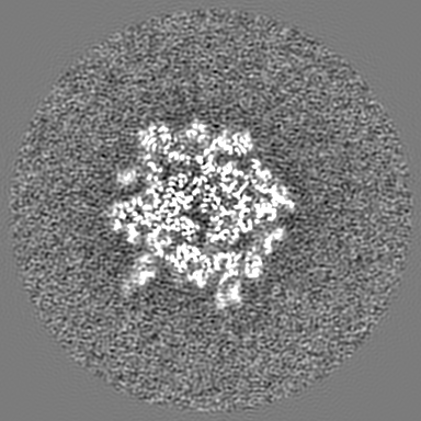
| Projections & slices | Image control
Images are generated by Spider. | ||||||||||||||||||||||||||||||||||||||||||||||||||||||||||||||||||||
| Voxel size | X=Y=Z: 1 Å | ||||||||||||||||||||||||||||||||||||||||||||||||||||||||||||||||||||
| Density |
| ||||||||||||||||||||||||||||||||||||||||||||||||||||||||||||||||||||
| Symmetry | Space group: 1 | ||||||||||||||||||||||||||||||||||||||||||||||||||||||||||||||||||||
| Details | EMDB XML:
CCP4 map header:
| ||||||||||||||||||||||||||||||||||||||||||||||||||||||||||||||||||||
 Z (Sec.)
 Y (Row.)
 X (Col.)


-Supplemental data
- Sample components
+Entire : 70S-dnaX complex
+Supramolecule #1: 70S-dnaX complex
+Macromolecule #1: 23S rRNA
+Macromolecule #2: 5S rRNA
+Macromolecule #32: 16S rRNA
+Macromolecule #53: P-site tRNA fMet
+Macromolecule #55: E-site tRNA Phe
+Macromolecule #56: messenger RNA
+Macromolecule #3: 50S ribosomal protein L2
+Macromolecule #4: 50S ribosomal protein L3
+Macromolecule #5: 50S ribosomal protein L4
+Macromolecule #6: 50S ribosomal protein L5
+Macromolecule #7: 50S ribosomal protein L6
+Macromolecule #8: 50S ribosomal protein L9
+Macromolecule #9: 50S ribosomal protein L13
+Macromolecule #10: 50S ribosomal protein L14
+Macromolecule #11: 50S ribosomal protein L15
+Macromolecule #12: 50S ribosomal protein L16
+Macromolecule #13: 50S ribosomal protein L17
+Macromolecule #14: 50S ribosomal protein L18
+Macromolecule #15: 50S ribosomal protein L19
+Macromolecule #16: 50S ribosomal protein L20
+Macromolecule #17: 50S ribosomal protein L21
+Macromolecule #18: 50S ribosomal protein L22
+Macromolecule #19: 50S ribosomal protein L23
+Macromolecule #20: 50S ribosomal protein L24
+Macromolecule #21: 50S ribosomal protein L25
+Macromolecule #22: 50S ribosomal protein L27
+Macromolecule #23: 50S ribosomal protein L28
+Macromolecule #24: 50S ribosomal protein L29
+Macromolecule #25: 50S ribosomal protein L30
+Macromolecule #26: 50S ribosomal protein L31
+Macromolecule #27: 50S ribosomal protein L32
+Macromolecule #28: 50S ribosomal protein L33
+Macromolecule #29: 50S ribosomal protein L34
+Macromolecule #30: 50S ribosomal protein L35
+Macromolecule #31: 50S ribosomal protein L36
+Macromolecule #33: 30S ribosomal protein S2
+Macromolecule #34: 30S ribosomal protein S3
+Macromolecule #35: 30S ribosomal protein S4
+Macromolecule #36: 30S ribosomal protein S5
+Macromolecule #37: 30S ribosomal protein S6
+Macromolecule #38: 30S ribosomal protein S7
+Macromolecule #39: 30S ribosomal protein S8
+Macromolecule #40: 30S ribosomal protein S9
+Macromolecule #41: 30S ribosomal protein S10
+Macromolecule #42: 30S ribosomal protein S11
+Macromolecule #43: 30S ribosomal protein S12
+Macromolecule #44: 30S ribosomal protein S13
+Macromolecule #45: 30S ribosomal protein S14 type Z
+Macromolecule #46: 30S ribosomal protein S15
+Macromolecule #47: 30S ribosomal protein S16
+Macromolecule #48: 30S ribosomal protein S17
+Macromolecule #49: 30S ribosomal protein S18
+Macromolecule #50: 30S ribosomal protein S19
+Macromolecule #51: 30S ribosomal protein S20
+Macromolecule #52: 30S ribosomal protein Thx
+Macromolecule #54: 50S ribosomal protein L1
+Macromolecule #57: ZINC ION
+Macromolecule #58: IRON/SULFUR CLUSTER

-Experimental details
-Structure determination
| Method | cryo EM |
|---|---|
 Processing Processing | single particle reconstruction |
| Aggregation state | particle |
-Sample preparation
| Buffer | pH: 7.5 |
|---|---|
| Vitrification | Cryogen name: ETHANE |
- Electron microscopy
| Microscope | FEI TITAN KRIOS |
|---|---|
| Image recording | Film or detector model: GATAN K2 SUMMIT (4k x 4k) / Detector mode: COUNTING / Average electron dose: 30.0 e/Å2 |
| Electron beam | Acceleration voltage: 300 kV / Electron source:  FIELD EMISSION GUN FIELD EMISSION GUN |
| Electron optics | Illumination mode: FLOOD BEAM / Imaging mode: BRIGHT FIELD |
| Experimental equipment |  Model: Titan Krios / Image courtesy: FEI Company |
-Image processing
| Final reconstruction | Algorithm: BACK PROJECTION / Resolution.type: BY AUTHOR / Resolution: 3.5 Å / Resolution method: FSC 0.143 CUT-OFF |
|---|
-Atomic model buiding 1
| Refinement | Space: REAL / Protocol: RIGID BODY FIT |
|---|---|
| Output model | PDB-5uq7: |
