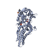
 Movie
Movie Controller


+ Open data

- Basic information
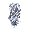
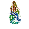
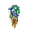
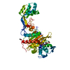
| Entry | Database: PDB / ID: 7nsl | ||||||
|---|---|---|---|---|---|---|---|
| Title | AL amyloid fibril from a lambda 1 light chain | ||||||
 Components Components | Amyloid lambda1 light chain | ||||||
 Keywords Keywords | IMMUNE SYSTEM / amyloid / antibody / systemic amyloidosis / light chain | ||||||
| Biological species |  Homo sapiens (human) Homo sapiens (human) | ||||||
| Method | ELECTRON MICROSCOPY / helical reconstruction / cryo EM / Resolution: 3.1 Å | ||||||
 Authors Authors | Karimi Farsijani, S. / Radamaker, L. / Fandrich, M. | ||||||
| Funding support |  Germany, 1items Germany, 1items
| ||||||
 Citation Citation | Journal: Nat Commun / Year: 2021 Title: Role of mutations and post-translational modifications in systemic AL amyloidosis studied by cryo-EM. Authors: Lynn Radamaker / Sara Karimi-Farsijani / Giada Andreotti / Julian Baur / Matthias Neumann / Sarah Schreiner / Natalie Berghaus / Raoul Motika / Christian Haupt / Paul Walther / Volker ...Authors: Lynn Radamaker / Sara Karimi-Farsijani / Giada Andreotti / Julian Baur / Matthias Neumann / Sarah Schreiner / Natalie Berghaus / Raoul Motika / Christian Haupt / Paul Walther / Volker Schmidt / Stefanie Huhn / Ute Hegenbart / Stefan O Schönland / Sebastian Wiese / Clarissa Read / Matthias Schmidt / Marcus Fändrich / Abstract: Systemic AL amyloidosis is a rare disease that is caused by the misfolding of immunoglobulin light chains (LCs). Potential drivers of amyloid formation in this disease are post-translational ...Systemic AL amyloidosis is a rare disease that is caused by the misfolding of immunoglobulin light chains (LCs). Potential drivers of amyloid formation in this disease are post-translational modifications (PTMs) and the mutational changes that are inserted into the LCs by somatic hypermutation. Here we present the cryo electron microscopy (cryo-EM) structure of an ex vivo λ1-AL amyloid fibril whose deposits disrupt the ordered cardiomyocyte structure in the heart. The fibril protein contains six mutational changes compared to the germ line and three PTMs (disulfide bond, N-glycosylation and pyroglutamylation). Our data imply that the disulfide bond, glycosylation and mutational changes contribute to determining the fibril protein fold and help to generate a fibril morphology that is able to withstand proteolytic degradation inside the body. | ||||||
| History |
|
- Structure visualization
| Movie |
 Movie viewer Movie viewer |
|---|---|
| Structure viewer | Molecule: MolmilJmol/JSmol |
- Downloads & links
-Download
| PDBx/mmCIF format | 7nsl.cif.gz | 180.7 KB | Display | PDBx/mmCIF format |
|---|---|---|---|---|
| PDB format | pdb7nsl.ent.gz | 147 KB | Display | PDB format |
| PDBx/mmJSON format | 7nsl.json.gz | Tree view | PDBx/mmJSON format | |
| Others |  Other downloads Other downloads |
-Validation report
| Arichive directory | https://data.pdbj.org/pub/pdb/validation_reports/ns/7nslftp://data.pdbj.org/pub/pdb/validation_reports/ns/7nsl | HTTPS FTP |
|---|
-Related structure data
| Related structure data |  12570MC M: map data used to model this data C: citing same article ( |
|---|---|
| Similar structure data | |
| EM raw data | EMPIAR-10730 (Title: AL amyloid fibril from a glycosylated lambda 1 light chain Data size: 495.9 Data #1: Unaligned multi-frame micrographs of AL amyloidosis extracted ex vivo amyloid fibrils patient FOR001 [micrographs - multiframe]) |


-Links
 PDBj
PDBj


- Assembly
| Deposited unit | 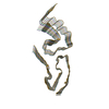
|
|---|---|
| 1 |
|
-Components
| #1: Antibody | Mass: 12122.354 Da / Num. of mol.: 7 / Source method: isolated from a natural source / Source: (natural) Homo sapiens (human) / Organ: Heart / Plasmid details: AL amyloidosis patient / Tissue: Heart muscle tissueHas protein modification | Y | |
|---|
-Experimental details
-Experiment
| Experiment | Method: ELECTRON MICROSCOPY |
|---|---|
| EM experiment | Aggregation state: FILAMENT / 3D reconstruction method: helical reconstruction |
- Sample preparation
| Component | Name: Amyloid fibril of an antibody lambda 1 immunoglobulin light chain Type: COMPLEX Details: Extracted fibrils from the explanted heart of a patient suffering from systemic AL amyloidosis Entity ID: all / Source: NATURAL |
|---|---|
| Molecular weight | Experimental value: NO |
| Source (natural) | Organism: Homo sapiens (human) / Organ: Heart / Tissue: Heart muscle |
| Buffer solution | pH: 7 |
| Buffer component | Name: Distilled water / Formula: H2O |
| Specimen | Embedding applied: NO / Shadowing applied: NO / Staining applied: NO / Vitrification applied: YES / Details: Sample in pure water, pH not determined |
| Specimen support | Grid material: COPPER / Grid type: C-flat-1.2/1.3 |
| Vitrification | Instrument: FEI VITROBOT MARK III / Cryogen name: ETHANE / Humidity: 95 % / Chamber temperature: 295 K / Details: blot for 9s before plunging |
- Electron microscopy imaging
| Experimental equipment |  Model: Titan Krios / Image courtesy: FEI Company |
|---|---|
| Microscopy | Model: FEI TITAN KRIOS |
| Electron gun | Electron source:  FIELD EMISSION GUN / Accelerating voltage: 300 kV / Illumination mode: FLOOD BEAM FIELD EMISSION GUN / Accelerating voltage: 300 kV / Illumination mode: FLOOD BEAM |
| Electron lens | Mode: BRIGHT FIELD / Cs: 2.7 mm |
| Specimen holder | Cryogen: NITROGEN / Specimen holder model: FEI TITAN KRIOS AUTOGRID HOLDER |
| Image recording | Electron dose: 40 e/Å2 / Detector mode: COUNTING / Film or detector model: GATAN K2 SUMMIT (4k x 4k) / Num. of grids imaged: 1 / Num. of real images: 3032 |
| EM imaging optics | Energyfilter slit width: 20 eV |
| Image scans | Width: 3838 / Height: 3710 / Movie frames/image: 40 |
- Processing
| EM software |
| ||||||||||||||||||||||||||||||
|---|---|---|---|---|---|---|---|---|---|---|---|---|---|---|---|---|---|---|---|---|---|---|---|---|---|---|---|---|---|---|---|
| Image processing | Details: Motion-corrected and dose-weighted movie frames | ||||||||||||||||||||||||||||||
| CTF correction | Details: CTF was estimated from the non-dose-weighted micrographs Type: PHASE FLIPPING AND AMPLITUDE CORRECTION | ||||||||||||||||||||||||||||||
| Helical symmerty | Angular rotation/subunit: -1.45566 ° / Axial rise/subunit: 4.76311 Å / Axial symmetry: C1 | ||||||||||||||||||||||||||||||
| Particle selection | Num. of particles selected: 43308 Details: manual particle picking helical start-end coordinates | ||||||||||||||||||||||||||||||
| 3D reconstruction | Resolution: 3.1 Å / Resolution method: FSC 0.143 CUT-OFF / Num. of particles: 43308 / Symmetry type: HELICAL | ||||||||||||||||||||||||||||||
| Atomic model building | Protocol: OTHER / Space: REAL Target criteria: REAL-SPACE (WEIGHTED MAP SUM AT ATOM CENTERS) Details: Secondary structure restraints and NCS were applied during refinement |
