| 登録情報 | データベース: PDB / ID: 1suj
|
|---|
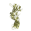
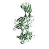
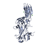
| タイトル | X-ray crystal structure of ambystoma tigrinum cone arrestin |
|---|
 要素 要素 | cone arrestin |
|---|
 キーワード キーワード | SIGNALING PROTEIN / sensory transduction |
|---|
| 機能・相同性 |  機能・相同性情報 機能・相同性情報
G protein-coupled receptor internalization / visual perception / G protein-coupled receptor binding / signal transduction類似検索 - 分子機能 Immunoglobulin-like - #840 / Immunoglobulin-like - #640 / Arrestin, conserved site / Arrestins signature. / Arrestin / Arrestin, N-terminal / Arrestin-like, N-terminal / Arrestin C-terminal-like domain / Arrestin (or S-antigen), N-terminal domain / Arrestin (or S-antigen), C-terminal domain ...Immunoglobulin-like - #840 / Immunoglobulin-like - #640 / Arrestin, conserved site / Arrestins signature. / Arrestin / Arrestin, N-terminal / Arrestin-like, N-terminal / Arrestin C-terminal-like domain / Arrestin (or S-antigen), N-terminal domain / Arrestin (or S-antigen), C-terminal domain / Arrestin (or S-antigen), C-terminal domain / Arrestin-like, C-terminal / Immunoglobulin E-set / Immunoglobulin-like / Sandwich / Mainly Beta類似検索 - ドメイン・相同性 |
|---|
| 生物種 | Ambystoma tigrinum (両生類) |
|---|
| 手法 |  X線回折 / シンクロトロン / 分子置換 / 解像度: 2.38 Å X線回折 / シンクロトロン / 分子置換 / 解像度: 2.38 Å |
|---|
 データ登録者 データ登録者 | Sutton, R.B. / Navarro, J. |
|---|
 引用 引用 | ジャーナル: J.Mol.Biol. / 年: 2005
タイトル: Crystal structure of cone arrestin at 2.3A: evolution of receptor specificity.
著者: Sutton, R.B. / Vishnivetskiy, S.A. / Robert, J. / Hanson, S.M. / Raman, D. / Knox, B.E. / Kono, M. / Navarro, J. / Gurevich, V.V. |
|---|
| 履歴 | | 登録 | 2004年3月26日 | 登録サイト: RCSB / 処理サイト: RCSB |
|---|
| 改定 1.0 | 2005年8月23日 | Provider: repository / タイプ: Initial release |
|---|
| 改定 1.1 | 2008年4月29日 | Group: Version format compliance |
|---|
| 改定 1.2 | 2011年7月13日 | Group: Version format compliance |
|---|
| 改定 1.3 | 2023年8月23日 | Group: Data collection / Database references / Refinement description
カテゴリ: chem_comp_atom / chem_comp_bond ...chem_comp_atom / chem_comp_bond / database_2 / pdbx_initial_refinement_model
Item: _database_2.pdbx_DOI / _database_2.pdbx_database_accession |
|---|
|
|---|
 ムービー
ムービー コントローラー


 データを開く
 基本情報
基本情報 構造の表示
構造の表示 ダウンロードとリンク
 その他のダウンロード


 PDBj
PDBj 集合体
集合体

 分子量: 18.015 Da / 分子数: 99 / 由来タイプ: 天然 / 式: H2O
 試料調製
試料調製 / ビームライン: F2 / 波長: 0.979 Å
 解析
解析