 Movie
Movie Controller


+ Open data

- Basic information
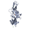
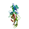
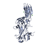
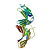
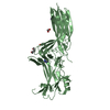
| Entry | Database: PDB / ID: 1g4m | ||||||
|---|---|---|---|---|---|---|---|
| Title | CRYSTAL STRUCTURE OF BOVINE BETA-ARRESTIN 1 | ||||||
 Components Components | BETA-ARRESTIN1 | ||||||
 Keywords Keywords | SIGNALING PROTEIN / Sensory transduction / alternative splicing | ||||||
| Function / homology |  Function and homology information Function and homology informationTGFBR3 regulates TGF-beta signaling / MAP2K and MAPK activation / Activation of SMO / Golgi Associated Vesicle Biogenesis / Lysosome Vesicle Biogenesis / AP-2 adaptor complex binding / Ub-specific processing proteases / clathrin coat of coated pit / Cargo recognition for clathrin-mediated endocytosis / clathrin heavy chain binding ...TGFBR3 regulates TGF-beta signaling / MAP2K and MAPK activation / Activation of SMO / Golgi Associated Vesicle Biogenesis / Lysosome Vesicle Biogenesis / AP-2 adaptor complex binding / Ub-specific processing proteases / clathrin coat of coated pit / Cargo recognition for clathrin-mediated endocytosis / clathrin heavy chain binding / Clathrin-mediated endocytosis / clathrin-dependent endocytosis / desensitization of G protein-coupled receptor signaling pathway / acetylcholine receptor binding / G protein-coupled receptor internalization / inositol hexakisphosphate binding / Thrombin signalling through proteinase activated receptors (PARs) / G alpha (s) signalling events / sensory perception / clathrin binding / small molecule binding / pseudopodium / phosphatidylinositol-3,4,5-trisphosphate binding / negative regulation of Notch signaling pathway / positive regulation of receptor internalization / receptor internalization / positive regulation of protein phosphorylation / G protein-coupled receptor binding / protein transport / cytoplasmic vesicle / molecular adaptor activity / ubiquitin-dependent protein catabolic process / positive regulation of ERK1 and ERK2 cascade / signal transduction / nucleus / plasma membrane / cytosol / cytoplasm Similarity search - Function | ||||||
| Biological species |  | ||||||
| Method |  X-RAY DIFFRACTION / SYNCHROTRON / MOLECULAR REPLACEMENT, SIRAS / Resolution: 1.9 Å X-RAY DIFFRACTION / SYNCHROTRON / MOLECULAR REPLACEMENT, SIRAS / Resolution: 1.9 Å | ||||||
 Authors Authors | Schubert, C. / Han, M. | ||||||
 Citation Citation | Journal: Structure / Year: 2001 Title: Crystal structure of beta-arrestin at 1.9 A: possible mechanism of receptor binding and membrane Translocation. Authors: Han, M. / Gurevich, V.V. / Vishnivetskiy, S.A. / Sigler, P.B. / Schubert, C. | ||||||
| History |
|
- Structure visualization
| Structure viewer | Molecule: MolmilJmol/JSmol |
|---|
- Downloads & links
-Download
| PDBx/mmCIF format | 1g4m.cif.gz | 156.5 KB | Display | PDBx/mmCIF format |
|---|---|---|---|---|
| PDB format | pdb1g4m.ent.gz | 124.3 KB | Display | PDB format |
| PDBx/mmJSON format | 1g4m.json.gz | Tree view | PDBx/mmJSON format | |
| Others |  Other downloads Other downloads |
-Validation report
| Arichive directory | https://data.pdbj.org/pub/pdb/validation_reports/g4/1g4mftp://data.pdbj.org/pub/pdb/validation_reports/g4/1g4m | HTTPS FTP |
|---|
-Related structure data
| Related structure data |  1g4rC  1cf1S S: Starting model for refinement C: citing same article ( |
|---|---|
| Similar structure data |


-Links
 PDBj
PDBj

- Assembly
| Deposited unit | 
| ||||||||
|---|---|---|---|---|---|---|---|---|---|
| 1 | 
| ||||||||
| 2 | 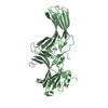
| ||||||||
| Unit cell |
|
-Components
| #1: Protein | Mass: 44309.578 Da / Num. of mol.: 2 / Fragment: TRUNCATION MUTANT: 1-393 Source method: isolated from a genetically manipulated source Source: (gene. exp.)  #2: Water | ChemComp-HOH / |  Mass: 18.015 Da / Num. of mol.: 269 / Source method: isolated from a natural source / Formula: H2O Mass: 18.015 Da / Num. of mol.: 269 / Source method: isolated from a natural source / Formula: H2O |
|---|
-Experimental details
-Experiment
| Experiment | Method: X-RAY DIFFRACTION / Number of used crystals: 3 |
|---|
- Sample preparation
| Crystal | Density Matthews: 2.98 Å3/Da / Density % sol: 58.33 % | ||||||||||||||||||||||||||||||||||||||||||||||||||||||||||||||||||||||
|---|---|---|---|---|---|---|---|---|---|---|---|---|---|---|---|---|---|---|---|---|---|---|---|---|---|---|---|---|---|---|---|---|---|---|---|---|---|---|---|---|---|---|---|---|---|---|---|---|---|---|---|---|---|---|---|---|---|---|---|---|---|---|---|---|---|---|---|---|---|---|---|
| Crystal grow | *PLUS Temperature: 19-20 ℃ / pH: 8.5 / Method: batch method | ||||||||||||||||||||||||||||||||||||||||||||||||||||||||||||||||||||||
| Components of the solutions | *PLUS
|
-Data collection
| Diffraction | Mean temperature: 100 K | |||||||||
|---|---|---|---|---|---|---|---|---|---|---|
| Diffraction source | Source: SYNCHROTRON / Site: NSLS  / Beamline: X25 / Wavelength: 0.979, 1.10 / Beamline: X25 / Wavelength: 0.979, 1.10 | |||||||||
| Detector | Type: BRANDEIS - B4 / Detector: CCD / Date: Apr 30, 2000 / Details: mirrors and Si(111) | |||||||||
| Radiation | Protocol: SINGLE WAVELENGTH / Monochromatic (M) / Laue (L): M / Scattering type: x-ray | |||||||||
| Radiation wavelength |
| |||||||||
| Reflection | Biso Wilson estimate: 22.3 Å2 | |||||||||
| Reflection | *PLUS Highest resolution: 1.96 Å / Lowest resolution: 35 Å / Num. obs: 49309 / % possible obs: 65.8 % / Redundancy: 3.2 % / Num. measured all: 158905 / Rmerge(I) obs: 0.047 | |||||||||
| Reflection shell | *PLUS Redundancy: 1.5 % / Rmerge(I) obs: 0.468 / Mean I/σ(I) obs: 1.1 |
- Processing
| Software |
| ||||||||||||||||||||||||||||||||||||||||
|---|---|---|---|---|---|---|---|---|---|---|---|---|---|---|---|---|---|---|---|---|---|---|---|---|---|---|---|---|---|---|---|---|---|---|---|---|---|---|---|---|---|
| Refinement | Method to determine structure: MOLECULAR REPLACEMENT, SIRAS Starting model: PDB ENTRY 1CF1 Resolution: 1.9→34.54 Å / Rfactor Rfree error: 0.004 / Data cutoff high absF: 2567021.55 / Data cutoff low absF: 0 / Isotropic thermal model: RESTRAINED / Cross valid method: THROUGHOUT / σ(F): 0 / σ(I): 0 / Stereochemistry target values: Engh & Huber
| ||||||||||||||||||||||||||||||||||||||||
| Solvent computation | Solvent model: FLAT MODEL / Bsol: 56.45 Å2 / ksol: 0.376 e/Å3 | ||||||||||||||||||||||||||||||||||||||||
| Displacement parameters | Biso mean: 55.1 Å2
| ||||||||||||||||||||||||||||||||||||||||
| Refine analyze |
| ||||||||||||||||||||||||||||||||||||||||
| Refinement step | Cycle: LAST / Resolution: 1.9→34.54 Å
| ||||||||||||||||||||||||||||||||||||||||
| Refine LS restraints |
| ||||||||||||||||||||||||||||||||||||||||
| LS refinement shell | Resolution: 1.9→1.97 Å / Rfactor Rfree error: 0.026 / Total num. of bins used: 10
| ||||||||||||||||||||||||||||||||||||||||
| Xplor file |
| ||||||||||||||||||||||||||||||||||||||||
| Software | *PLUS Name: CNS / Version: 1 / Classification: refinement | ||||||||||||||||||||||||||||||||||||||||
| Refinement | *PLUS σ(F): 0 / % reflection Rfree: 7.6 % | ||||||||||||||||||||||||||||||||||||||||
| Solvent computation | *PLUS | ||||||||||||||||||||||||||||||||||||||||
| Displacement parameters | *PLUS Biso mean: 55.1 Å2 | ||||||||||||||||||||||||||||||||||||||||
| Refine LS restraints | *PLUS
| ||||||||||||||||||||||||||||||||||||||||
| LS refinement shell | *PLUS Rfactor Rfree: 0.402 / % reflection Rfree: 8 % / Rfactor Rwork: 0.379 |
