- PDB-2r9v: Crystal structure of ATP synthase subunit alpha (TM1612) from The... -
+
Open data
ID or keywords:
Loading...
-
Basic information
Entry
Database: PDB / ID: 2r9v
Title
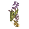
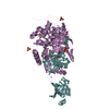
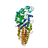
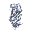
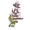
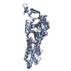
Crystal structure of ATP synthase subunit alpha (TM1612) from Thermotoga maritima at 2.10 A resolution
Components
ATP synthase subunit alpha
Keywords
HYDROLASE / TM1612 / ATP synthase subunit alpha / Structural Genomics / Joint Center for Structural Genomics / JCSG / Protein Structure Initiative / PSI-2 / ATP synthesis / ATP-binding / CF1 / Hydrogen ion transport / Inner membrane / Ion transport / Membrane / Nucleotide-binding / Transport
Function / homology
Function and homology information
proton motive force-driven ATP synthesis / H+-transporting two-sector ATPase / proton-transporting ATP synthase complex / proton-transporting ATP synthase activity, rotational mechanism / ADP binding / ATP binding / plasma membrane Similarity search - Function
ATP synthase alpha/beta chain, C-terminal domain / Lysin / Elongation Factor Tu (Ef-tu); domain 3 - #20 / ATP synthase, alpha subunit, C-terminal domain superfamily / ATP synthase, F1 complex, alpha subunit nucleotide-binding domain / ATP synthase, alpha subunit, C-terminal / ATP synthase, F1 complex, alpha subunit / ATP synthase alpha/beta chain, C terminal domain / ATPase, F1/V1/A1 complex, alpha/beta subunit, N-terminal domain superfamily / ATP synthase subunit alpha, N-terminal domain-like superfamily ...ATP synthase alpha/beta chain, C-terminal domain / Lysin / Elongation Factor Tu (Ef-tu); domain 3 - #20 / ATP synthase, alpha subunit, C-terminal domain superfamily / ATP synthase, F1 complex, alpha subunit nucleotide-binding domain / ATP synthase, alpha subunit, C-terminal / ATP synthase, F1 complex, alpha subunit / ATP synthase alpha/beta chain, C terminal domain / ATPase, F1/V1/A1 complex, alpha/beta subunit, N-terminal domain superfamily / ATP synthase subunit alpha, N-terminal domain-like superfamily / ATPase, F1/V1/A1 complex, alpha/beta subunit, N-terminal domain / ATP synthase alpha/beta family, beta-barrel domain / ATPase, alpha/beta subunit, nucleotide-binding domain, active site / ATP synthase alpha and beta subunits signature. / Elongation Factor Tu (Ef-tu); domain 3 / ATPase, F1/V1/A1 complex, alpha/beta subunit, nucleotide-binding domain / ATP synthase alpha/beta family, nucleotide-binding domain / P-loop containing nucleotide triphosphate hydrolases / Up-down Bundle / Beta Barrel / Rossmann fold / P-loop containing nucleoside triphosphate hydrolase / 3-Layer(aba) Sandwich / Mainly Beta / Mainly Alpha / Alpha Beta Similarity search - Domain/homology
BIOMOLECULE: 1 SEE REMARK 350 FOR THE AUTHOR PROVIDED AND PROGRAM GENERATED ASSEMBLY INFORMATION ... BIOMOLECULE: 1 SEE REMARK 350 FOR THE AUTHOR PROVIDED AND PROGRAM GENERATED ASSEMBLY INFORMATION FOR THE STRUCTURE IN THIS ENTRY. THE REMARK MAY ALSO PROVIDE INFORMATION ON BURIED SURFACE AREA. SIZE EXCLUSION CHROMATOGRAPHY AND CRYSTAL PACKING ANALYSIS SUPPORT THE ASSIGNMENT OF A MONOMER AS A SIGNIFICANT OLIGOMERIZATION STATE IN SOLUTION.
Remark 999
SEQUENCE THE CONSTRUCT WAS EXPRESSED WITH A PURIFICATION TAG MGSDKIHHHHHH FOLLOWED BY THE TARGET ... SEQUENCE THE CONSTRUCT WAS EXPRESSED WITH A PURIFICATION TAG MGSDKIHHHHHH FOLLOWED BY THE TARGET SEQUENCE. THIS GENE USES AN ALTERNATE INITIATION CODON THAT RESULTS IN A LEUCINE AT POSITION 1 WHEN EXPRESSED AS A FUSION.
Monochromator: Double crystal Si(111) / Protocol: SINGLE WAVELENGTH / Monochromatic (M) / Laue (L): M / Scattering type: x-ray
Radiation wavelength
Wavelength: 0.9796 Å / Relative weight: 1
Reflection
Resolution: 2.1→48.679 Å / Num. obs: 62330 / % possible obs: 99.8 % / Observed criterion σ(I): -3 / Redundancy: 7.17 % / Biso Wilson estimate: 27.14 Å2 / Rmerge(I) obs: 0.143 / Net I/σ(I): 11.44
Reflection shell
Diffraction-ID: 1
Resolution (Å)
Redundancy (%)
Rmerge(I) obs
Mean I/σ(I) obs
Num. measured obs
Num. unique obs
% possible all
2.1-2.19
7.29
0.897
2
52707
7233
99.8
2.19-2.29
0.845
2.5
49466
6789
99.8
2.29-2.41
0.579
3.2
48822
6701
99.7
2.41-2.56
0.447
4.1
49249
6776
99.8
2.56-2.75
0.325
5.6
47885
6575
99.7
2.75-3.03
0.206
8.7
50541
6977
99.8
3.03-3.47
0.105
15.7
49964
6951
99.7
3.47-4.36
0.055
26.8
49015
6945
99.9
4.36-48.679
0.043
32.3
49264
7383
99.8
-
Phasing
Phasing
Method: SAD
-
Processing
Software
Name
Version
Classification
NB
REFMAC
5.2.0019
refinement
PHENIX
refinement
SHELX
phasing
MolProbity
3beta29
modelbuilding
XSCALE
datascaling
PDB_EXTRACT
3
dataextraction
ADSC
Quantum
datacollection
XDS
datareduction
SHARP
phasing
Refinement
Method to determine structure: SAD / Resolution: 2.1→48.679 Å / Cor.coef. Fo:Fc: 0.964 / Cor.coef. Fo:Fc free: 0.946 / SU B: 8.152 / SU ML: 0.105 / TLS residual ADP flag: LIKELY RESIDUAL / Cross valid method: THROUGHOUT / σ(F): 0 / ESU R: 0.125 / ESU R Free: 0.125 Stereochemistry target values: MAXIMUM LIKELIHOOD WITH PHASES Details: 1. HYDROGENS HAVE BEEN ADDED IN THE RIDING POSITIONS. 2. ATOM RECORDS CONTAIN RESIDUAL B FACTORS ONLY. 3. A MET-INHIBITION PROTOCOL WAS USED FOR SELENOMETHIONINE INCORPORATION DURING PROTEIN ...Details: 1. HYDROGENS HAVE BEEN ADDED IN THE RIDING POSITIONS. 2. ATOM RECORDS CONTAIN RESIDUAL B FACTORS ONLY. 3. A MET-INHIBITION PROTOCOL WAS USED FOR SELENOMETHIONINE INCORPORATION DURING PROTEIN EXPRESSION. THE OCCUPANCY OF THE SE ATOMS IN THE MSE RESIDUES WAS REDUCED TO 0.75 FOR THE REDUCED SCATTERING POWER DUE TO PARTIAL S-MET INCORPORATION. 4. PG4 (PEG 200) AND CL IONS FROM THE CRYSTALLIZATION SOLUTION ARE MODELED. ATP AND MAGNESIUM WERE MODELED BASED ON HOMOLOGS AND DENSITY. RESIDUES 1-16 AS WELL AS PURIFICATION TAG ARE DISORDERED.
Rfactor
Num. reflection
% reflection
Selection details
Rfree
0.215
3176
5.1 %
RANDOM
Rwork
0.18
-
-
-
obs
0.182
62319
99.77 %
-
Solvent computation
Ion probe radii: 0.8 Å / Shrinkage radii: 0.8 Å / VDW probe radii: 1.2 Å / Solvent model: BABINET MODEL WITH MASK
Displacement parameters
Biso mean: 29.378 Å2
Baniso -1
Baniso -2
Baniso -3
1-
1.62 Å2
0 Å2
0 Å2
2-
-
1.62 Å2
0 Å2
3-
-
-
-3.23 Å2
Refinement step
Cycle: LAST / Resolution: 2.1→48.679 Å
Protein
Nucleic acid
Ligand
Solvent
Total
Num. atoms
3802
0
46
465
4313
Refine LS restraints
Refine-ID
Type
Dev ideal
Dev ideal target
Number
X-RAY DIFFRACTION
r_bond_refined_d
0.017
0.022
3971
X-RAY DIFFRACTION
r_bond_other_d
0.002
0.02
2770
X-RAY DIFFRACTION
r_angle_refined_deg
1.644
2
5383
X-RAY DIFFRACTION
r_angle_other_deg
0.978
3
6758
X-RAY DIFFRACTION
r_dihedral_angle_1_deg
4.831
5
504
X-RAY DIFFRACTION
r_dihedral_angle_2_deg
31.171
23.729
177
X-RAY DIFFRACTION
r_dihedral_angle_3_deg
13.231
15
718
X-RAY DIFFRACTION
r_dihedral_angle_4_deg
18.325
15
35
X-RAY DIFFRACTION
r_chiral_restr
0.099
0.2
611
X-RAY DIFFRACTION
r_gen_planes_refined
0.006
0.02
4386
X-RAY DIFFRACTION
r_gen_planes_other
0.001
0.02
788
X-RAY DIFFRACTION
r_nbd_refined
0.22
0.2
855
X-RAY DIFFRACTION
r_nbd_other
0.201
0.2
2985
X-RAY DIFFRACTION
r_nbtor_refined
0.18
0.2
1998
X-RAY DIFFRACTION
r_nbtor_other
0.088
0.2
2084
X-RAY DIFFRACTION
r_xyhbond_nbd_refined
0.156
0.2
297
X-RAY DIFFRACTION
r_metal_ion_refined
0.009
0.2
1
X-RAY DIFFRACTION
r_symmetry_vdw_refined
0.31
0.2
14
X-RAY DIFFRACTION
r_symmetry_vdw_other
0.285
0.2
58
X-RAY DIFFRACTION
r_symmetry_hbond_refined
0.196
0.2
26
X-RAY DIFFRACTION
r_mcbond_it
2.401
3
2565
X-RAY DIFFRACTION
r_mcbond_other
0.521
3
1002
X-RAY DIFFRACTION
r_mcangle_it
3.365
5
3955
X-RAY DIFFRACTION
r_scbond_it
4.311
6
1645
X-RAY DIFFRACTION
r_scangle_it
5.39
6
1419
LS refinement shell
Resolution: 2.1→2.154 Å / Total num. of bins used: 20
Rfactor
Num. reflection
% reflection
Rfree
0.322
237
-
Rwork
0.304
4282
-
all
-
4519
-
obs
-
-
99.71 %
Refinement TLS params.
Method: refined / Origin x: 51.8986 Å / Origin y: 15.8776 Å / Origin z: 81.6205 Å
11
12
13
21
22
23
31
32
33
T
-0.0956 Å2
-0.0223 Å2
0.0356 Å2
-
-0.0438 Å2
-0.0072 Å2
-
-
-0.0208 Å2
L
0.4867 °2
-0.0038 °2
0.4094 °2
-
0.2466 °2
0.2937 °2
-
-
1.5962 °2
S
0.0888 Å °
-0.021 Å °
0.0535 Å °
-0.0005 Å °
-0.1393 Å °
0.0076 Å °
-0.036 Å °
0.0062 Å °
0.0505 Å °
+
About Yorodumi
-
News
-
Feb 9, 2022. New format data for meta-information of EMDB entries
New format data for meta-information of EMDB entries
Version 3 of the EMDB header file is now the official format.
The previous official version 1.9 will be removed from the archive.
In the structure databanks used in Yorodumi, some data are registered as the other names, "COVID-19 virus" and "2019-nCoV". Here are the details of the virus and the list of structure data.
Jan 31, 2019. EMDB accession codes are about to change! (news from PDBe EMDB page)
EMDB accession codes are about to change! (news from PDBe EMDB page)
The allocation of 4 digits for EMDB accession codes will soon come to an end. Whilst these codes will remain in use, new EMDB accession codes will include an additional digit and will expand incrementally as the available range of codes is exhausted. The current 4-digit format prefixed with “EMD-” (i.e. EMD-XXXX) will advance to a 5-digit format (i.e. EMD-XXXXX), and so on. It is currently estimated that the 4-digit codes will be depleted around Spring 2019, at which point the 5-digit format will come into force.
The EM Navigator/Yorodumi systems omit the EMD- prefix.
Related info.:Q: What is EMD? / ID/Accession-code notation in Yorodumi/EM Navigator
Yorodumi is a browser for structure data from EMDB, PDB, SASBDB, etc.
This page is also the successor to EM Navigator detail page, and also detail information page/front-end page for Omokage search.
The word "yorodu" (or yorozu) is an old Japanese word meaning "ten thousand". "mi" (miru) is to see.
Related info.:EMDB / PDB / SASBDB / Comparison of 3 databanks / Yorodumi Search / Aug 31, 2016. New EM Navigator & Yorodumi / Yorodumi Papers / Jmol/JSmol / Function and homology information / Changes in new EM Navigator and Yorodumi
 Movie
Movie Controller

 Yorodumi
 Open data
 Basic information
 Components
 Keywords
 Function and homology information
 Thermotoga maritima MSB8 (bacteria)
 X-RAY DIFFRACTION /
  Authors
 Citation
 Structure visualization
 Downloads & links
 Other downloads
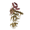


 PDBj
PDBj


 Assembly


 Mass: 24.305 Da / Num. of mol.: 1 / Source method: obtained synthetically / Formula: Mg
 Mass: 35.453 Da / Num. of mol.: 1 / Source method: obtained synthetically / Formula: Cl
 Mass: 507.181 Da / Num. of mol.: 1 / Source method: obtained synthetically / Formula: C10H16N5O13P3 / Comment: ATP, energy-carrying molecule*YM
 Mass: 194.226 Da / Num. of mol.: 1 / Source method: obtained synthetically / Formula: C8H18O5 / Comment: precipitant*YM
 Sample preparation
 / Beamline: 8.2.2 / Wavelength: 0.9796 Å
 Processing