 Movie
Movie Controller

[English] 日本語
 Yorodumi
- PDB-7mrw: Native RhopH complex of the malaria parasite Plasmodium falciparum -
+ Open data

- Basic information
| Entry | Database: PDB / ID: 7mrw | |||||||||||||||||||||||||||||||||
|---|---|---|---|---|---|---|---|---|---|---|---|---|---|---|---|---|---|---|---|---|---|---|---|---|---|---|---|---|---|---|---|---|---|---|
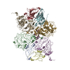
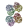
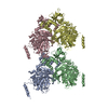
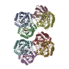
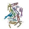
| Title | Native RhopH complex of the malaria parasite Plasmodium falciparum | |||||||||||||||||||||||||||||||||
 Components Components |
| |||||||||||||||||||||||||||||||||
 Keywords Keywords | MEMBRANE PROTEIN / malaria / rhoptry / complex / soluble | |||||||||||||||||||||||||||||||||
| Function / homology |  Function and homology information Function and homology informationrhoptry / adhesion of symbiont to microvasculature / symbiont-containing vacuole membrane / cytoplasmic vesicle / host cell cytoplasm / host cell plasma membrane Similarity search - Function | |||||||||||||||||||||||||||||||||
| Biological species |  | |||||||||||||||||||||||||||||||||
| Method | ELECTRON MICROSCOPY / single particle reconstruction / cryo EM / Resolution: 3.72 Å | |||||||||||||||||||||||||||||||||
 Authors Authors | Ho, C.M. / Jih, J. / Lai, M. / Li, X.R. / Goldberg, D.E. / Beck, J.R. / Zhou, Z.H. | |||||||||||||||||||||||||||||||||
| Funding support |  United States, 10items United States, 10items
| |||||||||||||||||||||||||||||||||
 Citation Citation | Journal: Proc Natl Acad Sci U S A / Year: 2021 Title: Native structure of the RhopH complex, a key determinant of malaria parasite nutrient acquisition. Authors: Chi-Min Ho / Jonathan Jih / Mason Lai / Xiaorun Li / Daniel E Goldberg / Josh R Beck / Z Hong Zhou / Abstract: The RhopH complex is implicated in malaria parasites' ability to invade and create new permeability pathways in host erythrocytes, but its mechanisms remain poorly understood. Here, we enrich the ...The RhopH complex is implicated in malaria parasites' ability to invade and create new permeability pathways in host erythrocytes, but its mechanisms remain poorly understood. Here, we enrich the endogenous RhopH complex in a native soluble form, comprising RhopH2, CLAG3.1, and RhopH3, directly from parasite cell lysates and determine its atomic structure using cryo-electron microscopy (cryo-EM), mass spectrometry, and the cryoID program. CLAG3.1 is positioned between RhopH2 and RhopH3, which both share substantial binding interfaces with CLAG3.1 but make minimal contacts with each other. The forces stabilizing individual subunits include 13 intramolecular disulfide bonds. Notably, CLAG3.1 residues 1210 to 1223, previously predicted to constitute a transmembrane helix, are embedded within a helical bundle formed by residues 979 to 1289 near the C terminus of CLAG3.1. Buried in the core of the RhopH complex and largely shielded from solvent, insertion of this putative transmembrane helix into the erythrocyte membrane would likely require a large conformational rearrangement. Given the unusually high disulfide content of the complex, it is possible that such a rearrangement could be initiated by the breakage of allosteric disulfide bonds, potentially triggered by interactions at the erythrocyte membrane. This first direct observation of an exported transmembrane protein-in a soluble, trafficking state and with atomic details of buried putative membrane-insertion helices-offers insights into the assembly and trafficking of RhopH and other parasite-derived complexes to the erythrocyte membrane. Our study demonstrates the potential the endogenous structural proteomics approach holds for elucidating the molecular mechanisms of hard-to-isolate complexes in their native, functional forms. | |||||||||||||||||||||||||||||||||
| History |
|
- Structure visualization
| Movie |
Movie viewer |
|---|---|
| Structure viewer | Molecule: MolmilJmol/JSmol |
- Downloads & links
-Download
| PDBx/mmCIF format | 7mrw.cif.gz | 558 KB | Display | PDBx/mmCIF format |
|---|---|---|---|---|
| PDB format | pdb7mrw.ent.gz | 434.5 KB | Display | PDB format |
| PDBx/mmJSON format | 7mrw.json.gz | Tree view | PDBx/mmJSON format | |
| Others |  Other downloads Other downloads |
-Validation report
| Arichive directory | https://data.pdbj.org/pub/pdb/validation_reports/mr/7mrwftp://data.pdbj.org/pub/pdb/validation_reports/mr/7mrw | HTTPS FTP |
|---|
-Related structure data
| Related structure data | 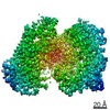 23959MC M: map data used to model this data C: citing same article ( |
|---|---|
| Similar structure data |


-Links
 PDBj
PDBj- Assembly
| Deposited unit | 
|
|---|---|
| 1 |
|
-Components
| #1: Protein | Mass: 167444.219 Da / Num. of mol.: 1 / Source method: isolated from a natural source Details: CLAG3.1, subunit of the soluble form of the RhopH complex Source: (natural) |
|---|---|
| #2: Protein | Mass: 162886.344 Da / Num. of mol.: 1 / Source method: isolated from a natural source Details: RhopH2, subunit of the soluble form of the RhopH complex Source: (natural) |
| #3: Protein | Mass: 104997.406 Da / Num. of mol.: 1 / Source method: isolated from a natural source Details: RhopH3, subunit of the soluble form of the RhopH complex Source: (natural) |
| Has protein modification | Y |
-Experimental details
-Experiment
| Experiment | Method: ELECTRON MICROSCOPY |
|---|---|
| EM experiment | Aggregation state: PARTICLE / 3D reconstruction method: single particle reconstruction |
- Sample preparation
| Component | Name: High molecular mass rhoptry protein complex (RhopH complex) Type: COMPLEX / Entity ID: all / Source: NATURAL |
|---|---|
| Molecular weight | Experimental value: NO |
| Source (natural) | Organism: |
| Buffer solution | pH: 7.4 |
| Specimen | Embedding applied: NO / Shadowing applied: NO / Staining applied: NO / Vitrification applied: YES |
| Vitrification | Instrument: FEI VITROBOT MARK IV / Cryogen name: ETHANE |
- Electron microscopy imaging
| Experimental equipment |  Model: Titan Krios / Image courtesy: FEI Company |
|---|---|
| Microscopy | Model: FEI TITAN KRIOS |
| Electron gun | Electron source:  FIELD EMISSION GUN / Accelerating voltage: 300 kV / Illumination mode: FLOOD BEAM FIELD EMISSION GUN / Accelerating voltage: 300 kV / Illumination mode: FLOOD BEAM |
| Electron lens | Mode: BRIGHT FIELD |
| Image recording | Electron dose: 60 e/Å2 / Film or detector model: GATAN K2 SUMMIT (4k x 4k) |
- Processing
| Software |
| ||||||||||||||||||||||||
|---|---|---|---|---|---|---|---|---|---|---|---|---|---|---|---|---|---|---|---|---|---|---|---|---|---|
| EM software |
| ||||||||||||||||||||||||
| CTF correction | Type: PHASE FLIPPING AND AMPLITUDE CORRECTION | ||||||||||||||||||||||||
| Particle selection | Num. of particles selected: 2900167 | ||||||||||||||||||||||||
| Symmetry | Point symmetry: C1 (asymmetric) | ||||||||||||||||||||||||
| 3D reconstruction | Resolution: 3.72 Å / Resolution method: FSC 0.143 CUT-OFF / Num. of particles: 108872 / Symmetry type: POINT | ||||||||||||||||||||||||
| Atomic model building | Protocol: AB INITIO MODEL / Space: REAL | ||||||||||||||||||||||||
| Refinement | Stereochemistry target values: GeoStd + Monomer Library | ||||||||||||||||||||||||
| Refine LS restraints |
|
