Movie
Movie Controller


+ Open data

- Basic information
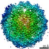
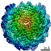
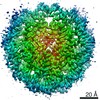
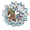
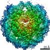
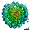
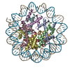
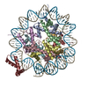
| Entry | Database: PDB / ID: 7lv8 | |||||||||
|---|---|---|---|---|---|---|---|---|---|---|
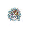
| Title | Structure of the Marseillevirus nucleosome | |||||||||
 Components Components |
| |||||||||
 Keywords Keywords | STRUCTURAL PROTEIN/DNA / STRUCTURAL PROTEIN / STRUCTURAL PROTEIN-DNA complex | |||||||||
| Function / homology |  Function and homology information Function and homology informationstructural constituent of chromatin / protein heterodimerization activity / DNA binding Similarity search - Function | |||||||||
| Biological species |  Marseillevirus marseillevirus Marseillevirus marseillevirussynthetic construct (others) | |||||||||
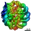
| Method | ELECTRON MICROSCOPY / single particle reconstruction / cryo EM / Resolution: 3.4 Å | |||||||||
 Authors Authors | Valencia-Sanchez, M.I. / Abini-Agbomson, S. / Armache, K.-J. | |||||||||
| Funding support |  United States, 2items United States, 2items
| |||||||||
 Citation Citation | Journal: Nat Struct Mol Biol / Year: 2021 Title: The structure of a virus-encoded nucleosome. Authors: Marco Igor Valencia-Sánchez / Stephen Abini-Agbomson / Miao Wang / Rachel Lee / Nikita Vasilyev / Jenny Zhang / Pablo De Ioannes / Bernard La Scola / Paul Talbert / Steve Henikoff / Evgeny ...Authors: Marco Igor Valencia-Sánchez / Stephen Abini-Agbomson / Miao Wang / Rachel Lee / Nikita Vasilyev / Jenny Zhang / Pablo De Ioannes / Bernard La Scola / Paul Talbert / Steve Henikoff / Evgeny Nudler / Albert Erives / Karim-Jean Armache /  Abstract: Certain large DNA viruses, including those in the Marseilleviridae family, encode histones. Here we show that fused histone pairs Hβ-Hα and Hδ-Hγ from Marseillevirus are structurally analogous to ...Certain large DNA viruses, including those in the Marseilleviridae family, encode histones. Here we show that fused histone pairs Hβ-Hα and Hδ-Hγ from Marseillevirus are structurally analogous to the eukaryotic histone pairs H2B-H2A and H4-H3. These viral histones form 'forced' heterodimers, and a heterotetramer of four such heterodimers assembles DNA to form structures virtually identical to canonical eukaryotic nucleosomes. | |||||||||
| History |
|
- Structure visualization
| Movie |
Movie viewer |
|---|---|
| Structure viewer | Molecule: MolmilJmol/JSmol |
- Downloads & links
-Download
| PDBx/mmCIF format | 7lv8.cif.gz | 256.1 KB | Display | PDBx/mmCIF format |
|---|---|---|---|---|
| PDB format | pdb7lv8.ent.gz | 190.1 KB | Display | PDB format |
| PDBx/mmJSON format | 7lv8.json.gz | Tree view | PDBx/mmJSON format | |
| Others |  Other downloads Other downloads |
-Validation report
| Arichive directory | https://data.pdbj.org/pub/pdb/validation_reports/lv/7lv8ftp://data.pdbj.org/pub/pdb/validation_reports/lv/7lv8 | HTTPS FTP |
|---|
-Related structure data
| Related structure data | 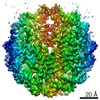 23529MC 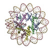 7lv9C C: citing same article ( M: map data used to model this data |
|---|---|
| Similar structure data |

-Links
 PDBj
PDBj


- Assembly
| Deposited unit | 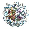
|
|---|---|
| 1 |
|
-Components
-Histone doublet Delta-Gamma ... , 2 types, 4 molecules BFAE
| #1: Protein | Mass: 10462.416 Da / Num. of mol.: 2 Source method: isolated from a genetically manipulated source Source: (gene. exp.) Marseillevirus marseillevirus / Gene: MAR_ORF413 / Production host:  #2: Protein | Mass: 11767.600 Da / Num. of mol.: 2 Source method: isolated from a genetically manipulated source Source: (gene. exp.) Marseillevirus marseillevirus / Gene: MAR_ORF413 / Production host: |
|---|
-Histone doublet Beta-Alpha ... , 2 types, 4 molecules DHCG
| #3: Protein | Mass: 11228.021 Da / Num. of mol.: 2 Source method: isolated from a genetically manipulated source Source: (gene. exp.) Marseillevirus marseillevirus / Production host: #4: Protein | Mass: 17675.625 Da / Num. of mol.: 2 Source method: isolated from a genetically manipulated source Source: (gene. exp.) Marseillevirus marseillevirus / Gene: MAR_ORF414 / Production host: |
|---|
-DNA chain , 2 types, 2 molecules IJ
| #5: DNA chain | Mass: 37530.910 Da / Num. of mol.: 1 / Source method: obtained synthetically / Source: (synth.) synthetic construct (others) |
|---|---|
| #6: DNA chain | Mass: 37152.672 Da / Num. of mol.: 1 / Source method: obtained synthetically / Source: (synth.) synthetic construct (others) |
-Details
| Has protein modification | Y |
|---|
-Experimental details
-Experiment
| Experiment | Method: ELECTRON MICROSCOPY |
|---|---|
| EM experiment | Aggregation state: PARTICLE / 3D reconstruction method: single particle reconstruction |
- Sample preparation
| Component | Name: Structure of the Marseillevirus nucleosome / Type: COMPLEX / Details: virus-encoded histone doublets Marseillevirus / Entity ID: all / Source: RECOMBINANT | ||||||||||||||||||||
|---|---|---|---|---|---|---|---|---|---|---|---|---|---|---|---|---|---|---|---|---|---|
| Molecular weight | Experimental value: NO | ||||||||||||||||||||
| Source (natural) |
| ||||||||||||||||||||
| Source (recombinant) | Organism: | ||||||||||||||||||||
| Buffer solution | pH: 7.5 | ||||||||||||||||||||
| Buffer component |
| ||||||||||||||||||||
| Specimen | Conc.: 3.3 mg/ml / Embedding applied: NO / Shadowing applied: NO / Staining applied: NO / Vitrification applied: YES | ||||||||||||||||||||
| Specimen support | Grid material: GOLD / Grid mesh size: 400 divisions/in. / Grid type: Quantifoil R1.2/1.3 | ||||||||||||||||||||
| Vitrification | Instrument: FEI VITROBOT MARK IV / Cryogen name: ETHANE / Humidity: 100 % / Chamber temperature: 297 K |
- Electron microscopy imaging
| Experimental equipment |  Model: Titan Krios / Image courtesy: FEI Company |
|---|---|
| Microscopy | Model: FEI TITAN KRIOS |
| Electron gun | Electron source:  FIELD EMISSION GUN / Accelerating voltage: 300 kV / Illumination mode: OTHER FIELD EMISSION GUN / Accelerating voltage: 300 kV / Illumination mode: OTHER |
| Electron lens | Mode: BRIGHT FIELD / Nominal magnification: 64000 X / Nominal defocus max: 2400 nm / Nominal defocus min: 1000 nm / Cs: 2.7 mm / Alignment procedure: COMA FREE |
| Specimen holder | Cryogen: NITROGEN / Specimen holder model: FEI TITAN KRIOS AUTOGRID HOLDER |
| Image recording | Average exposure time: 2.5 sec. / Electron dose: 65 e/Å2 / Film or detector model: GATAN K3 (6k x 4k) / Num. of grids imaged: 1 / Num. of real images: 4503 |
| EM imaging optics | Energyfilter slit width: 20 eV |
- Processing
| Software | Name: PHENIX / Version: 1.19.1_4122: / Classification: refinement | ||||||||||||||||||||||||||||||||||||||||
|---|---|---|---|---|---|---|---|---|---|---|---|---|---|---|---|---|---|---|---|---|---|---|---|---|---|---|---|---|---|---|---|---|---|---|---|---|---|---|---|---|---|
| EM software |
| ||||||||||||||||||||||||||||||||||||||||
| CTF correction | Type: PHASE FLIPPING AND AMPLITUDE CORRECTION | ||||||||||||||||||||||||||||||||||||||||
| Particle selection | Num. of particles selected: 3017414 | ||||||||||||||||||||||||||||||||||||||||
| Symmetry | Point symmetry: C1 (asymmetric) | ||||||||||||||||||||||||||||||||||||||||
| 3D reconstruction | Resolution: 3.4 Å / Resolution method: FSC 0.143 CUT-OFF / Num. of particles: 146506 / Algorithm: FOURIER SPACE / Num. of class averages: 1 / Symmetry type: POINT | ||||||||||||||||||||||||||||||||||||||||
| Atomic model building | Protocol: OTHER / Space: REAL | ||||||||||||||||||||||||||||||||||||||||
| Atomic model building | 3D fitting-ID: 1 / Source name: PDB / Type: experimental model
| ||||||||||||||||||||||||||||||||||||||||
| Refine LS restraints |
|