 Movie
Movie Controller


+ Open data

- Basic information
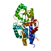
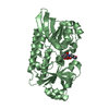
| Entry | Database: PDB / ID: 7kxw | ||||||
|---|---|---|---|---|---|---|---|
| Title | Crystal structure of DCLK1-KD in complex with DCLK1-IN-1 | ||||||
 Components Components | (Serine/threonine-protein kinase ...) x 2 | ||||||
 Keywords Keywords | TRANSFERASE / KINASE / DOUBLECORTIN-LIKE | ||||||
| Function / homology |  Function and homology information Function and homology informationaxon extension / endosomal transport / protein localization to nucleus / central nervous system development / response to virus / nervous system development / protein phosphorylation / protein kinase activity / non-specific serine/threonine protein kinase / intracellular signal transduction ...axon extension / endosomal transport / protein localization to nucleus / central nervous system development / response to virus / nervous system development / protein phosphorylation / protein kinase activity / non-specific serine/threonine protein kinase / intracellular signal transduction / protein serine kinase activity / protein serine/threonine kinase activity / ATP binding / plasma membrane / cytoplasm Similarity search - Function | ||||||
| Biological species |  Homo sapiens (human) Homo sapiens (human) | ||||||
| Method |  X-RAY DIFFRACTION / SYNCHROTRON / MOLECULAR REPLACEMENT / Resolution: 3.002 Å X-RAY DIFFRACTION / SYNCHROTRON / MOLECULAR REPLACEMENT / Resolution: 3.002 Å | ||||||
 Authors Authors | Patel, O. / Lucet, I. | ||||||
| Funding support |  Australia, 1items Australia, 1items
| ||||||
 Citation Citation | Journal: Commun Biol / Year: 2021 Title: Structural basis for small molecule targeting of Doublecortin Like Kinase 1 with DCLK1-IN-1. Authors: Patel, O. / Roy, M.J. / Kropp, A. / Hardy, J.M. / Dai, W. / Lucet, I.S. | ||||||
| History |
|
- Structure visualization
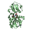
| Structure viewer | Molecule: MolmilJmol/JSmol |
|---|
- Downloads & links
-Download
| PDBx/mmCIF format | 7kxw.cif.gz | 116.4 KB | Display | PDBx/mmCIF format |
|---|---|---|---|---|
| PDB format | pdb7kxw.ent.gz | 86.7 KB | Display | PDB format |
| PDBx/mmJSON format | 7kxw.json.gz | Tree view | PDBx/mmJSON format | |
| Others |  Other downloads Other downloads |
-Validation report
| Arichive directory | https://data.pdbj.org/pub/pdb/validation_reports/kx/7kxwftp://data.pdbj.org/pub/pdb/validation_reports/kx/7kxw | HTTPS FTP |
|---|
-Related structure data
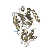
| Related structure data |  7kx6C  7kx8C  5jzjS S: Starting model for refinement C: citing same article ( |
|---|---|
| Similar structure data |


-Links
 PDBj
PDBj- Assembly
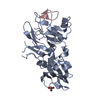
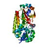
| Deposited unit | 
| |||||||||||||||||||||||||||||||||||||||||||||||||||||||||||||||||||||||||
|---|---|---|---|---|---|---|---|---|---|---|---|---|---|---|---|---|---|---|---|---|---|---|---|---|---|---|---|---|---|---|---|---|---|---|---|---|---|---|---|---|---|---|---|---|---|---|---|---|---|---|---|---|---|---|---|---|---|---|---|---|---|---|---|---|---|---|---|---|---|---|---|---|---|---|
| 1 | 
| |||||||||||||||||||||||||||||||||||||||||||||||||||||||||||||||||||||||||
| 2 | 
| |||||||||||||||||||||||||||||||||||||||||||||||||||||||||||||||||||||||||
| Unit cell |
| |||||||||||||||||||||||||||||||||||||||||||||||||||||||||||||||||||||||||
| Noncrystallographic symmetry (NCS) | NCS domain:
NCS domain segments:
|