 Movie
Movie Controller


+ Open data

- Basic information
| Entry | Database: EMDB / ID: EMD-7327 | |||||||||
|---|---|---|---|---|---|---|---|---|---|---|
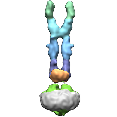
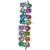
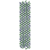
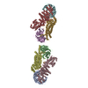
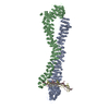
| Title | Structure of a membrane protein complex | |||||||||
 Map data Map data | primary map | |||||||||
 Sample Sample |
| |||||||||
 Keywords Keywords | PCDH15 / LHFPL5 / protocadherin / tip link / hair cell / TMHS / hearing / MEMBRANE PROTEIN | |||||||||
| Function / homology |  Function and homology information Function and homology informationdetection of mechanical stimulus involved in equilibrioception / equilibrioception / sensory perception of light stimulus / inner ear receptor cell stereocilium organization / righting reflex / inner ear auditory receptor cell differentiation / stereocilium bundle / detection of mechanical stimulus involved in sensory perception of sound / stereocilium / photoreceptor cell maintenance ...detection of mechanical stimulus involved in equilibrioception / equilibrioception / sensory perception of light stimulus / inner ear receptor cell stereocilium organization / righting reflex / inner ear auditory receptor cell differentiation / stereocilium bundle / detection of mechanical stimulus involved in sensory perception of sound / stereocilium / photoreceptor cell maintenance / non-motile cilium assembly / auditory receptor cell stereocilium organization / adult walking behavior / homophilic cell-cell adhesion / inner ear development / startle response / actin filament bundle assembly / photoreceptor outer segment / visual perception / cell adhesion molecule binding / morphogenesis of an epithelium / actin filament organization / locomotory behavior / sensory perception of sound / response to calcium ion / multicellular organism growth / cell adhesion / calcium ion binding / synapse / : / membrane / plasma membrane / cytoplasm Similarity search - Function | |||||||||
| Biological species |  | |||||||||
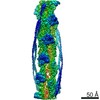
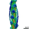
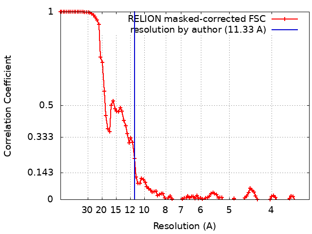
| Method | single particle reconstruction / cryo EM / Resolution: 11.33 Å | |||||||||
 Authors Authors | Gouaux E / Ge J | |||||||||
 Citation Citation | Journal: Elife / Year: 2018 Title: Structure of mouse protocadherin 15 of the stereocilia tip link in complex with LHFPL5. Authors: Jingpeng Ge / Johannes Elferich / April Goehring / Huaying Zhao / Peter Schuck / Eric Gouaux /  Abstract: Hearing and balance involve the transduction of mechanical stimuli into electrical signals by deflection of bundles of stereocilia linked together by protocadherin 15 (PCDH15) and cadherin 23 'tip ...Hearing and balance involve the transduction of mechanical stimuli into electrical signals by deflection of bundles of stereocilia linked together by protocadherin 15 (PCDH15) and cadherin 23 'tip links'. PCDH15 transduces tip link tension into opening of a mechano-electrical transduction (MET) ion channel. PCDH15 also interacts with LHFPL5, a candidate subunit of the MET channel. Here we illuminate the PCDH15-LHFPL5 structure, showing how the complex is composed of PCDH15 and LHFPL5 subunit pairs related by a 2-fold axis. The extracellular cadherin domains define a mobile tether coupled to a rigid, 2-fold symmetric 'collar' proximal to the membrane bilayer. LHFPL5 forms extensive interactions with the PCDH15 transmembrane helices and stabilizes the overall PCDH15-LHFPL5 assembly. Our studies illuminate the architecture of the PCDH15-LHFPL5 complex, localize mutations associated with deafness, and shed new light on how forces in the PCDH15 tether may be transduced into the stereocilia membrane. | |||||||||
| History |
|
- Structure visualization
| Movie |
Movie viewer |
|---|---|
| Structure viewer | EM map: SurfViewMolmilJmol/JSmol |
| Supplemental images |
- Downloads & links
-EMDB archive
| Map data | emd_7327.map.gz | 78.2 MB | EMDB map data format | |
|---|---|---|---|---|
| Header (meta data) | emd-7327-v30.xmlemd-7327.xml | 11.5 KB 11.5 KB | Display Display | EMDB header |
| FSC (resolution estimation) | emd_7327_fsc.xml | 10.1 KB | Display | FSC data file |
| Images |  emd_7327.png emd_7327.png | 53.9 KB | ||
| Filedesc metadata | emd-7327.cif.gz | 6 KB | ||
| Archive directory |  http://ftp.pdbj.org/pub/emdb/structures/EMD-7327ftp://ftp.pdbj.org/pub/emdb/structures/EMD-7327 http://ftp.pdbj.org/pub/emdb/structures/EMD-7327ftp://ftp.pdbj.org/pub/emdb/structures/EMD-7327 | HTTPS FTP |
-Related structure data
| Related structure data |  6c13MC  7328C  6c10C 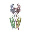 6c14C C: citing same article ( M: atomic model generated by this map |
|---|---|
| Similar structure data |

-Links
| EMDB pages | EMDB (EBI/PDBe) / EMDataResource |
|---|---|
| Related items in Molecule of the Month |

-Map
| File | Download / File: emd_7327.map.gz / Format: CCP4 / Size: 83.7 MB / Type: IMAGE STORED AS FLOATING POINT NUMBER (4 BYTES) | ||||||||||||||||||||||||||||||||||||||||||||||||||||||||||||||||||||
|---|---|---|---|---|---|---|---|---|---|---|---|---|---|---|---|---|---|---|---|---|---|---|---|---|---|---|---|---|---|---|---|---|---|---|---|---|---|---|---|---|---|---|---|---|---|---|---|---|---|---|---|---|---|---|---|---|---|---|---|---|---|---|---|---|---|---|---|---|---|
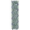
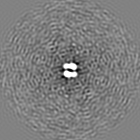
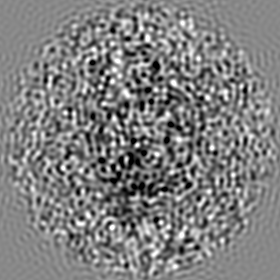
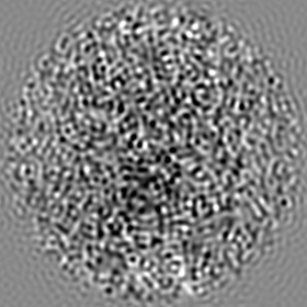
| Annotation | primary map | ||||||||||||||||||||||||||||||||||||||||||||||||||||||||||||||||||||
| Projections & slices | Image control
Images are generated by Spider. | ||||||||||||||||||||||||||||||||||||||||||||||||||||||||||||||||||||
| Voxel size | X=Y=Z: 1.72 Å | ||||||||||||||||||||||||||||||||||||||||||||||||||||||||||||||||||||
| Density |
| ||||||||||||||||||||||||||||||||||||||||||||||||||||||||||||||||||||
| Symmetry | Space group: 1 | ||||||||||||||||||||||||||||||||||||||||||||||||||||||||||||||||||||
| Details | EMDB XML:
CCP4 map header:
| ||||||||||||||||||||||||||||||||||||||||||||||||||||||||||||||||||||
 Z (Sec.)
 Y (Row.)
 X (Col.)


-Supplemental data
- Sample components
-Entire : Complex of membrane proteins
| Entire | Name: Complex of membrane proteins |
|---|---|
| Components |
|
-Supramolecule #1: Complex of membrane proteins
| Supramolecule | Name: Complex of membrane proteins / type: complex / ID: 1 / Parent: 0 / Macromolecule list: all |
|---|---|
| Source (natural) | Organism: |
-Macromolecule #1: Protocadherin-15
| Macromolecule | Name: Protocadherin-15 / type: protein_or_peptide / ID: 1 / Number of copies: 2 / Enantiomer: LEVO |
|---|---|
| Source (natural) | Organism: |
| Molecular weight | Theoretical: 72.989023 KDa |
| Recombinant expression | Organism:  Homo sapiens (human) Homo sapiens (human) |
| Sequence | String: QYDDSPVFTN STYTVVVEEN LPAGTSFLQI EAKDVDLGAN VSYRIRSPEV KHLFALHPFT GELSLLRSLD YEAFPDQEAS ITFLAEAFD IYGTMPPGIA TVTVIVKDMN DYPPVFSKRI YKGMVAPDAV KGTPITTVYA EDADPPGMPA SRVRYRVDDV Q FPYPASIF ...String: QYDDSPVFTN STYTVVVEEN LPAGTSFLQI EAKDVDLGAN VSYRIRSPEV KHLFALHPFT GELSLLRSLD YEAFPDQEAS ITFLAEAFD IYGTMPPGIA TVTVIVKDMN DYPPVFSKRI YKGMVAPDAV KGTPITTVYA EDADPPGMPA SRVRYRVDDV Q FPYPASIF DVEEDSGRVV TRVNLNEEPT TIFKLVVVAF DDGEPVMSSS ATVRILVLHP GEIPRFTQEE YRPPPVSELA AR GTVVGVI SAAAINQSIV YSIVAGNEED KFGINNVTGV IYVNSPLDYE TRTSYVLRVQ ADSLEVVLAN LRVPSKSNTA KVY IEIQDE NDHPPVFQKK FYIGGVSEDA RMFASVLRVK ATDRDTGNYS AMAYRLIIPP IKEGKEGFVV ETYTGLIKTA MLFH NMRRS YFKFQVIATD DYGKGLSGKA DVLVSVVNQL DMQVIVSNVP PTLVEKKIED LTEILDRYVQ EQIPGAKVVV ESIGA RRHG DAYSLEDYSK CDLTVYAIDP QTNRAIDRNE LFKFLDGKLL DINKDFQPYY GEGGRILEIR TPEAVTSIKK RGESLG YTE GALLALAFII ILCCIPAILV VLVSYRQFKV RQAECTKTAR IQSAMPAAKP AAPVPAAPAP PPPPPPPPPG AHLYEEL GE SAMHKYETAL FESRLVPR UniProtKB: Protocadherin-15 |
-Experimental details
-Structure determination
| Method | cryo EM |
|---|---|
 Processing Processing | single particle reconstruction |
| Aggregation state | particle |
-Sample preparation
| Concentration | 1.5 mg/mL | ||||||||||||
|---|---|---|---|---|---|---|---|---|---|---|---|---|---|
| Buffer | pH: 8 Component:
| ||||||||||||
| Grid | Model: Quantifoil R1.2/1.3 / Material: GOLD / Mesh: 300 / Pretreatment - Type: GLOW DISCHARGE | ||||||||||||
| Vitrification | Cryogen name: ETHANE / Chamber humidity: 100 % / Chamber temperature: 285 K / Instrument: FEI VITROBOT MARK III |
- Electron microscopy
| Microscope | FEI TITAN KRIOS |
|---|---|
| Specialist optics | Phase plate: VOLTA PHASE PLATE |
| Image recording | Film or detector model: GATAN K2 SUMMIT (4k x 4k) / Detector mode: SUPER-RESOLUTION / Average exposure time: 10.0 sec. / Average electron dose: 27.0 e/Å2 |
| Electron beam | Acceleration voltage: 300 kV / Electron source:  FIELD EMISSION GUN FIELD EMISSION GUN |
| Electron optics | Illumination mode: FLOOD BEAM / Imaging mode: BRIGHT FIELD |
| Experimental equipment |  Model: Titan Krios / Image courtesy: FEI Company |
+Image processing
-Atomic model buiding 1
| Refinement | Protocol: FLEXIBLE FIT |
|---|---|
| Output model | PDB-6c13: |
