ムービー
ムービー コントローラー


+ データを開く

- 基本情報
基本情報
| 登録情報 | データベース: PDB / ID: 6z7p | |||||||||
|---|---|---|---|---|---|---|---|---|---|---|
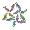
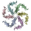
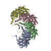
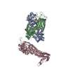
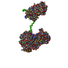
| タイトル | Composite model of the Caulobacter crescentus S-layer bound to the O-antigen of lipopolysaccharide | |||||||||
 要素 要素 | S-layer protein S層 S層 | |||||||||
 キーワード キーワード | STRUCTURAL PROTEIN (タンパク質) / RsaA S-layer sub-tomogram averaging Caulobacter lipopolysaccharide O-antigen | |||||||||
| 機能・相同性 | RsaA N-terminal domain / S層 / RTX calcium-binding nonapeptide repeat / RTX calcium-binding nonapeptide repeat (4 copies) / Serralysin-like metalloprotease, C-terminal / calcium ion binding / extracellular region / S-layer protein 機能・相同性情報 機能・相同性情報 | |||||||||
| 生物種 |  Caulobacter vibrioides (カウロバクター・クレセンタス) Caulobacter vibrioides (カウロバクター・クレセンタス) | |||||||||
| 手法 | 電子顕微鏡法 / サブトモグラム平均法 / クライオ電子顕微鏡法 / 解像度: 4.8 Å | |||||||||
 データ登録者 データ登録者 | Bharat, T.A.M. / von Kugelgen, A. | |||||||||
| 資金援助 |  英国, 1件 英国, 1件
| |||||||||
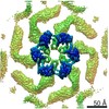
 引用 引用 | ジャーナル: Cell / 年: 2020 タイトル: In Situ Structure of an Intact Lipopolysaccharide-Bound Bacterial Surface Layer. 著者: Andriko von Kügelgen / Haiping Tang / Gail G Hardy / Danguole Kureisaite-Ciziene / Yves V Brun / Phillip J Stansfeld / Carol V Robinson / Tanmay A M Bharat /   要旨: Most bacterial and all archaeal cells are encapsulated by a paracrystalline, protective, and cell-shape-determining proteinaceous surface layer (S-layer). On Gram-negative bacteria, S-layers are ...Most bacterial and all archaeal cells are encapsulated by a paracrystalline, protective, and cell-shape-determining proteinaceous surface layer (S-layer). On Gram-negative bacteria, S-layers are anchored to cells via lipopolysaccharide. Here, we report an electron cryomicroscopy structure of the Caulobacter crescentus S-layer bound to the O-antigen of lipopolysaccharide. Using native mass spectrometry and molecular dynamics simulations, we deduce the length of the O-antigen on cells and show how lipopolysaccharide binding and S-layer assembly is regulated by calcium. Finally, we present a near-atomic resolution in situ structure of the complete S-layer using cellular electron cryotomography, showing S-layer arrangement at the tip of the O-antigen. A complete atomic structure of the S-layer shows the power of cellular tomography for in situ structural biology and sheds light on a very abundant class of self-assembling molecules with important roles in prokaryotic physiology with marked potential for synthetic biology and surface-display applications. #1: ジャーナル: Nat Microbiol / 年: 2017タイトル: Structure of the hexagonal surface layer on Caulobacter crescentus cells. 著者: Tanmay A M Bharat / Danguole Kureisaite-Ciziene / Gail G Hardy / Ellen W Yu / Jessica M Devant / Wim J H Hagen / Yves V Brun / John A G Briggs / Jan Löwe /  要旨: Many prokaryotic cells are encapsulated by a surface layer (S-layer) consisting of repeating units of S-layer proteins. S-layer proteins are a diverse class of molecules found in Gram-positive and ...Many prokaryotic cells are encapsulated by a surface layer (S-layer) consisting of repeating units of S-layer proteins. S-layer proteins are a diverse class of molecules found in Gram-positive and Gram-negative bacteria and most archaea. S-layers protect cells from the outside, provide mechanical stability and also play roles in pathogenicity. In situ structural information about this highly abundant class of proteins is scarce, so atomic details of how S-layers are arranged on the surface of cells have remained elusive. Here, using purified Caulobacter crescentus' sole S-layer protein RsaA, we obtained a 2.7 Å X-ray structure that shows the hexameric S-layer lattice. We also solved a 7.4 Å structure of the S-layer through electron cryotomography and sub-tomogram averaging of cell stalks. The X-ray structure was docked unambiguously into the electron cryotomography map, resulting in a pseudo-atomic-level description of the in vivo S-layer, which agrees completely with the atomic X-ray lattice model. The cellular S-layer atomic structure shows that the S-layer is porous, with a largest gap dimension of 27 Å, and is stabilized by multiple Ca ions bound near the interfaces. This study spans different spatial scales from atoms to cells by combining X-ray crystallography with electron cryotomography and sub-nanometre-resolution sub-tomogram averaging. | |||||||||
| 履歴 |
|
- 構造の表示
構造の表示
| ムービー |
ムービービューア |
|---|---|
| 構造ビューア | 分子: MolmilJmol/JSmol |
- ダウンロードとリンク
-ダウンロード
| PDBx/mmCIF形式 | 6z7p.cif.gz | 173.1 KB | 表示 | PDBx/mmCIF形式 |
|---|---|---|---|---|
| PDB形式 | pdb6z7p.ent.gz | 126.8 KB | 表示 | PDB形式 |
| PDBx/mmJSON形式 | 6z7p.json.gz | ツリー表示 | PDBx/mmJSON形式 | |
| その他 |  その他のダウンロード その他のダウンロード |
-検証レポート
| アーカイブディレクトリ | https://data.pdbj.org/pub/pdb/validation_reports/z7/6z7pftp://data.pdbj.org/pub/pdb/validation_reports/z7/6z7p | HTTPS FTP |
|---|
-関連構造データ


-リンク
 PDBj
PDBj- 集合体
集合体
| 登録構造単位 | 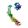
|
|---|---|
| 1 |
|
-要素
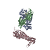
| #1: タンパク質 | S層 / Paracrystalline surface layer protein 分子量: 98022.703 Da / 分子数: 1 / 由来タイプ: 天然 由来: (天然) Caulobacter vibrioides (strain ATCC 19089 / CB15) (カウロバクター・クレセンタス)株: ATCC 19089 / CB15 / 参照: UniProt: P35828 | ||
|---|---|---|---|
| #2: 多糖 | 4-acetamido-4,6-dideoxy-alpha-D-mannopyranose-(1-3)-4-acetamido-4,6-dideoxy-alpha-D-mannopyranose- ...4-acetamido-4,6-dideoxy-alpha-D-mannopyranose-(1-3)-4-acetamido-4,6-dideoxy-alpha-D-mannopyranose-(1-3)-beta-D-mannopyranose-(1-3)-4-acetamido-4,6-dideoxy-alpha-D-mannopyranose-(1-3)-4-acetamido-4,6-dideoxy-alpha-D-mannopyranose-(1-3)-beta-D-mannopyranose-(1-3)-4-acetamido-4,6-dideoxy-alpha-D-mannopyranose-(1-3)-4-acetamido-4,6-dideoxy-alpha-D-mannopyranose-(1-3)-beta-D-mannopyranoseオリゴ糖 / 分子量: 1627.594 Da / 分子数: 1 / 由来タイプ: 合成 | ||
| #3: 化合物 | ChemComp-CA /   分子量: 40.078 Da / 分子数: 22 / 由来タイプ: 合成 / 式: Ca / タイプ: SUBJECT OF INVESTIGATION 分子量: 40.078 Da / 分子数: 22 / 由来タイプ: 合成 / 式: Ca / タイプ: SUBJECT OF INVESTIGATION研究の焦点であるリガンドがあるか | Y | |
-実験情報
-実験
| 実験 | 手法: 電子顕微鏡法 |
|---|---|
| EM実験 | 試料の集合状態: CELL / 3次元再構成法: サブトモグラム平均法 |
- 試料調製
試料調製
| 構成要素 | 名称: S-layerS層 / タイプ: COMPLEX / Entity ID: #1 / 由来: NATURAL |
|---|---|
| 由来(天然) | 生物種: Caulobacter vibrioides NA1000 (カウロバクター・クレセンタス) 株: YB2811 / 細胞内の位置: extra-cellular |
| 緩衝液 | pH: 7 / 詳細: PYE |
| 試料 | 包埋: NO / シャドウイング: NO / 染色: NO / 凍結: YES / 詳細: Caulobacter crescentus stalk |
| 試料支持 | 詳細: 15 mA / グリッドの材料: COPPER/RHODIUM / グリッドのサイズ: 200 divisions/in. / グリッドのタイプ: Quantifoil R2/2 |
| 急速凍結 | 装置: FEI VITROBOT MARK IV / 凍結剤: NITROGEN / 湿度: 100 % / 凍結前の試料温度: 283.15 K / 詳細: 1.5 s blot |
- 電子顕微鏡撮影
| 実験機器 |  モデル: Titan Krios / 画像提供: FEI Company |
|---|---|
| 顕微鏡 | モデル: FEI TITAN KRIOS |
| 電子銃 | 電子線源: FIELD EMISSION GUN / 加速電圧: 300 kV / 照射モード: FLOOD BEAM |
| 電子レンズ | モード: BRIGHT FIELDBright-field microscopy / 倍率(公称値): 105000 X / 倍率(補正後): 105000 X / 最大 デフォーカス(公称値): -5000 nm / 最小 デフォーカス(公称値): -2000 nm / Calibrated defocus min: -2000 nm / 最大 デフォーカス(補正後): -5000 nm / Cs: 2.7 mm / C2レンズ絞り径: 70 µm / アライメント法: COMA FREE |
| 試料ホルダ | 凍結剤: NITROGEN 試料ホルダーモデル: FEI TITAN KRIOS AUTOGRID HOLDER 最高温度: 70 K / 最低温度: 70 K |
| 撮影 | 平均露光時間: 1 sec. / 電子線照射量: 3.4 e/Å2 / 検出モード: SUPER-RESOLUTION フィルム・検出器のモデル: GATAN K2 QUANTUM (4k x 4k) 撮影したグリッド数: 1 / 詳細: Dose symmetric tilt scheme (Hagen et al, JSB) |
| 電子光学装置 | エネルギーフィルター名称: GIF Quantum LS / 色収差補正装置: not used / エネルギーフィルタースリット幅: 20 eV / 球面収差補正装置: not used |
- 解析
解析
| EMソフトウェア |
| |||||||||||||||||||||||||
|---|---|---|---|---|---|---|---|---|---|---|---|---|---|---|---|---|---|---|---|---|---|---|---|---|---|---|
| CTF補正 | 詳細: Following Turonova and Briggs NovaCTF / タイプ: PHASE FLIPPING AND AMPLITUDE CORRECTION | |||||||||||||||||||||||||
| 対称性 | 点対称性: C1 (非対称) | |||||||||||||||||||||||||
| 3次元再構成 | 解像度: 4.8 Å / 解像度の算出法: FSC 0.143 CUT-OFF / 粒子像の数: 51866 / アルゴリズム: FOURIER SPACE 詳細: Tomogram generation in IMOD using the NovaCTF implementation of Turonova and Briggs. Sub-tomogram averaging performed using the AV3 package (Friedrich Foerster) applied to tubular specimens ...詳細: Tomogram generation in IMOD using the NovaCTF implementation of Turonova and Briggs. Sub-tomogram averaging performed using the AV3 package (Friedrich Foerster) applied to tubular specimens (Bharat et al, PLoS Biology, 2011) クラス平均像の数: 1 / 対称性のタイプ: POINT | |||||||||||||||||||||||||
| EM volume selection | 手法: RELIONList of Walmart brands / 詳細: RELION subtomogram averaging / Num. of tomograms: 110 / Num. of volumes extracted: 51866 / Reference model: Ab initio | |||||||||||||||||||||||||
| 原子モデル構築 | プロトコル: RIGID BODY FIT / 空間: REAL / Target criteria: USCF Chimera 詳細: Initial rigid body fit of the C-terminal X-ray structure (PDB: 5N8P, amino acids 249-1046) and the N-terminal cryo-EM structure (PDB: 6T72, amino acids 2-243) was performed in USCF Chimera. ...詳細: Initial rigid body fit of the C-terminal X-ray structure (PDB: 5N8P, amino acids 249-1046) and the N-terminal cryo-EM structure (PDB: 6T72, amino acids 2-243) was performed in USCF Chimera. The missing amino acid linker was manually added suing Coot. The model was not refined against the map and all B-factors were set to zero due to resolution anisotropy. | |||||||||||||||||||||||||
| 原子モデル構築 | 3D fitting-ID: 1 / PDB chain-ID: A / Source name: PDB / タイプ: experimental model
|