National Institutes of Health/National Institute of General Medical Sciences (NIH/NIGMS)
GM082946
United States
Citation
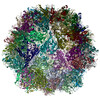
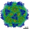
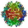
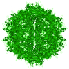
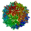
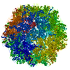
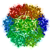
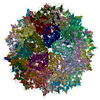
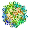
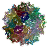
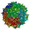
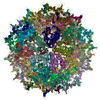
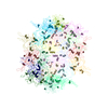
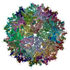
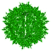
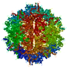
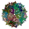
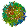
Journal: Viruses / Year: 2020 Title: Structural Characterization of Cuta- and Tusavirus: Insight into Protoparvoviruses Capsid Morphology. Authors: Mario Mietzsch / Robert McKenna / Elina Väisänen / Jennifer C Yu / Maria Ilyas / Joshua A Hull / Justin Kurian / J Kennon Smith / Paul Chipman / Yi Lasanajak / David Smith / Maria ...Authors: Mario Mietzsch / Robert McKenna / Elina Väisänen / Jennifer C Yu / Maria Ilyas / Joshua A Hull / Justin Kurian / J Kennon Smith / Paul Chipman / Yi Lasanajak / David Smith / Maria Söderlund-Venermo / Mavis Agbandje-McKenna / Abstract: Several members of the genus, capable of infecting humans, have been recently discovered, including cutavirus (CuV) and tusavirus (TuV). To begin the characterization of these viruses, we have used ...Several members of the genus, capable of infecting humans, have been recently discovered, including cutavirus (CuV) and tusavirus (TuV). To begin the characterization of these viruses, we have used cryo-electron microscopy and image reconstruction to determine their capsid structures to ~2.9 Å resolution, and glycan array and cell-based assays to identify glycans utilized for cellular entry. Structural comparisons show that the CuV and TuV capsids share common features with other parvoviruses, including an eight-stranded anti-parallel β-barrel, depressions at the icosahedral 2-fold and surrounding the 5-fold axes, and a channel at the 5-fold axes. However, the viruses exhibit significant topological differences in their viral protein surface loops. These result in three separated 3-fold protrusions, similar to the bufaviruses also infecting humans, suggesting a host-driven structure evolution. The surface loops contain residues involved in receptor binding, cellular trafficking, and antigenic reactivity in other parvoviruses. In addition, terminal sialic acid was identified as the glycan potentially utilized by both CuV and TuV for cellular entry, with TuV showing additional recognition of poly-sialic acid and sialylated Lewis X (sLeXLeXLeX) motifs reported to be upregulated in neurotropic and cancer cells, respectively. These structures provide a platform for annotating the cellular interactions of these human pathogens.
History
Deposition
May 20, 2020
Deposition site: RCSB / Processing site: RCSB
Revision 1.0
Jul 1, 2020
Provider: repository / Type: Initial release
Revision 1.0
Jul 1, 2020
Data content type: EM metadata / Data content type: EM metadata / Provider: repository / Type: Initial release
Revision 1.0
Jul 1, 2020
Data content type: Image / Data content type: Image / Provider: repository / Type: Initial release
Revision 1.0
Jul 1, 2020
Data content type: Primary map / Data content type: Primary map / Provider: repository / Type: Initial release
Revision 1.0
Jul 1, 2020
Data content type: Image / Data content type: Image / Provider: repository / Type: Initial release
Revision 1.0
Jul 1, 2020
Data content type: Primary map / Data content type: Primary map / Provider: repository / Type: Initial release
Data content type: EM metadata / Data content type: EM metadata / EM metadata / Group: Data processing / Experimental summary / Data content type: EM metadata / EM metadata / Category: em_admin / em_software / Data content type: EM metadata / EM metadata / Item: _em_admin.last_update / _em_software.name
In the structure databanks used in Yorodumi, some data are registered as the other names, "COVID-19 virus" and "2019-nCoV". Here are the details of the virus and the list of structure data.
Jan 31, 2019. EMDB accession codes are about to change! (news from PDBe EMDB page)
EMDB accession codes are about to change! (news from PDBe EMDB page)
The allocation of 4 digits for EMDB accession codes will soon come to an end. Whilst these codes will remain in use, new EMDB accession codes will include an additional digit and will expand incrementally as the available range of codes is exhausted. The current 4-digit format prefixed with “EMD-” (i.e. EMD-XXXX) will advance to a 5-digit format (i.e. EMD-XXXXX), and so on. It is currently estimated that the 4-digit codes will be depleted around Spring 2019, at which point the 5-digit format will come into force.
The EM Navigator/Yorodumi systems omit the EMD- prefix.
Related info.:Q: What is EMD? / ID/Accession-code notation in Yorodumi/EM Navigator
Yorodumi is a browser for structure data from EMDB, PDB, SASBDB, etc.
This page is also the successor to EM Navigator detail page, and also detail information page/front-end page for Omokage search.
The word "yorodu" (or yorozu) is an old Japanese word meaning "ten thousand". "mi" (miru) is to see.
Related info.:EMDB / PDB / SASBDB / Comparison of 3 databanks / Yorodumi Search / Aug 31, 2016. New EM Navigator & Yorodumi / Yorodumi Papers / Jmol/JSmol / Function and homology information / Changes in new EM Navigator and Yorodumi
 Movie
Movie Controller


 Open data
 Basic information
 Components
 Keywords
 Function and homology information
 Cutavirus
 Authors
 United States, 1items
  Citation
 Structure visualization
 Downloads & links
 Other downloads
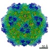


 PDBj
PDBj


 Assembly
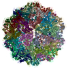

 Spodoptera frugiperda (fall armyworm) / References: UniProt: A0A1B0VEZ1
 Sample preparation
 Electron microscopy imaging
 FIELD EMISSION GUN / Accelerating voltage: 300 kV / Illumination mode: FLOOD BEAM
 Processing