 Movie
Movie Controller


+ Open data

- Basic information
| Entry | Database: PDB / ID: 6vi3 | |||||||||
|---|---|---|---|---|---|---|---|---|---|---|
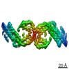
| Title | Straight Filament from Alzheimer's Disease Human Brain Tissue | |||||||||
 Components Components | Microtubule-associated protein tau | |||||||||
 Keywords Keywords | PROTEIN FIBRIL / Pathological amyloid fibril cross-beta fold parallel beta-sheets | |||||||||
| Function / homology |  Function and homology information Function and homology informationplus-end-directed organelle transport along microtubule / histone-dependent DNA binding / negative regulation of protein localization to mitochondrion / neurofibrillary tangle / microtubule lateral binding / axonal transport / tubulin complex / positive regulation of protein localization to synapse / phosphatidylinositol bisphosphate binding / generation of neurons ...plus-end-directed organelle transport along microtubule / histone-dependent DNA binding / negative regulation of protein localization to mitochondrion / neurofibrillary tangle / microtubule lateral binding / axonal transport / tubulin complex / positive regulation of protein localization to synapse / phosphatidylinositol bisphosphate binding / generation of neurons / rRNA metabolic process / axonal transport of mitochondrion / regulation of mitochondrial fission / axon development / regulation of microtubule-based movement / intracellular distribution of mitochondria / regulation of chromosome organization / central nervous system neuron development / minor groove of adenine-thymine-rich DNA binding / lipoprotein particle binding / microtubule polymerization / negative regulation of mitochondrial membrane potential / regulation of microtubule polymerization / dynactin binding / apolipoprotein binding / protein polymerization / main axon / Caspase-mediated cleavage of cytoskeletal proteins / regulation of microtubule polymerization or depolymerization / negative regulation of mitochondrial fission / axolemma / glial cell projection / neurofibrillary tangle assembly / positive regulation of axon extension / regulation of cellular response to heat / positive regulation of microtubule polymerization / positive regulation of protein localization / Activation of AMPK downstream of NMDARs / positive regulation of superoxide anion generation / cellular response to brain-derived neurotrophic factor stimulus / regulation of long-term synaptic depression / supramolecular fiber organization / cytoplasmic microtubule organization / regulation of calcium-mediated signaling / axon cytoplasm / somatodendritic compartment / synapse assembly / phosphatidylinositol binding / nuclear periphery / astrocyte activation / enzyme inhibitor activity / protein phosphatase 2A binding / stress granule assembly / regulation of microtubule cytoskeleton organization / regulation of autophagy / cellular response to reactive oxygen species / microglial cell activation / cellular response to nerve growth factor stimulus / Hsp90 protein binding / protein homooligomerization / SH3 domain binding / PKR-mediated signaling / synapse organization / regulation of synaptic plasticity / response to lead ion / microtubule cytoskeleton organization / memory / neuron projection development / cytoplasmic ribonucleoprotein granule / cell-cell signaling / single-stranded DNA binding / cellular response to heat / growth cone / protein-folding chaperone binding / microtubule cytoskeleton / actin binding / cell body / double-stranded DNA binding / sequence-specific DNA binding / microtubule binding / amyloid fibril formation / dendritic spine / microtubule / protein-macromolecule adaptor activity / learning or memory / neuron projection / membrane raft / negative regulation of gene expression / axon / neuronal cell body / DNA damage response / dendrite / protein kinase binding / enzyme binding / mitochondrion / DNA binding / RNA binding / extracellular region / identical protein binding / nucleus Similarity search - Function | |||||||||
| Biological species |  Homo sapiens (human) Homo sapiens (human) | |||||||||
| Method | ELECTRON MICROSCOPY / helical reconstruction / cryo EM / Resolution: 3.3 Å | |||||||||
 Authors Authors | Arakhamia, T. / Lee, C.E. / Carlomagno, Y. / Duong, D.M. / Kundinger, S.R. / Wang, K. / Williams, D. / DeTure, M. / Dickson, D.W. / Cook, C.N. ...Arakhamia, T. / Lee, C.E. / Carlomagno, Y. / Duong, D.M. / Kundinger, S.R. / Wang, K. / Williams, D. / DeTure, M. / Dickson, D.W. / Cook, C.N. / Seyfried, N.T. / Petrucelli, L. / Fitzpatrick, A.W.P. | |||||||||
| Funding support |  United States, 2items United States, 2items
| |||||||||
 Citation Citation | Journal: Acta Neuropathol / Year: 2018 Title: Tau filaments from multiple cases of sporadic and inherited Alzheimer's disease adopt a common fold. Authors: Benjamin Falcon / Wenjuan Zhang / Manuel Schweighauser / Alexey G Murzin / Ruben Vidal / Holly J Garringer / Bernardino Ghetti / Sjors H W Scheres / Michel Goedert /  Abstract: The ordered assembly of tau protein into abnormal filaments is a defining characteristic of Alzheimer's disease (AD) and other neurodegenerative disorders. It is not known if the structures of tau ...The ordered assembly of tau protein into abnormal filaments is a defining characteristic of Alzheimer's disease (AD) and other neurodegenerative disorders. It is not known if the structures of tau filaments vary within, or between, the brains of individuals with AD. We used a combination of electron cryo-microscopy (cryo-EM) and immuno-gold negative-stain electron microscopy (immuno-EM) to determine the structures of paired helical filaments (PHFs) and straight filaments (SFs) from the frontal cortex of 17 cases of AD (15 sporadic and 2 inherited) and 2 cases of atypical AD (posterior cortical atrophy). The high-resolution structures of PHFs and SFs from the frontal cortex of 3 cases of AD, 2 sporadic and 1 inherited, were determined by cryo-EM. We also used immuno-EM to study the PHFs and SFs from a number of cortical and subcortical brain regions. PHFs outnumbered SFs in all AD cases. By cryo-EM, PHFs and SFs were made of two C-shaped protofilaments with a combined cross-β/β-helix structure, as described previously for one case of AD. The higher resolution structures obtained here showed two additional amino acids at each end of the protofilament. The immuno-EM findings, which indicated the presence of repeats 3 and 4, but not of the N-terminal regions of repeats 1 and 2, of tau in the filament cores of all AD cases, were consistent with the cryo-EM results. These findings show that there is no significant variation in tau filament structures between individuals with AD. This knowledge will be crucial for understanding the mechanisms that underlie tau filament formation and for developing novel diagnostics and therapies. | |||||||||
| History |
| |||||||||
| Remark 0 | THIS ENTRY 6VI3 REFLECTS AN ALTERNATIVE MODELING OF THE ORIGINAL DATA IN EMD-0260, DETERMINED BY ...THIS ENTRY 6VI3 REFLECTS AN ALTERNATIVE MODELING OF THE ORIGINAL DATA IN EMD-0260, DETERMINED BY Falcon, B. et al. |
- Structure visualization
| Movie |
Movie viewer |
|---|---|
| Structure viewer | Molecule: MolmilJmol/JSmol |
- Downloads & links
-Download
| PDBx/mmCIF format | 6vi3.cif.gz | 36.5 KB | Display | PDBx/mmCIF format |
|---|---|---|---|---|
| PDB format | pdb6vi3.ent.gz | 24.9 KB | Display | PDB format |
| PDBx/mmJSON format | 6vi3.json.gz | Tree view | PDBx/mmJSON format | |
| Others |  Other downloads Other downloads |
-Validation report
| Arichive directory | https://data.pdbj.org/pub/pdb/validation_reports/vi/6vi3ftp://data.pdbj.org/pub/pdb/validation_reports/vi/6vi3 | HTTPS FTP |
|---|
-Related structure data
| Related structure data |  0260M  6vh7C  6vhaC  6vhlC M: map data used to model this data C: citing same article ( |
|---|---|
| Similar structure data |


-Links
 PDBj
PDBj


- Assembly
| Deposited unit | 
|
|---|---|
| 1 |
|
-Components
| #1: Protein | Mass: 8370.578 Da / Num. of mol.: 2 Source method: isolated from a genetically manipulated source Source: (gene. exp.) Homo sapiens (human) / Gene: MAPT, MAPTL, MTBT1, TAU / Production host: Homo sapiens (human) / References: UniProt: P10636#2: Chemical |   Type: peptide linking / Mass: 75.067 Da / Num. of mol.: 2 / Source method: obtained synthetically / Formula: C2H5NO2 / Feature type: SUBJECT OF INVESTIGATION Type: peptide linking / Mass: 75.067 Da / Num. of mol.: 2 / Source method: obtained synthetically / Formula: C2H5NO2 / Feature type: SUBJECT OF INVESTIGATIONHas ligand of interest | Y | |
|---|
-Experimental details
-Experiment
| Experiment | Method: ELECTRON MICROSCOPY |
|---|---|
| EM experiment | Aggregation state: FILAMENT / 3D reconstruction method: helical reconstruction |
- Sample preparation
| Component | Name: Straight Filament from Alzheimer's Disease Human Brain Tissue Type: TISSUE / Entity ID: #1 / Source: NATURAL |
|---|---|
| Source (natural) | Organism: Homo sapiens (human) |
| Buffer solution | pH: 7.4 |
| Specimen | Embedding applied: NO / Shadowing applied: NO / Staining applied: NO / Vitrification applied: YES |
| Specimen support | Details: unspecified |
| Vitrification | Cryogen name: ETHANE |
- Electron microscopy imaging
| Experimental equipment |  Model: Titan Krios / Image courtesy: FEI Company |
|---|---|
| Microscopy | Model: FEI TITAN KRIOS |
| Electron gun | Electron source:  FIELD EMISSION GUN / Accelerating voltage: 300 kV / Illumination mode: FLOOD BEAM FIELD EMISSION GUN / Accelerating voltage: 300 kV / Illumination mode: FLOOD BEAM |
| Electron lens | Mode: BRIGHT FIELD |
| Image recording | Electron dose: 60 e/Å2 / Film or detector model: GATAN K2 SUMMIT (4k x 4k) |
- Processing
| CTF correction | Type: PHASE FLIPPING AND AMPLITUDE CORRECTION |
|---|---|
| Helical symmerty | Angular rotation/subunit: -1.04 ° / Axial rise/subunit: 4.76 Å / Axial symmetry: C1 |
| 3D reconstruction | Resolution: 3.3 Å / Resolution method: FSC 0.143 CUT-OFF / Num. of particles: 62782 / Symmetry type: HELICAL |
