+ Open data
- Basic information
| Entry | Database: PDB / ID: 6uh7 | ||||||
|---|---|---|---|---|---|---|---|
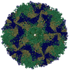
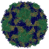
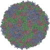
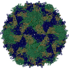
| Title | EV-A71 strain 11316 complexed with MADAL compound 30 | ||||||
 Components Components |
| ||||||
 Keywords Keywords | VIRUS / Enterovirus / inhibitor | ||||||
| Function / homology |  Function and homology information Function and homology informationsymbiont-mediated suppression of host cytoplasmic pattern recognition receptor signaling pathway via inhibition of MDA-5 activity / picornain 2A / symbiont-mediated suppression of host mRNA export from nucleus / symbiont genome entry into host cell via pore formation in plasma membrane / picornain 3C / T=pseudo3 icosahedral viral capsid / host cell cytoplasmic vesicle membrane / viral capsid / nucleoside-triphosphate phosphatase / channel activity ...symbiont-mediated suppression of host cytoplasmic pattern recognition receptor signaling pathway via inhibition of MDA-5 activity / picornain 2A / symbiont-mediated suppression of host mRNA export from nucleus / symbiont genome entry into host cell via pore formation in plasma membrane / picornain 3C / T=pseudo3 icosahedral viral capsid / host cell cytoplasmic vesicle membrane / viral capsid / nucleoside-triphosphate phosphatase / channel activity / monoatomic ion transmembrane transport / host cell cytoplasm / DNA replication / RNA helicase activity / endocytosis involved in viral entry into host cell / symbiont-mediated suppression of host gene expression / symbiont-mediated activation of host autophagy / RNA-directed RNA polymerase / cysteine-type endopeptidase activity / viral RNA genome replication / RNA-directed RNA polymerase activity / DNA-templated transcription / virion attachment to host cell / host cell nucleus / structural molecule activity / ATP hydrolysis activity / proteolysis / RNA binding / zinc ion binding / ATP binding / membrane Similarity search - Function | ||||||
| Biological species |   Enterovirus A71 Enterovirus A71 | ||||||
| Method | ELECTRON MICROSCOPY / single particle reconstruction / cryo EM / Resolution: 2.87 Å | ||||||
 Authors Authors | Lee, H. / Hafenstein, S. | ||||||
 Citation Citation |  Journal: J Med Chem / Year: 2020 Journal: J Med Chem / Year: 2020Title: Scaffold Simplification Strategy Leads to a Novel Generation of Dual Human Immunodeficiency Virus and Enterovirus-A71 Entry Inhibitors. Authors: Belén Martínez-Gualda / Liang Sun / Olaia Martí-Marí / Sam Noppen / Rana Abdelnabi / Carol M Bator / Ernesto Quesada / Leen Delang / Carmen Mirabelli / Hyunwook Lee / Dominique Schols / ...Authors: Belén Martínez-Gualda / Liang Sun / Olaia Martí-Marí / Sam Noppen / Rana Abdelnabi / Carol M Bator / Ernesto Quesada / Leen Delang / Carmen Mirabelli / Hyunwook Lee / Dominique Schols / Johan Neyts / Susan Hafenstein / María-José Camarasa / Federico Gago / Ana San-Félix /    Abstract: Currently, there are only three FDA-approved drugs that inhibit human immunodeficiency virus (HIV) entry-fusion into host cells. The situation is even worse for enterovirus EV71 infection for which ...Currently, there are only three FDA-approved drugs that inhibit human immunodeficiency virus (HIV) entry-fusion into host cells. The situation is even worse for enterovirus EV71 infection for which no antiviral therapies are available. We describe here the discovery of potent entry dual inhibitors of HIV and EV71. These compounds contain in their structure three or four tryptophan (Trp) residues linked to a central scaffold. Critical for anti-HIV/EV71 activity is the presence of extra phenyl rings, bearing one or two carboxylates, at the C2 position of the indole ring of each Trp residue. The most potent derivatives, and , inhibit early steps of the replicative cycles of HIV-1 and EV-A71 by interacting with their respective viral surfaces (glycoprotein gp120 of HIV and the fivefold axis of the EV-A71 capsid). The high potency, low toxicity, facile chemical synthesis, and great opportunities for chemical optimization make them useful prototypes for future medicinal chemistry studies. | ||||||
| History |
|
- Structure visualization
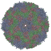
| Movie |
 Movie viewer Movie viewer |
|---|---|
| Structure viewer | Molecule:  Molmil Molmil Jmol/JSmol Jmol/JSmol |
- Downloads & links
- Download
| PDBx/mmCIF format |  6uh7.cif.gz 6uh7.cif.gz | 155 KB | Display |  PDBx/mmCIF format PDBx/mmCIF format |
|---|---|---|---|---|
| PDB format |  pdb6uh7.ent.gz pdb6uh7.ent.gz | 122.1 KB | Display |  PDB format PDB format |
| PDBx/mmJSON format |  6uh7.json.gz 6uh7.json.gz | Tree view |  PDBx/mmJSON format PDBx/mmJSON format | |
| Others |  Other downloads Other downloads |
-Validation report
| Summary document |  6uh7_validation.pdf.gz 6uh7_validation.pdf.gz | 998.5 KB | Display |  wwPDB validaton report wwPDB validaton report |
|---|---|---|---|---|
| Full document |  6uh7_full_validation.pdf.gz 6uh7_full_validation.pdf.gz | 1003.4 KB | Display | |
| Data in XML |  6uh7_validation.xml.gz 6uh7_validation.xml.gz | 29.7 KB | Display | |
| Data in CIF |  6uh7_validation.cif.gz 6uh7_validation.cif.gz | 43.3 KB | Display | |
| Arichive directory |  https://data.pdbj.org/pub/pdb/validation_reports/uh/6uh7 https://data.pdbj.org/pub/pdb/validation_reports/uh/6uh7 ftp://data.pdbj.org/pub/pdb/validation_reports/uh/6uh7 ftp://data.pdbj.org/pub/pdb/validation_reports/uh/6uh7 | HTTPS FTP |
-Related structure data
| Related structure data |  20769MC  6uh1C  6uh6C M: map data used to model this data C: citing same article ( |
|---|---|
| Similar structure data |
- Links
Links
- Assembly
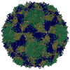
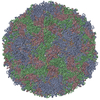
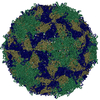
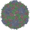
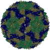
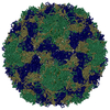
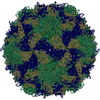
| Deposited unit | 
|
|---|---|
| 1 | x 60
|
| 2 |
|
| 3 | x 5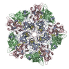
|
| 4 | x 6
|
| 5 | 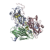
|
| Symmetry | Point symmetry: (Schoenflies symbol: I (icosahedral)) |
- Components
| #1: Protein | Mass: 32748.754 Da / Num. of mol.: 1 / Source method: isolated from a natural source / Source: (natural)   Enterovirus A71 / References: UniProt: D4QGA8 Enterovirus A71 / References: UniProt: D4QGA8 |
|---|---|
| #2: Protein | Mass: 27812.250 Da / Num. of mol.: 1 / Source method: isolated from a natural source / Source: (natural)   Enterovirus A71 / References: UniProt: I6W7A3 Enterovirus A71 / References: UniProt: I6W7A3 |
| #3: Protein | Mass: 26540.332 Da / Num. of mol.: 1 / Source method: isolated from a natural source / Source: (natural)   Enterovirus A71 Enterovirus A71References: UniProt: A0A0E3SXU7, picornain 2A, nucleoside-triphosphate phosphatase, picornain 3C, RNA-directed RNA polymerase |
| #4: Protein | Mass: 7501.162 Da / Num. of mol.: 1 / Source method: isolated from a natural source / Source: (natural)   Enterovirus A71 Enterovirus A71References: UniProt: E9RGA0, picornain 2A, nucleoside-triphosphate phosphatase, picornain 3C, RNA-directed RNA polymerase |
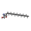
| #5: Chemical | ChemComp-SPH / |
| Has ligand of interest | N |
-Experimental details
-Experiment
| Experiment | Method: ELECTRON MICROSCOPY |
|---|---|
| EM experiment | Aggregation state: PARTICLE / 3D reconstruction method: single particle reconstruction |
- Sample preparation
| Component | Name: Enterovirus A71 / Type: VIRUS / Entity ID: #1-#4 / Source: NATURAL |
|---|---|
| Source (natural) | Organism:   Enterovirus A71 Enterovirus A71 |
| Details of virus | Empty: NO / Enveloped: NO / Isolate: STRAIN / Type: VIRION |
| Buffer solution | pH: 7.5 |
| Specimen | Embedding applied: NO / Shadowing applied: NO / Staining applied: NO / Vitrification applied: YES |
| Vitrification | Cryogen name: ETHANE |
- Electron microscopy imaging
| Experimental equipment |  Model: Titan Krios / Image courtesy: FEI Company |
|---|---|
| Microscopy | Model: FEI TITAN KRIOS |
| Electron gun | Electron source:  FIELD EMISSION GUN / Accelerating voltage: 300 kV / Illumination mode: FLOOD BEAM FIELD EMISSION GUN / Accelerating voltage: 300 kV / Illumination mode: FLOOD BEAM |
| Electron lens | Mode: BRIGHT FIELD |
| Image recording | Electron dose: 44 e/Å2 / Film or detector model: FEI FALCON III (4k x 4k) |
- Processing
| CTF correction | Type: PHASE FLIPPING AND AMPLITUDE CORRECTION |
|---|---|
| 3D reconstruction | Resolution: 2.87 Å / Resolution method: FSC 0.143 CUT-OFF / Num. of particles: 30452 / Symmetry type: POINT |
 Movie
Movie Controller








 PDBj
PDBj

