 Movie
Movie Controller


+ Open data

- Basic information
| Entry | Database: EMDB / ID: EMD-20768 | |||||||||
|---|---|---|---|---|---|---|---|---|---|---|
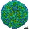
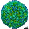
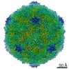
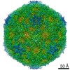
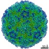
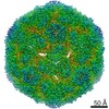
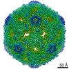
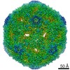
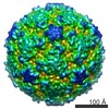
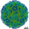
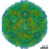
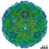
| Title | EV-A71 strain 11316 complexed with MADAL compound 22 | |||||||||
 Map data Map data | Cryo EM map of enterovirus A71 strain 11316 complexed with MADAL compound 22 | |||||||||
 Sample Sample |
| |||||||||
 Keywords Keywords | Enterovirus / VIRUS / inhibitor | |||||||||
| Function / homology |  Function and homology information Function and homology informationsymbiont-mediated suppression of host cytoplasmic pattern recognition receptor signaling pathway via inhibition of MDA-5 activity / picornain 2A / symbiont-mediated suppression of host mRNA export from nucleus / symbiont genome entry into host cell via pore formation in plasma membrane / picornain 3C / T=pseudo3 icosahedral viral capsid / host cell cytoplasmic vesicle membrane / viral capsid / ribonucleoside triphosphate phosphatase activity / nucleoside-triphosphate phosphatase ...symbiont-mediated suppression of host cytoplasmic pattern recognition receptor signaling pathway via inhibition of MDA-5 activity / picornain 2A / symbiont-mediated suppression of host mRNA export from nucleus / symbiont genome entry into host cell via pore formation in plasma membrane / picornain 3C / T=pseudo3 icosahedral viral capsid / host cell cytoplasmic vesicle membrane / viral capsid / ribonucleoside triphosphate phosphatase activity / nucleoside-triphosphate phosphatase / channel activity / monoatomic ion transmembrane transport / host cell cytoplasm / DNA replication / RNA helicase activity / endocytosis involved in viral entry into host cell / symbiont-mediated suppression of host gene expression / symbiont-mediated activation of host autophagy / RNA-directed RNA polymerase / cysteine-type endopeptidase activity / viral RNA genome replication / RNA-directed RNA polymerase activity / virion attachment to host cell / host cell nucleus / DNA-templated transcription / structural molecule activity / proteolysis / RNA binding / zinc ion binding / ATP binding Similarity search - Function | |||||||||
| Biological species |   Enterovirus A71 Enterovirus A71 | |||||||||
| Method | single particle reconstruction / cryo EM / Resolution: 2.98 Å | |||||||||
 Authors Authors | Lee H / Hafenstein S | |||||||||
 Citation Citation | Journal: J Med Chem / Year: 2020 Title: Scaffold Simplification Strategy Leads to a Novel Generation of Dual Human Immunodeficiency Virus and Enterovirus-A71 Entry Inhibitors. Authors: Belén Martínez-Gualda / Liang Sun / Olaia Martí-Marí / Sam Noppen / Rana Abdelnabi / Carol M Bator / Ernesto Quesada / Leen Delang / Carmen Mirabelli / Hyunwook Lee / Dominique Schols / ...Authors: Belén Martínez-Gualda / Liang Sun / Olaia Martí-Marí / Sam Noppen / Rana Abdelnabi / Carol M Bator / Ernesto Quesada / Leen Delang / Carmen Mirabelli / Hyunwook Lee / Dominique Schols / Johan Neyts / Susan Hafenstein / María-José Camarasa / Federico Gago / Ana San-Félix /    Abstract: Currently, there are only three FDA-approved drugs that inhibit human immunodeficiency virus (HIV) entry-fusion into host cells. The situation is even worse for enterovirus EV71 infection for which ...Currently, there are only three FDA-approved drugs that inhibit human immunodeficiency virus (HIV) entry-fusion into host cells. The situation is even worse for enterovirus EV71 infection for which no antiviral therapies are available. We describe here the discovery of potent entry dual inhibitors of HIV and EV71. These compounds contain in their structure three or four tryptophan (Trp) residues linked to a central scaffold. Critical for anti-HIV/EV71 activity is the presence of extra phenyl rings, bearing one or two carboxylates, at the C2 position of the indole ring of each Trp residue. The most potent derivatives, and , inhibit early steps of the replicative cycles of HIV-1 and EV-A71 by interacting with their respective viral surfaces (glycoprotein gp120 of HIV and the fivefold axis of the EV-A71 capsid). The high potency, low toxicity, facile chemical synthesis, and great opportunities for chemical optimization make them useful prototypes for future medicinal chemistry studies. | |||||||||
| History |
|
- Structure visualization
| Movie |
Movie viewer |
|---|---|
| Structure viewer | EM map: SurfViewMolmilJmol/JSmol |
| Supplemental images |

- Downloads & links
-EMDB archive
| Map data | emd_20768.map.gz | 217.8 MB | EMDB map data format | |
|---|---|---|---|---|
| Header (meta data) | emd-20768-v30.xmlemd-20768.xml | 14.9 KB 14.9 KB | Display Display | EMDB header |
| Images |  emd_20768.png emd_20768.png | 88.8 KB | ||
| Filedesc metadata | emd-20768.cif.gz | 5.5 KB | ||
| Others | emd_20768_additional.map.gz | 261.4 MB | ||
| Archive directory |  http://ftp.pdbj.org/pub/emdb/structures/EMD-20768ftp://ftp.pdbj.org/pub/emdb/structures/EMD-20768 http://ftp.pdbj.org/pub/emdb/structures/EMD-20768ftp://ftp.pdbj.org/pub/emdb/structures/EMD-20768 | HTTPS FTP |
-Related structure data
| Related structure data |  6uh6MC  6uh1C  6uh7C M: atomic model generated by this map C: citing same article ( |
|---|---|
| Similar structure data |

-Links
| EMDB pages | EMDB (EBI/PDBe) / EMDataResource |
|---|---|
| Related items in Molecule of the Month |


-Map
| File | Download / File: emd_20768.map.gz / Format: CCP4 / Size: 282.6 MB / Type: IMAGE STORED AS FLOATING POINT NUMBER (4 BYTES) | ||||||||||||||||||||||||||||||||||||||||||||||||||||||||||||
|---|---|---|---|---|---|---|---|---|---|---|---|---|---|---|---|---|---|---|---|---|---|---|---|---|---|---|---|---|---|---|---|---|---|---|---|---|---|---|---|---|---|---|---|---|---|---|---|---|---|---|---|---|---|---|---|---|---|---|---|---|---|
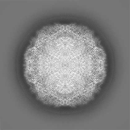
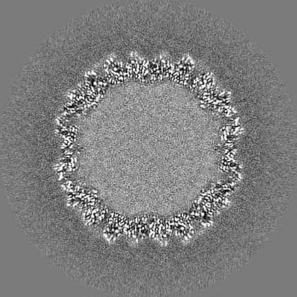
| Annotation | Cryo EM map of enterovirus A71 strain 11316 complexed with MADAL compound 22 | ||||||||||||||||||||||||||||||||||||||||||||||||||||||||||||
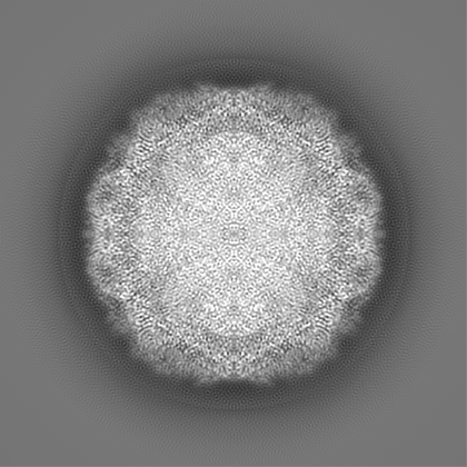
| Projections & slices | Image control
Images are generated by Spider. | ||||||||||||||||||||||||||||||||||||||||||||||||||||||||||||
| Voxel size | X=Y=Z: 1.1 Å | ||||||||||||||||||||||||||||||||||||||||||||||||||||||||||||
| Density |
| ||||||||||||||||||||||||||||||||||||||||||||||||||||||||||||
| Symmetry | Space group: 1 | ||||||||||||||||||||||||||||||||||||||||||||||||||||||||||||
| Details | EMDB XML:
CCP4 map header:
| ||||||||||||||||||||||||||||||||||||||||||||||||||||||||||||
 Z (Sec.)
 Y (Row.)
 X (Col.)


-Supplemental data
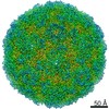
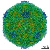
-Additional map: Sharpened map of the EV-A71-MADAL 22 complex
| File | emd_20768_additional.map | ||||||||||||
|---|---|---|---|---|---|---|---|---|---|---|---|---|---|
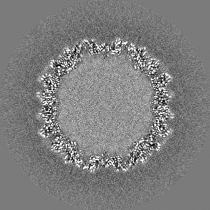
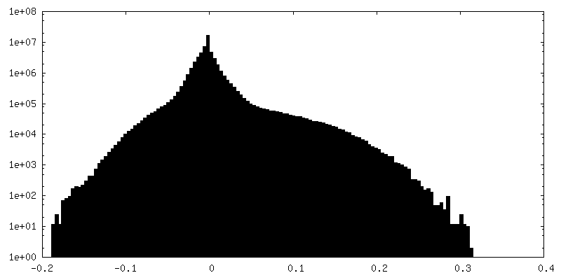
| Annotation | Sharpened map of the EV-A71-MADAL 22 complex | ||||||||||||
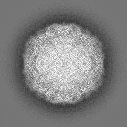
| Projections & Slices |
| ||||||||||||
| Density Histograms |


- Sample components
-Entire : Enterovirus A71
| Entire | Name: Enterovirus A71 |
|---|---|
| Components |
|
-Supramolecule #1: Enterovirus A71
| Supramolecule | Name: Enterovirus A71 / type: virus / ID: 1 / Parent: 0 / Macromolecule list: #1-#4 / NCBI-ID: 39054 / Sci species name: Enterovirus A71 / Virus type: VIRION / Virus isolate: STRAIN / Virus enveloped: No / Virus empty: No |
|---|
-Macromolecule #1: VP1
| Macromolecule | Name: VP1 / type: protein_or_peptide / ID: 1 / Number of copies: 1 / Enantiomer: LEVO |
|---|---|
| Source (natural) | Organism: Enterovirus A71 |
| Molecular weight | Theoretical: 32.748754 KDa |
| Sequence | String: GDRVADVIES SIGDSVSRAL TQALPAPTGQ NTQVSSHRLD TGEVPALQAA EIGASSNTSD ESMIETRCVL NSHSTAETTL DSFFSRAGL VGEIDLPLEG TTNPNGYANW DIDITGYAQM RRKVELFTYM RFDAEFTFVA CTPTGQVVPQ LLQYMFVPPG A PKPESRES ...String: GDRVADVIES SIGDSVSRAL TQALPAPTGQ NTQVSSHRLD TGEVPALQAA EIGASSNTSD ESMIETRCVL NSHSTAETTL DSFFSRAGL VGEIDLPLEG TTNPNGYANW DIDITGYAQM RRKVELFTYM RFDAEFTFVA CTPTGQVVPQ LLQYMFVPPG A PKPESRES LAWQTATNPS VFVKLTDPPA QVSVPFMSPA SAYQWFYDGY PTFGEHKQEK DLEYGACPNN MMGTFSVRTV GS SKSKYPL VVRIYMRMKH VRAWIPRPMR NQNYLFKANP NYAGNSIKPT GTSRSAITTL UniProtKB: Genome polyprotein |
-Macromolecule #2: VP2
| Macromolecule | Name: VP2 / type: protein_or_peptide / ID: 2 / Number of copies: 1 / Enantiomer: LEVO |
|---|---|
| Source (natural) | Organism: Enterovirus A71 |
| Molecular weight | Theoretical: 26.946342 KDa |
| Sequence | String: SDRVAQLTIG NSTITTQEAA NIIVGYGEWP SYCSDDDATA VDKPTRPDVS VNRFYTLDTK LWEKSSKGWY WKFPDVLTET GVFGQNAQF HYLYRSGFCI HVQCNASKFH QGALLVAILP EYVIGTVAGG TGTEDSHPPY KQTQPGADGF ELQHPYVLDA G IPISQLTV ...String: SDRVAQLTIG NSTITTQEAA NIIVGYGEWP SYCSDDDATA VDKPTRPDVS VNRFYTLDTK LWEKSSKGWY WKFPDVLTET GVFGQNAQF HYLYRSGFCI HVQCNASKFH QGALLVAILP EYVIGTVAGG TGTEDSHPPY KQTQPGADGF ELQHPYVLDA G IPISQLTV CHHQRINLRT NNCATIIVPY MNTLPFDSAL NHCNFGLLVV PISPLDFDQG ATPVIPITIT LAPMCSEFAG LR QAVTQ UniProtKB: Genome polyprotein |
-Macromolecule #3: VP3
| Macromolecule | Name: VP3 / type: protein_or_peptide / ID: 3 / Number of copies: 1 / Enantiomer: LEVO / EC number: picornain 2A |
|---|---|
| Source (natural) | Organism: Enterovirus A71 |
| Molecular weight | Theoretical: 26.540332 KDa |
| Sequence | String: GFPTELKPGT NQFLTTDDGV SAPILPNFHP TPCIHIPGEV RNLLELCQVE TILEVNNVPT NATSLMERLR FPVSAQAGKG ELCAVFRAD PGRDGPWQST MLGQLCGYYT QWSGSLEVTF MFTGSFMATG KMLIAYTPPG GPLPKDRATA MLGTHVIWDF G LQSSVTLV ...String: GFPTELKPGT NQFLTTDDGV SAPILPNFHP TPCIHIPGEV RNLLELCQVE TILEVNNVPT NATSLMERLR FPVSAQAGKG ELCAVFRAD PGRDGPWQST MLGQLCGYYT QWSGSLEVTF MFTGSFMATG KMLIAYTPPG GPLPKDRATA MLGTHVIWDF G LQSSVTLV IPWISNTHYR AHARDGVFDY YTTGLVSIWY QTNYVVPIGA PNTAYIIALA AAQKNFTMKL CKDTSHILQT AS IQ UniProtKB: Genome polyprotein |
-Macromolecule #4: VP4
| Macromolecule | Name: VP4 / type: protein_or_peptide / ID: 4 / Number of copies: 1 / Enantiomer: LEVO / EC number: picornain 2A |
|---|---|
| Source (natural) | Organism: Enterovirus A71 |
| Molecular weight | Theoretical: 7.501162 KDa |
| Sequence | String: MGSQVSTQRS GSHENSNSAT EGSTINYTTI NYYKDSYAAT AGKQSLKQDP DKFANPVKDI FTEMAAPLK UniProtKB: Genome polyprotein |
-Macromolecule #5: SPHINGOSINE
| Macromolecule | Name: SPHINGOSINE / type: ligand / ID: 5 / Number of copies: 1 / Formula: SPH |
|---|---|
| Molecular weight | Theoretical: 299.492 Da |
| Chemical component information |  ChemComp-SPH: |
-Experimental details
-Structure determination
| Method | cryo EM |
|---|---|
 Processing Processing | single particle reconstruction |
| Aggregation state | particle |
-Sample preparation
| Buffer | pH: 7.5 |
|---|---|
| Vitrification | Cryogen name: ETHANE |
- Electron microscopy
| Microscope | FEI TITAN KRIOS |
|---|---|
| Image recording | Film or detector model: FEI FALCON III (4k x 4k) / Average electron dose: 44.0 e/Å2 |
| Electron beam | Acceleration voltage: 300 kV / Electron source:  FIELD EMISSION GUN FIELD EMISSION GUN |
| Electron optics | Illumination mode: FLOOD BEAM / Imaging mode: BRIGHT FIELD |
| Experimental equipment |  Model: Titan Krios / Image courtesy: FEI Company |
-Image processing
| Startup model | Type of model: OTHER |
|---|---|
| Final reconstruction | Resolution.type: BY AUTHOR / Resolution: 2.98 Å / Resolution method: FSC 0.143 CUT-OFF / Number images used: 42926 |
| Initial angle assignment | Type: RANDOM ASSIGNMENT |
| Final angle assignment | Type: PROJECTION MATCHING |
