 Movie
Movie Controller


+ Open data

- Basic information
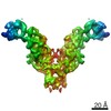
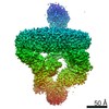
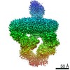
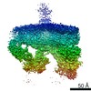
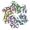
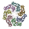
| Entry | Database: PDB / ID: 6sgx | ||||||||||||||||||
|---|---|---|---|---|---|---|---|---|---|---|---|---|---|---|---|---|---|---|---|
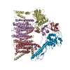
| Title | Structure of protomer 1 of the ESX-3 core complex | ||||||||||||||||||
 Components Components |
| ||||||||||||||||||
 Keywords Keywords | MEMBRANE PROTEIN / Type VII Secretion System ESX-3 secretion system T7SS ESX-3 Mycobacterium smegmatis | ||||||||||||||||||
| Function / homology |  Function and homology information Function and homology informationHydrolases; Acting on acid anhydrides / hydrolase activity / DNA binding / extracellular region / ATP binding / plasma membrane Similarity search - Function | ||||||||||||||||||
| Biological species |  Mycobacterium smegmatis (bacteria) Mycobacterium smegmatis (bacteria) | ||||||||||||||||||
| Method | ELECTRON MICROSCOPY / single particle reconstruction / cryo EM / Resolution: 3.7 Å | ||||||||||||||||||
 Authors Authors | Famelis, N. / Rivera-Calzada, A. / Llorca, O. / Geibel, S. | ||||||||||||||||||
| Funding support |  Spain, Spain,  Germany, 5items Germany, 5items
| ||||||||||||||||||
 Citation Citation | Journal: Nature / Year: 2019 Title: Architecture of the mycobacterial type VII secretion system. Authors: Nikolaos Famelis / Angel Rivera-Calzada / Gianluca Degliesposti / Maria Wingender / Nicole Mietrach / J Mark Skehel / Rafael Fernandez-Leiro / Bettina Böttcher / Andreas Schlosser / Oscar ...Authors: Nikolaos Famelis / Angel Rivera-Calzada / Gianluca Degliesposti / Maria Wingender / Nicole Mietrach / J Mark Skehel / Rafael Fernandez-Leiro / Bettina Böttcher / Andreas Schlosser / Oscar Llorca / Sebastian Geibel /  Abstract: Host infection by pathogenic mycobacteria, such as Mycobacterium tuberculosis, is facilitated by virulence factors that are secreted by type VII secretion systems. A molecular understanding of the ...Host infection by pathogenic mycobacteria, such as Mycobacterium tuberculosis, is facilitated by virulence factors that are secreted by type VII secretion systems. A molecular understanding of the type VII secretion mechanism has been hampered owing to a lack of three-dimensional structures of the fully assembled secretion apparatus. Here we report the cryo-electron microscopy structure of a membrane-embedded core complex of the ESX-3/type VII secretion system from Mycobacterium smegmatis. The core of the ESX-3 secretion machine consists of four protein components-EccB3, EccC3, EccD3 and EccE3, in a 1:1:2:1 stoichiometry-which form two identical protomers. The EccC3 coupling protein comprises a flexible array of four ATPase domains, which are linked to the membrane through a stalk domain. The domain of unknown function (DUF) adjacent to the stalk is identified as an ATPase domain that is essential for secretion. EccB3 is predominantly periplasmatic, but a small segment crosses the membrane and contacts the stalk domain. This suggests that conformational changes in the stalk domain-triggered by substrate binding at the distal end of EccC3 and subsequent ATP hydrolysis in the DUF-could be coupled to substrate secretion to the periplasm. Our results reveal that the architecture of type VII secretion systems differs markedly from that of other known secretion machines, and provide a structural understanding of these systems that will be useful for the design of antimicrobial strategies that target bacterial virulence. | ||||||||||||||||||
| History |
|
- Structure visualization
| Movie |
Movie viewer |
|---|---|
| Structure viewer | Molecule: MolmilJmol/JSmol |
- Downloads & links
-Download
| PDBx/mmCIF format | 6sgx.cif.gz | 258.3 KB | Display | PDBx/mmCIF format |
|---|---|---|---|---|
| PDB format | pdb6sgx.ent.gz | 204.5 KB | Display | PDB format |
| PDBx/mmJSON format | 6sgx.json.gz | Tree view | PDBx/mmJSON format | |
| Others |  Other downloads Other downloads |
-Validation report
| Arichive directory | https://data.pdbj.org/pub/pdb/validation_reports/sg/6sgxftp://data.pdbj.org/pub/pdb/validation_reports/sg/6sgx | HTTPS FTP |
|---|
-Related structure data
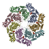
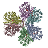
| Related structure data |  10187MC 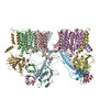 6sgwC 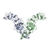 6sgyC 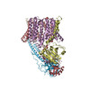 6sgzC M: map data used to model this data C: citing same article ( |
|---|---|
| Similar structure data |

-Links
 PDBj
PDBj- Assembly
| Deposited unit | 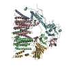
|
|---|---|
| 1 |
|
-Components
| #1: Protein | Mass: 8875.269 Da / Num. of mol.: 1 Source method: isolated from a genetically manipulated source Source: (gene. exp.) Mycobacterium smegmatis (strain ATCC 700084 / mc(2)155) (bacteria)Gene: eccB3, MSMEG_0616, MSMEI_0600 Production host: Mycolicibacterium smegmatis MC2 155 (bacteria)References: UniProt: A0QQ39, Hydrolases; Acting on acid anhydrides | ||||
|---|---|---|---|---|---|
| #2: Protein | Mass: 47114.125 Da / Num. of mol.: 2 Source method: isolated from a genetically manipulated source Source: (gene. exp.) Mycobacterium smegmatis (strain ATCC 700084 / mc(2)155) (bacteria)Gene: eccD3, snm, MSMEG_0623, MSMEI_0607 Production host: Mycolicibacterium smegmatis MC2 155 (bacteria)References: UniProt: A0QQ46 #3: Protein | | Mass: 44653.078 Da / Num. of mol.: 1 Source method: isolated from a genetically manipulated source Source: (gene. exp.) Mycobacterium smegmatis (strain ATCC 700084 / mc(2)155) (bacteria)Gene: eccC3, MSMEG_0617, MSMEI_0601 Production host: Mycolicibacterium smegmatis MC2 155 (bacteria)References: UniProt: A0QQ40 #4: Protein | | Mass: 30813.355 Da / Num. of mol.: 1 Source method: isolated from a genetically manipulated source Source: (gene. exp.) Mycobacterium smegmatis (strain ATCC 700084 / mc(2)155) (bacteria)Gene: eccE3, MSMEG_0626, MSMEI_0609 Production host: Mycolicibacterium smegmatis MC2 155 (bacteria)References: UniProt: A0QQ48 |
-Experimental details
-Experiment
| Experiment | Method: ELECTRON MICROSCOPY |
|---|---|
| EM experiment | Aggregation state: PARTICLE / 3D reconstruction method: single particle reconstruction |
- Sample preparation
| Component | Name: Protomer1 of the ESX-3 core complex / Type: COMPLEX Details: ESX-3 core complex The sample consists of four protein components, EccB3:EccC3:EccD3:EccE3 in a 1:1:2:1 stoichiometry Molecular weight of the complex without the amphipol micelle: 0.65 MDa Entity ID: all / Source: RECOMBINANT |
|---|---|
| Molecular weight | Value: 0.65 MDa / Experimental value: YES |
| Source (natural) | Organism: Mycolicibacterium smegmatis MC2 155 (bacteria) |
| Source (recombinant) | Organism: Mycolicibacterium smegmatis MC2 155 (bacteria) / Plasmid: pMyNT |
| Buffer solution | pH: 8 / Details: 30 mM Hepes pH 8.0, 150 mM NaCl |
| Specimen | Conc.: 0.3 mg/ml / Embedding applied: NO / Shadowing applied: NO / Staining applied: NO / Vitrification applied: YES / Details: bound to Amphipol A8-35 |
| Vitrification | Instrument: FEI VITROBOT MARK IV / Cryogen name: ETHANE / Humidity: 100 % / Chamber temperature: 278 K / Details: Blotting time: 3s Blotting force: -10 |
- Electron microscopy imaging
| Experimental equipment |  Model: Titan Krios / Image courtesy: FEI Company |
|---|---|
| Microscopy | Model: FEI TITAN KRIOS |
| Electron gun | Electron source:  FIELD EMISSION GUN / Accelerating voltage: 300 kV / Illumination mode: SPOT SCAN FIELD EMISSION GUN / Accelerating voltage: 300 kV / Illumination mode: SPOT SCAN |
| Electron lens | Mode: BRIGHT FIELD / Nominal magnification: 75000 X / Nominal defocus max: 2600 nm / Nominal defocus min: 1600 nm / Cs: 2.7 mm |
| Specimen holder | Cryogen: NITROGEN / Specimen holder model: FEI TITAN KRIOS AUTOGRID HOLDER |
| Image recording | Electron dose: 50 e/Å2 / Detector mode: COUNTING / Film or detector model: FEI FALCON III (4k x 4k) / Num. of real images: 11903 Details: Images acquired as 55 frames movies at a calibrated magnification of 1.0635 Angs/px |
- Processing
| Software | Name: PHENIX / Version: 1.15.2_3472: / Classification: refinement | |||||||||||||||||||||||||||||||||||||||||||||
|---|---|---|---|---|---|---|---|---|---|---|---|---|---|---|---|---|---|---|---|---|---|---|---|---|---|---|---|---|---|---|---|---|---|---|---|---|---|---|---|---|---|---|---|---|---|---|
| EM software |
| |||||||||||||||||||||||||||||||||||||||||||||
| Image processing | Details: Images were collected in counting mode | |||||||||||||||||||||||||||||||||||||||||||||
| CTF correction | Type: PHASE FLIPPING AND AMPLITUDE CORRECTION | |||||||||||||||||||||||||||||||||||||||||||||
| Particle selection | Num. of particles selected: 2066007 | |||||||||||||||||||||||||||||||||||||||||||||
| Symmetry | Point symmetry: C1 (asymmetric) | |||||||||||||||||||||||||||||||||||||||||||||
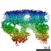
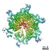
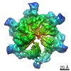
| 3D reconstruction | Resolution: 3.7 Å / Resolution method: FSC 0.143 CUT-OFF / Num. of particles: 126308 Details: In order to get the best possible resolution for protomer 1, the extra density not corresponding to this protomer was subtracted and masked out during the refinement. Symmetry type: POINT | |||||||||||||||||||||||||||||||||||||||||||||
| Atomic model building | B value: 29.4 / Protocol: AB INITIO MODEL / Space: REAL / Target criteria: Map correlation coefficient | |||||||||||||||||||||||||||||||||||||||||||||
| Refine LS restraints |
|
