 Movie
Movie Controller

[English] 日本語
 Yorodumi
- PDB-6bfz: Crystal structure of enolase from E. coli with a mixture of apo f... -
+ Open data

- Basic information
| Entry | Database: PDB / ID: 6bfz | ||||||
|---|---|---|---|---|---|---|---|
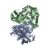
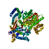
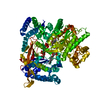
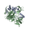
| Title | Crystal structure of enolase from E. coli with a mixture of apo form, substrate, and product form | ||||||
 Components Components | Enolase | ||||||
 Keywords Keywords | LYASE / Enolase / Escherichia coli / Apo / substrate / product | ||||||
| Function / homology |  Function and homology information Function and homology informationbacterial degradosome / phosphopyruvate hydratase / phosphopyruvate hydratase complex / phosphopyruvate hydratase activity / RNA catabolic process / RNA processing / glycolytic process / cytoskeleton / magnesium ion binding / cell surface ...bacterial degradosome / phosphopyruvate hydratase / phosphopyruvate hydratase complex / phosphopyruvate hydratase activity / RNA catabolic process / RNA processing / glycolytic process / cytoskeleton / magnesium ion binding / cell surface / protein homodimerization activity / extracellular region / membrane / identical protein binding / cytosol Similarity search - Function | ||||||
| Biological species |  | ||||||
| Method |  X-RAY DIFFRACTION / SYNCHROTRON / MOLECULAR REPLACEMENT / Resolution: 2.21 Å X-RAY DIFFRACTION / SYNCHROTRON / MOLECULAR REPLACEMENT / Resolution: 2.21 Å | ||||||
 Authors Authors | Erlandsen, H. / Wright, D. / Krucinska, J. | ||||||
 Citation Citation | Journal: Biochemistry / Year: 2019 Title: Structural and Functional Studies of Bacterial Enolase, a Potential Target against Gram-Negative Pathogens. Authors: Krucinska, J. / Falcone, E. / Erlandsen, H. / Hazeen, A. / Lombardo, M.N. / Estrada, A. / Robinson, V.L. / Anderson, A.C. / Wright, D.L. | ||||||
| History |
|
- Structure visualization
| Structure viewer | Molecule: MolmilJmol/JSmol |
|---|
- Downloads & links
-Download
| PDBx/mmCIF format | 6bfz.cif.gz | 504.1 KB | Display | PDBx/mmCIF format |
|---|---|---|---|---|
| PDB format | pdb6bfz.ent.gz | 408.5 KB | Display | PDB format |
| PDBx/mmJSON format | 6bfz.json.gz | Tree view | PDBx/mmJSON format | |
| Others |  Other downloads Other downloads |
-Validation report
| Arichive directory | https://data.pdbj.org/pub/pdb/validation_reports/bf/6bfzftp://data.pdbj.org/pub/pdb/validation_reports/bf/6bfz | HTTPS FTP |
|---|
-Related structure data
| Related structure data | 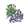 6bfyC 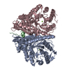 2fymS C: citing same article ( S: Starting model for refinement |
|---|---|
| Similar structure data |


-Links
 PDBj
PDBj
- Assembly
| Deposited unit | 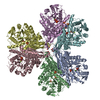
| ||||||||
|---|---|---|---|---|---|---|---|---|---|
| 1 | 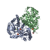
| ||||||||
| 2 | 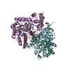
| ||||||||
| 3 | 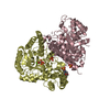
| ||||||||
| Unit cell |
|
-Components
-Protein , 1 types, 6 molecules ABEFDC
| #1: Protein | Mass: 47373.672 Da / Num. of mol.: 6 Source method: isolated from a genetically manipulated source Source: (gene. exp.) References: UniProt: B7MLA0, UniProt: P0A6P9*PLUS, phosphopyruvate hydratase |
|---|
-Non-polymers , 7 types, 705 molecules 


| #2: Chemical | ChemComp-MG /  Mass: 24.305 Da / Num. of mol.: 6 / Source method: obtained synthetically / Formula: Mg Mass: 24.305 Da / Num. of mol.: 6 / Source method: obtained synthetically / Formula: Mg#3: Chemical | ChemComp-SO4 /  Mass: 96.063 Da / Num. of mol.: 13 / Source method: obtained synthetically / Formula: SO4 Mass: 96.063 Da / Num. of mol.: 13 / Source method: obtained synthetically / Formula: SO4#4: Chemical |  Mass: 92.094 Da / Num. of mol.: 3 / Source method: obtained synthetically / Formula: C3H8O3 Mass: 92.094 Da / Num. of mol.: 3 / Source method: obtained synthetically / Formula: C3H8O3#5: Chemical | ChemComp-PEP / |  Mass: 168.042 Da / Num. of mol.: 1 / Source method: obtained synthetically / Formula: C3H5O6P Mass: 168.042 Da / Num. of mol.: 1 / Source method: obtained synthetically / Formula: C3H5O6P#6: Chemical | ChemComp-2PG /  Mass: 186.057 Da / Num. of mol.: 4 / Source method: obtained synthetically / Formula: C3H7O7P Mass: 186.057 Da / Num. of mol.: 4 / Source method: obtained synthetically / Formula: C3H7O7P#7: Chemical |  Mass: 195.237 Da / Num. of mol.: 2 / Source method: obtained synthetically / Formula: C6H13NO4S / Comment: pH buffer*YM Mass: 195.237 Da / Num. of mol.: 2 / Source method: obtained synthetically / Formula: C6H13NO4S / Comment: pH buffer*YM#8: Water | ChemComp-HOH / | Mass: 18.015 Da / Num. of mol.: 676 / Source method: isolated from a natural source / Formula: H2O |
|---|
-Experimental details
-Experiment
| Experiment | Method: X-RAY DIFFRACTION / Number of used crystals: 1 |
|---|
- Sample preparation
| Crystal | Density Matthews: 2.9 Å3/Da / Density % sol: 57.63 % |
|---|---|
| Crystal grow | Temperature: 293 K / Method: vapor diffusion, hanging drop / pH: 6 Details: 2.0 M ammonium sulfate, 0.1 M MES, pH 6.0, 0.1 M sodium/potassium tartrate |
-Data collection
| Diffraction | Mean temperature: 100 K |
|---|---|
| Diffraction source | Source: SYNCHROTRON / Site: NSLS-II  / Beamline: 17-ID-1 / Wavelength: 0.979186 Å / Beamline: 17-ID-1 / Wavelength: 0.979186 Å |
| Detector | Type: DECTRIS EIGER X 9M / Detector: PIXEL / Date: Feb 1, 2017 Details: White beam slits, double crystal Si(111) monochromator with horizontal theta-axis, tandem flat beam deflecting silicon mirrors (Pd and Si lanes), Kirkpatrick-Baez focusing silica mirrors, Pd- ...Details: White beam slits, double crystal Si(111) monochromator with horizontal theta-axis, tandem flat beam deflecting silicon mirrors (Pd and Si lanes), Kirkpatrick-Baez focusing silica mirrors, Pd-coated, each bent adaptively with 16 piezo actuators |
| Radiation | Monochromator: double crystal Si(111) / Protocol: SINGLE WAVELENGTH / Monochromatic (M) / Laue (L): M / Scattering type: x-ray |
| Radiation wavelength | Wavelength: 0.979186 Å / Relative weight: 1 |
| Reflection | Resolution: 2.21→29.69 Å / Num. obs: 165432 / % possible obs: 99.3 % / Redundancy: 1.9 % / Biso Wilson estimate: 30.4 Å2 / CC1/2: 0.998 / Rmerge(I) obs: 0.036 / Rpim(I) all: 0.036 / Rrim(I) all: 0.051 / Net I/σ(I): 13.8 |
| Reflection shell | Resolution: 2.21→2.24 Å / Redundancy: 1.9 % / Rmerge(I) obs: 0.332 / Mean I/σ(I) obs: 2.2 / Num. unique obs: 7139 / CC1/2: 0.786 / Rpim(I) all: 0.332 / Rrim(I) all: 0.47 / % possible all: 87.4 |
- Processing
| Software |
| ||||||||||||||||||||||||||||||||||||||||||||||||||||||||||||||||||||||||||||||||||||||||||||||||||||||||||||||||||||||||||||||||||||||||||||||||||||||||||||||||||||||||||||||||||||||
|---|---|---|---|---|---|---|---|---|---|---|---|---|---|---|---|---|---|---|---|---|---|---|---|---|---|---|---|---|---|---|---|---|---|---|---|---|---|---|---|---|---|---|---|---|---|---|---|---|---|---|---|---|---|---|---|---|---|---|---|---|---|---|---|---|---|---|---|---|---|---|---|---|---|---|---|---|---|---|---|---|---|---|---|---|---|---|---|---|---|---|---|---|---|---|---|---|---|---|---|---|---|---|---|---|---|---|---|---|---|---|---|---|---|---|---|---|---|---|---|---|---|---|---|---|---|---|---|---|---|---|---|---|---|---|---|---|---|---|---|---|---|---|---|---|---|---|---|---|---|---|---|---|---|---|---|---|---|---|---|---|---|---|---|---|---|---|---|---|---|---|---|---|---|---|---|---|---|---|---|---|---|---|---|
| Refinement | Method to determine structure: MOLECULAR REPLACEMENT Starting model: PDB entry 2FYM Resolution: 2.21→29.69 Å / Cor.coef. Fo:Fc: 0.964 / Cor.coef. Fo:Fc free: 0.943 / SU B: 5.078 / SU ML: 0.126 / Cross valid method: THROUGHOUT / ESU R: 0.2 / ESU R Free: 0.176 / Details: HYDROGENS HAVE BEEN ADDED IN THE RIDING POSITIONS
| ||||||||||||||||||||||||||||||||||||||||||||||||||||||||||||||||||||||||||||||||||||||||||||||||||||||||||||||||||||||||||||||||||||||||||||||||||||||||||||||||||||||||||||||||||||||
| Solvent computation | Ion probe radii: 0.8 Å / Shrinkage radii: 0.8 Å / VDW probe radii: 1.2 Å | ||||||||||||||||||||||||||||||||||||||||||||||||||||||||||||||||||||||||||||||||||||||||||||||||||||||||||||||||||||||||||||||||||||||||||||||||||||||||||||||||||||||||||||||||||||||
| Displacement parameters | Biso mean: 40.171 Å2
| ||||||||||||||||||||||||||||||||||||||||||||||||||||||||||||||||||||||||||||||||||||||||||||||||||||||||||||||||||||||||||||||||||||||||||||||||||||||||||||||||||||||||||||||||||||||
| Refinement step | Cycle: LAST / Resolution: 2.21→29.69 Å
| ||||||||||||||||||||||||||||||||||||||||||||||||||||||||||||||||||||||||||||||||||||||||||||||||||||||||||||||||||||||||||||||||||||||||||||||||||||||||||||||||||||||||||||||||||||||
| Refine LS restraints |
|