DNA-binding transcription activator activity / phosphorelay signal transduction system / cis-regulatory region sequence-specific DNA binding / protein-DNA complex / positive regulation of DNA-templated transcription / ATP binding / metal ion binding / identical protein binding / cytoplasm Similarity search - Function
Sigma-54 interaction domain ATP-binding region A signature. / Sigma-54 interaction domain, ATP-binding site 1 / Sigma-54 interaction domain, ATP-binding site 2 / Sigma-54 interaction domain ATP-binding region B signature. / : / AAA+ ATPase lid domain / Sigma-54 interaction domain profile. / Sigma-54 interaction domain / RNA polymerase sigma factor 54 interaction domain / DNA binding HTH domain, Fis-type ...Sigma-54 interaction domain ATP-binding region A signature. / Sigma-54 interaction domain, ATP-binding site 1 / Sigma-54 interaction domain, ATP-binding site 2 / Sigma-54 interaction domain ATP-binding region B signature. / : / AAA+ ATPase lid domain / Sigma-54 interaction domain profile. / Sigma-54 interaction domain / RNA polymerase sigma factor 54 interaction domain / DNA binding HTH domain, Fis-type / Bacterial regulatory protein, Fis family / Helicase, Ruva Protein; domain 3 - #60 / Response regulator receiver domain / cheY-homologous receiver domain / Signal transduction response regulator, receiver domain / Response regulatory domain profile. / CheY-like superfamily / Helicase, Ruva Protein; domain 3 / Homeobox-like domain superfamily / P-loop containing nucleotide triphosphate hydrolases / ATPases associated with a variety of cellular activities / AAA+ ATPase domain / Rossmann fold / P-loop containing nucleoside triphosphate hydrolase / Orthogonal Bundle / 3-Layer(aba) Sandwich / Mainly Alpha / Alpha Beta Similarity search - Domain/homology
Resolution: 2.63→47.637 Å / SU ML: 0.48 / σ(F): 1.33 / Phase error: 24.89 / Stereochemistry target values: ML Details: CNS_Solve 1.2 and Coot were also used in refinement.
Rfactor
Num. reflection
% reflection
Rfree
0.2407
7580
9.98 %
Rwork
0.2055
-
-
obs
0.2091
75935
99.95 %
all
-
75935
-
Solvent computation
Shrinkage radii: 0.9 Å / VDW probe radii: 1.11 Å / Solvent model: FLAT BULK SOLVENT MODEL / Bsol: 40.117 Å2 / ksol: 0.322 e/Å3
Refinement step
Cycle: LAST / Resolution: 2.63→47.637 Å
Protein
Nucleic acid
Ligand
Solvent
Total
Num. atoms
13825
0
224
391
14440
Refine LS restraints
Refine-ID
Type
Dev ideal
Number
X-RAY DIFFRACTION
f_bond_d
0.01
14294
X-RAY DIFFRACTION
f_angle_d
1.34
19229
X-RAY DIFFRACTION
f_dihedral_angle_d
17.148
5453
X-RAY DIFFRACTION
f_chiral_restr
0.093
2114
X-RAY DIFFRACTION
f_plane_restr
0.004
2415
Refine LS restraints NCS
Ens-ID
Dom-ID
Auth asym-ID
Number
Refine-ID
Type
Rms dev position (Å)
1
1
A
1948
X-RAY DIFFRACTION
POSITIONAL
1
2
B
1948
X-RAY DIFFRACTION
POSITIONAL
0.052
1
3
C
1948
X-RAY DIFFRACTION
POSITIONAL
0.051
1
4
D
1948
X-RAY DIFFRACTION
POSITIONAL
0.048
1
5
E
1948
X-RAY DIFFRACTION
POSITIONAL
0.04
1
6
F
1948
X-RAY DIFFRACTION
POSITIONAL
0.049
1
7
G
1948
X-RAY DIFFRACTION
POSITIONAL
0.05
LS refinement shell
Resolution (Å)
Rfactor Rfree
Num. reflection Rfree
Rfactor Rwork
Num. reflection Rwork
Refine-ID
% reflection obs (%)
2.63-2.6599
0.3872
237
0.3099
2264
X-RAY DIFFRACTION
100
2.6599-2.6912
0.3356
253
0.2997
2261
X-RAY DIFFRACTION
100
2.6912-2.724
0.3519
239
0.2953
2273
X-RAY DIFFRACTION
100
2.724-2.7585
0.3633
237
0.2946
2205
X-RAY DIFFRACTION
100
2.7585-2.7948
0.3352
243
0.2738
2212
X-RAY DIFFRACTION
100
2.7948-2.833
0.3277
244
0.2694
2237
X-RAY DIFFRACTION
100
2.833-2.8735
0.301
254
0.2652
2277
X-RAY DIFFRACTION
100
2.8735-2.9164
0.3466
241
0.2787
2284
X-RAY DIFFRACTION
100
2.9164-2.962
0.3342
255
0.2491
2194
X-RAY DIFFRACTION
100
2.962-3.0105
0.2761
237
0.2422
2220
X-RAY DIFFRACTION
100
3.0105-3.0624
0.2827
259
0.2462
2250
X-RAY DIFFRACTION
100
3.0624-3.1181
0.3014
251
0.2435
2323
X-RAY DIFFRACTION
100
3.1181-3.1781
0.2839
240
0.2244
2203
X-RAY DIFFRACTION
100
3.1781-3.2429
0.29
247
0.2266
2236
X-RAY DIFFRACTION
100
3.2429-3.3134
0.2782
252
0.2161
2275
X-RAY DIFFRACTION
100
3.3134-3.3905
0.2595
261
0.2103
2309
X-RAY DIFFRACTION
100
3.3905-3.4752
0.2193
252
0.1974
2192
X-RAY DIFFRACTION
100
3.4752-3.5692
0.2279
254
0.1842
2252
X-RAY DIFFRACTION
100
3.5692-3.6741
0.2178
264
0.1791
2312
X-RAY DIFFRACTION
100
3.6741-3.7927
0.2361
237
0.1885
2227
X-RAY DIFFRACTION
100
3.7927-3.9282
0.1989
269
0.1588
2261
X-RAY DIFFRACTION
100
3.9282-4.0854
0.1916
231
0.1712
2355
X-RAY DIFFRACTION
100
4.0854-4.2712
0.1975
264
0.1599
2223
X-RAY DIFFRACTION
100
4.2712-4.4962
0.1689
257
0.1484
2341
X-RAY DIFFRACTION
100
4.4962-4.7777
0.2087
258
0.1445
2242
X-RAY DIFFRACTION
100
4.7777-5.1462
0.1692
259
0.1447
2362
X-RAY DIFFRACTION
100
5.1462-5.6633
0.1813
281
0.15
2287
X-RAY DIFFRACTION
100
5.6633-6.4811
0.2001
255
0.1656
2347
X-RAY DIFFRACTION
100
6.4811-8.1588
0.171
273
0.156
2414
X-RAY DIFFRACTION
100
8.1588-47.6447
0.1759
276
0.17
2517
X-RAY DIFFRACTION
99
+
About Yorodumi
-
News
-
Feb 9, 2022. New format data for meta-information of EMDB entries
New format data for meta-information of EMDB entries
Version 3 of the EMDB header file is now the official format.
The previous official version 1.9 will be removed from the archive.
In the structure databanks used in Yorodumi, some data are registered as the other names, "COVID-19 virus" and "2019-nCoV". Here are the details of the virus and the list of structure data.
Jan 31, 2019. EMDB accession codes are about to change! (news from PDBe EMDB page)
EMDB accession codes are about to change! (news from PDBe EMDB page)
The allocation of 4 digits for EMDB accession codes will soon come to an end. Whilst these codes will remain in use, new EMDB accession codes will include an additional digit and will expand incrementally as the available range of codes is exhausted. The current 4-digit format prefixed with “EMD-” (i.e. EMD-XXXX) will advance to a 5-digit format (i.e. EMD-XXXXX), and so on. It is currently estimated that the 4-digit codes will be depleted around Spring 2019, at which point the 5-digit format will come into force.
The EM Navigator/Yorodumi systems omit the EMD- prefix.
Related info.:Q: What is EMD? / ID/Accession-code notation in Yorodumi/EM Navigator
Yorodumi is a browser for structure data from EMDB, PDB, SASBDB, etc.
This page is also the successor to EM Navigator detail page, and also detail information page/front-end page for Omokage search.
The word "yorodu" (or yorozu) is an old Japanese word meaning "ten thousand". "mi" (miru) is to see.
Related info.:EMDB / PDB / SASBDB / Comparison of 3 databanks / Yorodumi Search / Aug 31, 2016. New EM Navigator & Yorodumi / Yorodumi Papers / Jmol/JSmol / Function and homology information / Changes in new EM Navigator and Yorodumi
 Movie
Movie Controller

 Yorodumi
 Open data
 Basic information
 Components
 Keywords
 Function and homology information
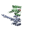
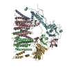
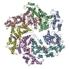
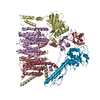
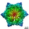
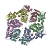
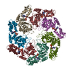
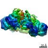
 Aquifex aeolicus (bacteria)
 X-RAY DIFFRACTION /
  Authors
 Citation
 Structure visualization
 Downloads & links
 Other downloads


 PDBj
PDBj

 Assembly
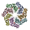

 Mass: 507.181 Da / Num. of mol.: 7 / Source method: obtained synthetically / Formula: C10H16N5O13P3 / Comment: ATP, energy-carrying molecule*YM
 Mass: 24.305 Da / Num. of mol.: 7 / Source method: obtained synthetically / Formula: Mg
 Mass: 18.015 Da / Num. of mol.: 391 / Source method: isolated from a natural source / Formula: H2O
 Sample preparation
 / Beamline: 8.3.1 / Wavelength: 1.1159 Å
 Processing