 Movie
Movie Controller

[English] 日本語
 Yorodumi
- PDB-6rqf: 3.6 Angstrom cryo-EM structure of the dimeric cytochrome b6f comp... -
+ Open data

- Basic information
| Entry | Database: PDB / ID: 6rqf | |||||||||||||||||||||||||||||||||||||||||||||||||||||||||||||||||||||||||||||||||||||||||||||
|---|---|---|---|---|---|---|---|---|---|---|---|---|---|---|---|---|---|---|---|---|---|---|---|---|---|---|---|---|---|---|---|---|---|---|---|---|---|---|---|---|---|---|---|---|---|---|---|---|---|---|---|---|---|---|---|---|---|---|---|---|---|---|---|---|---|---|---|---|---|---|---|---|---|---|---|---|---|---|---|---|---|---|---|---|---|---|---|---|---|---|---|---|---|---|
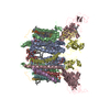
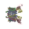
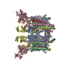
| Title | 3.6 Angstrom cryo-EM structure of the dimeric cytochrome b6f complex from Spinacia oleracea with natively bound thylakoid lipids and plastoquinone molecules | |||||||||||||||||||||||||||||||||||||||||||||||||||||||||||||||||||||||||||||||||||||||||||||
 Components Components |
| |||||||||||||||||||||||||||||||||||||||||||||||||||||||||||||||||||||||||||||||||||||||||||||
 Keywords Keywords | PHOTOSYNTHESIS / Plastoquinol-plastocyanin oxido-reductase Cytochrome b6f Photosynthesis Electron transport | |||||||||||||||||||||||||||||||||||||||||||||||||||||||||||||||||||||||||||||||||||||||||||||
| Function / homology |  Function and homology information Function and homology informationcytochrome b6f complex / plastoquinol-plastocyanin reductase / plastoquinol--plastocyanin reductase activity / cytochrome complex assembly / photosynthetic electron transport chain / chloroplast thylakoid membrane / photosynthesis / respiratory electron transport chain / 2 iron, 2 sulfur cluster binding / electron transfer activity ...cytochrome b6f complex / plastoquinol-plastocyanin reductase / plastoquinol--plastocyanin reductase activity / cytochrome complex assembly / photosynthetic electron transport chain / chloroplast thylakoid membrane / photosynthesis / respiratory electron transport chain / 2 iron, 2 sulfur cluster binding / electron transfer activity / oxidoreductase activity / iron ion binding / heme binding / membrane / metal ion binding / plasma membrane Similarity search - Function | |||||||||||||||||||||||||||||||||||||||||||||||||||||||||||||||||||||||||||||||||||||||||||||
| Biological species |  Spinacia oleracea (spinach) Spinacia oleracea (spinach) | |||||||||||||||||||||||||||||||||||||||||||||||||||||||||||||||||||||||||||||||||||||||||||||
| Method | ELECTRON MICROSCOPY / single particle reconstruction / cryo EM / Resolution: 3.58 Å | |||||||||||||||||||||||||||||||||||||||||||||||||||||||||||||||||||||||||||||||||||||||||||||
 Authors Authors | Malone, L.A. / Qian, P. / Mayneord, G.E. / Hitchcock, A. / Farmer, D. / Thompson, R. / Swainsbury, D.J.K. / Ranson, N. / Hunter, C.N. / Johnson, M.P. | |||||||||||||||||||||||||||||||||||||||||||||||||||||||||||||||||||||||||||||||||||||||||||||
| Funding support |  United Kingdom, 3items United Kingdom, 3items
| |||||||||||||||||||||||||||||||||||||||||||||||||||||||||||||||||||||||||||||||||||||||||||||
 Citation Citation | Journal: Nature / Year: 2019 Title: Cryo-EM structure of the spinach cytochrome bf complex at 3.6 Å resolution. Authors: Lorna A Malone / Pu Qian / Guy E Mayneord / Andrew Hitchcock / David A Farmer / Rebecca F Thompson / David J K Swainsbury / Neil A Ranson / C Neil Hunter / Matthew P Johnson / Abstract: The cytochrome b f (cytb f ) complex has a central role in oxygenic photosynthesis, linking electron transfer between photosystems I and II and converting solar energy into a transmembrane ...The cytochrome b f (cytb f ) complex has a central role in oxygenic photosynthesis, linking electron transfer between photosystems I and II and converting solar energy into a transmembrane proton gradient for ATP synthesis. Electron transfer within cytb f occurs via the quinol (Q) cycle, which catalyses the oxidation of plastoquinol (PQH) and the reduction of both plastocyanin (PC) and plastoquinone (PQ) at two separate sites via electron bifurcation. In higher plants, cytb f also acts as a redox-sensing hub, pivotal to the regulation of light harvesting and cyclic electron transfer that protect against metabolic and environmental stresses. Here we present a 3.6 Å resolution cryo-electron microscopy (cryo-EM) structure of the dimeric cytb f complex from spinach, which reveals the structural basis for operation of the Q cycle and its redox-sensing function. The complex contains up to three natively bound PQ molecules. The first, PQ1, is located in one cytb f monomer near the PQ oxidation site (Q) adjacent to haem b and chlorophyll a. Two conformations of the chlorophyll a phytyl tail were resolved, one that prevents access to the Q site and another that permits it, supporting a gating function for the chlorophyll a involved in redox sensing. PQ2 straddles the intermonomer cavity, partially obstructing the PQ reduction site (Q) on the PQ1 side and committing the electron transfer network to turnover at the occupied Q site in the neighbouring monomer. A conformational switch involving the haem c propionate promotes two-electron, two-proton reduction at the Q site and avoids formation of the reactive intermediate semiquinone. The location of a tentatively assigned third PQ molecule is consistent with a transition between the Q and Q sites in opposite monomers during the Q cycle. The spinach cytb f structure therefore provides new insights into how the complex fulfils its catalytic and regulatory roles in photosynthesis. | |||||||||||||||||||||||||||||||||||||||||||||||||||||||||||||||||||||||||||||||||||||||||||||
| History |
|
- Structure visualization
| Movie |
Movie viewer |
|---|---|
| Structure viewer | Molecule: MolmilJmol/JSmol |
- Downloads & links
-Download
| PDBx/mmCIF format | 6rqf.cif.gz | 380.4 KB | Display | PDBx/mmCIF format |
|---|---|---|---|---|
| PDB format | pdb6rqf.ent.gz | 306 KB | Display | PDB format |
| PDBx/mmJSON format | 6rqf.json.gz | Tree view | PDBx/mmJSON format | |
| Others |  Other downloads Other downloads |
-Validation report
| Arichive directory | https://data.pdbj.org/pub/pdb/validation_reports/rq/6rqfftp://data.pdbj.org/pub/pdb/validation_reports/rq/6rqf | HTTPS FTP |
|---|
-Related structure data
| Related structure data |  4981MC M: map data used to model this data C: citing same article ( |
|---|---|
| Similar structure data |


-Links
 PDBj
PDBj


- Assembly
| Deposited unit | 
|
|---|---|
| 1 |
|
-Components
-Protein , 3 types, 6 molecules IAKCLD
| #1: Protein | Mass: 24186.504 Da / Num. of mol.: 2 / Source method: isolated from a natural source / Source: (natural) Spinacia oleracea (spinach) / References: UniProt: P00165#3: Protein | Mass: 31357.020 Da / Num. of mol.: 2 / Source method: isolated from a natural source / Source: (natural) Spinacia oleracea (spinach) / References: UniProt: P16013#4: Protein | Mass: 18956.480 Da / Num. of mol.: 2 / Source method: isolated from a natural source / Source: (natural) Spinacia oleracea (spinach)References: UniProt: P08980, plastoquinol-plastocyanin reductase |
|---|
-Cytochrome b6-f complex subunit ... , 5 types, 10 molecules JBMENFOGPH
| #2: Protein | Mass: 17456.662 Da / Num. of mol.: 2 / Source method: isolated from a natural source / Source: (natural) Spinacia oleracea (spinach) / References: UniProt: P00166#5: Protein/peptide | Mass: 3452.200 Da / Num. of mol.: 2 / Source method: isolated from a natural source / Source: (natural) Spinacia oleracea (spinach) / References: UniProt: Q9M3L0#6: Protein/peptide | Mass: 3774.431 Da / Num. of mol.: 2 / Source method: isolated from a natural source / Source: (natural) Spinacia oleracea (spinach) / References: UniProt: P80883#7: Protein/peptide | Mass: 4171.985 Da / Num. of mol.: 2 / Source method: isolated from a natural source / Source: (natural) Spinacia oleracea (spinach) / References: UniProt: P69461#8: Protein/peptide | Mass: 3170.809 Da / Num. of mol.: 2 / Source method: isolated from a natural source / Source: (natural) Spinacia oleracea (spinach) / References: UniProt: P61045 |
|---|
-Non-polymers , 10 types, 29 molecules 


| #9: Chemical | ChemComp-HEM /  Mass: 616.487 Da / Num. of mol.: 4 / Source method: obtained synthetically / Formula: C34H32FeN4O4 Mass: 616.487 Da / Num. of mol.: 4 / Source method: obtained synthetically / Formula: C34H32FeN4O4#10: Chemical | ChemComp-HEC /  Mass: 618.503 Da / Num. of mol.: 4 / Source method: obtained synthetically / Formula: C34H34FeN4O4 Mass: 618.503 Da / Num. of mol.: 4 / Source method: obtained synthetically / Formula: C34H34FeN4O4#11: Chemical |  Mass: 893.489 Da / Num. of mol.: 2 / Source method: obtained synthetically / Formula: C55H72MgN4O5 Mass: 893.489 Da / Num. of mol.: 2 / Source method: obtained synthetically / Formula: C55H72MgN4O5#12: Chemical |  Mass: 536.873 Da / Num. of mol.: 2 / Source method: obtained synthetically / Formula: C40H56 Mass: 536.873 Da / Num. of mol.: 2 / Source method: obtained synthetically / Formula: C40H56#13: Chemical |  Mass: 749.201 Da / Num. of mol.: 3 / Source method: obtained synthetically / Formula: C53H80O2 Mass: 749.201 Da / Num. of mol.: 3 / Source method: obtained synthetically / Formula: C53H80O2#14: Chemical |  Mass: 763.100 Da / Num. of mol.: 3 / Source method: obtained synthetically / Formula: C42H85NO8P / Comment: phospholipid*YM Mass: 763.100 Da / Num. of mol.: 3 / Source method: obtained synthetically / Formula: C42H85NO8P / Comment: phospholipid*YM#15: Chemical |  Mass: 787.158 Da / Num. of mol.: 2 / Source method: obtained synthetically / Formula: C45H86O10 Mass: 787.158 Da / Num. of mol.: 2 / Source method: obtained synthetically / Formula: C45H86O10#16: Chemical | ChemComp-PGV / (  Mass: 749.007 Da / Num. of mol.: 4 / Source method: obtained synthetically / Formula: C40H77O10P / Comment: phospholipid*YM Mass: 749.007 Da / Num. of mol.: 4 / Source method: obtained synthetically / Formula: C40H77O10P / Comment: phospholipid*YM#17: Chemical |  Mass: 175.820 Da / Num. of mol.: 2 / Source method: obtained synthetically / Formula: Fe2S2 Mass: 175.820 Da / Num. of mol.: 2 / Source method: obtained synthetically / Formula: Fe2S2#18: Chemical |  Mass: 795.116 Da / Num. of mol.: 3 / Source method: obtained synthetically / Formula: C41H78O12S Mass: 795.116 Da / Num. of mol.: 3 / Source method: obtained synthetically / Formula: C41H78O12S |
|---|
-Details
| Has ligand of interest | N |
|---|---|
| Has protein modification | Y |
-Experimental details
-Experiment
| Experiment | Method: ELECTRON MICROSCOPY |
|---|---|
| EM experiment | Aggregation state: PARTICLE / 3D reconstruction method: single particle reconstruction |
- Sample preparation
| Component | Name: Spinach Cytochrome b6f complex with native bound plastoquinone and thylakoid lipids Type: COMPLEX / Entity ID: #1-#8 / Source: NATURAL |
|---|---|
| Source (natural) | Organism: Spinacia oleracea (spinach) |
| Buffer solution | pH: 8 |
| Specimen | Embedding applied: NO / Shadowing applied: NO / Staining applied: NO / Vitrification applied: YES |
| Specimen support | Grid material: COPPER / Grid mesh size: 400 divisions/in. / Grid type: Quantifoil R1.2/1.3 |
| Vitrification | Instrument: LEICA EM GP / Cryogen name: ETHANE / Humidity: 90 % / Chamber temperature: 281 K |
- Electron microscopy imaging
| Experimental equipment |  Model: Titan Krios / Image courtesy: FEI Company |
|---|---|
| Microscopy | Model: FEI TITAN KRIOS |
| Electron gun | Electron source:  FIELD EMISSION GUN / Accelerating voltage: 300 kV / Illumination mode: FLOOD BEAM FIELD EMISSION GUN / Accelerating voltage: 300 kV / Illumination mode: FLOOD BEAM |
| Electron lens | Mode: BRIGHT FIELD / Nominal magnification: 130000 X / Nominal defocus max: 2500 nm / Nominal defocus min: 1500 nm / Cs: 2.7 mm / C2 aperture diameter: 70 µm / Alignment procedure: COMA FREE |
| Specimen holder | Cryogen: NITROGEN / Specimen holder model: FEI TITAN KRIOS AUTOGRID HOLDER |
| Image recording | Electron dose: 1.15 e/Å2 / Detector mode: COUNTING / Film or detector model: GATAN K2 SUMMIT (4k x 4k) |
| EM imaging optics | Energyfilter name: GIF Bioquantum / Energyfilter slit width: 20 eV |
- Processing
| Software | Name: PHENIX / Version: (1.14_3260: phenix.real_space_refine) / Classification: refinement | ||||||||||||||||||||||||||||
|---|---|---|---|---|---|---|---|---|---|---|---|---|---|---|---|---|---|---|---|---|---|---|---|---|---|---|---|---|---|
| EM software |
| ||||||||||||||||||||||||||||
| CTF correction | Type: PHASE FLIPPING AND AMPLITUDE CORRECTION | ||||||||||||||||||||||||||||
| Particle selection | Num. of particles selected: 422660 | ||||||||||||||||||||||||||||
| Symmetry | Point symmetry: C1 (asymmetric) | ||||||||||||||||||||||||||||
| 3D reconstruction | Resolution: 3.58 Å / Resolution method: FSC 0.143 CUT-OFF / Num. of particles: 108560 / Symmetry type: POINT |
