 Movie
Movie Controller


+ Open data

- Basic information
| Entry | Database: PDB / ID: 6njn | ||||||||||||
|---|---|---|---|---|---|---|---|---|---|---|---|---|---|
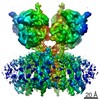
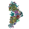
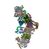
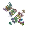
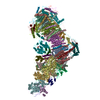
| Title | Architecture and subunit arrangement of native AMPA receptors | ||||||||||||
 Components Components |
| ||||||||||||
 Keywords Keywords | MEMBRANE PROTEIN / AMPA receptor / ligand gated ion channel / neurotransmitter / synapse | ||||||||||||
| Function / homology |  Function and homology information Function and homology informationCargo concentration in the ER / Presynaptic depolarization and calcium channel opening / axonal spine / positive regulation of locomotion involved in locomotory behavior / COPII-mediated vesicle transport / positive regulation of membrane potential / eye blink reflex / positive regulation of protein localization to basolateral plasma membrane / cellular response to ammonium ion / response to sucrose ...Cargo concentration in the ER / Presynaptic depolarization and calcium channel opening / axonal spine / positive regulation of locomotion involved in locomotory behavior / COPII-mediated vesicle transport / positive regulation of membrane potential / eye blink reflex / positive regulation of protein localization to basolateral plasma membrane / cellular response to ammonium ion / response to sucrose / myosin V binding / cerebellar mossy fiber / LGI-ADAM interactions / neuron spine / Trafficking of AMPA receptors / postsynaptic neurotransmitter receptor diffusion trapping / proximal dendrite / regulation of AMPA receptor activity / response to arsenic-containing substance / regulation of monoatomic ion transmembrane transport / channel regulator activity / cellular response to L-glutamate / membrane hyperpolarization / ligand-gated calcium channel activity / cellular response to dsRNA / dendritic spine membrane / nervous system process / beta-2 adrenergic receptor binding / Synaptic adhesion-like molecules / long-term synaptic depression / protein targeting to membrane / cellular response to peptide hormone stimulus / voltage-gated calcium channel complex / spine synapse / dendritic spine neck / dendritic spine cytoplasm / dendritic spine head / cellular response to amine stimulus / peptide hormone receptor binding / response to psychosocial stress / Activation of AMPA receptors / neurotransmitter receptor localization to postsynaptic specialization membrane / ligand-gated monoatomic cation channel activity / protein heterotetramerization / response to morphine / perisynaptic space / spinal cord development / neuronal cell body membrane / neuromuscular junction development / protein kinase A binding / Trafficking of GluR2-containing AMPA receptors / response to lithium ion / AMPA glutamate receptor activity / parallel fiber to Purkinje cell synapse / AMPA glutamate receptor clustering / transmission of nerve impulse / kainate selective glutamate receptor activity / immunoglobulin binding / adenylate cyclase binding / behavioral response to pain / asymmetric synapse / AMPA glutamate receptor complex / regulation of receptor recycling / response to electrical stimulus / extracellularly glutamate-gated ion channel activity / cellular response to glycine / membrane depolarization / ionotropic glutamate receptor complex / Unblocking of NMDA receptors, glutamate binding and activation / G-protein alpha-subunit binding / conditioned place preference / glutamate receptor binding / positive regulation of synaptic transmission / regulation of postsynaptic membrane neurotransmitter receptor levels / voltage-gated calcium channel activity / long-term memory / postsynaptic density, intracellular component / regulation of synaptic transmission, glutamatergic / synaptic cleft / response to fungicide / neuronal action potential / positive regulation of synaptic transmission, glutamatergic / extracellular ligand-gated monoatomic ion channel activity / cytoskeletal protein binding / glutamate-gated receptor activity / cellular response to brain-derived neurotrophic factor stimulus / regulation of long-term synaptic depression / somatodendritic compartment / glutamate-gated calcium ion channel activity / synapse assembly / presynaptic active zone membrane / excitatory synapse / ionotropic glutamate receptor signaling pathway / ionotropic glutamate receptor binding / dendrite membrane / dendrite cytoplasm / ligand-gated monoatomic ion channel activity involved in regulation of presynaptic membrane potential / positive regulation of excitatory postsynaptic potential / hippocampal mossy fiber to CA3 synapse / dendritic shaft Similarity search - Function | ||||||||||||
| Biological species |  | ||||||||||||
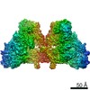
| Method | ELECTRON MICROSCOPY / single particle reconstruction / cryo EM / Resolution: 6.5 Å | ||||||||||||
 Authors Authors | Gouaux, E. / Zhao, Y. | ||||||||||||
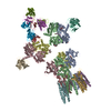
 Citation Citation | Journal: Science / Year: 2019 Title: Architecture and subunit arrangement of native AMPA receptors elucidated by cryo-EM. Authors: Yan Zhao / Shanshuang Chen / Adam C Swensen / Wei-Jun Qian / Eric Gouaux /  Abstract: Glutamate-gated AMPA receptors mediate the fast component of excitatory signal transduction at chemical synapses throughout all regions of the mammalian brain. AMPA receptors are tetrameric ...Glutamate-gated AMPA receptors mediate the fast component of excitatory signal transduction at chemical synapses throughout all regions of the mammalian brain. AMPA receptors are tetrameric assemblies composed of four subunits, GluA1-GluA4. Despite decades of study, the subunit composition, subunit arrangement, and molecular structure of native AMPA receptors remain unknown. Here we elucidate the structures of 10 distinct native AMPA receptor complexes by single-particle cryo-electron microscopy (cryo-EM). We find that receptor subunits are arranged nonstochastically, with the GluA2 subunit preferentially occupying the B and D positions of the tetramer and with triheteromeric assemblies comprising a major population of native AMPA receptors. Cryo-EM maps define the structure for S2-M4 linkers between the ligand-binding and transmembrane domains, suggesting how neurotransmitter binding is coupled to ion channel gating. | ||||||||||||
| History |
|
- Structure visualization
| Movie |
Movie viewer |
|---|---|
| Structure viewer | Molecule: MolmilJmol/JSmol |
- Downloads & links
-Download
| PDBx/mmCIF format | 6njn.cif.gz | 883 KB | Display | PDBx/mmCIF format |
|---|---|---|---|---|
| PDB format | pdb6njn.ent.gz | 675.4 KB | Display | PDB format |
| PDBx/mmJSON format | 6njn.json.gz | Tree view | PDBx/mmJSON format | |
| Others |  Other downloads Other downloads |
-Validation report
| Arichive directory | https://data.pdbj.org/pub/pdb/validation_reports/nj/6njnftp://data.pdbj.org/pub/pdb/validation_reports/nj/6njn | HTTPS FTP |
|---|
-Related structure data
| Related structure data | 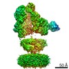 9389MC 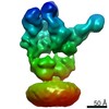 0426C 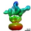 0427C 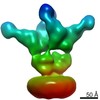 0428C 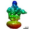 0429C 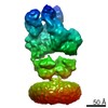 0430C 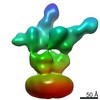 0431C 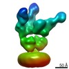 0432C 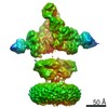 9387C 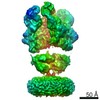 9388C 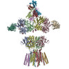 6njlC 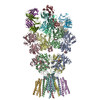 6njmC M: map data used to model this data C: citing same article ( |
|---|---|
| Similar structure data |
-Links
 PDBj
PDBj


- Assembly
| Deposited unit | 
|
|---|---|
| 1 |
|
-Components
-Glutamate receptor ... , 3 types, 4 molecules ABDC
| #1: Protein | Mass: 101518.773 Da / Num. of mol.: 1 / Source method: isolated from a natural source / Source: (natural) | ||
|---|---|---|---|
| #2: Protein | Mass: 98783.805 Da / Num. of mol.: 2 / Source method: isolated from a natural source / Source: (natural) #3: Protein | | Mass: 100556.680 Da / Num. of mol.: 1 / Source method: isolated from a natural source / Source: (natural) |
-Antibody , 5 types, 9 molecules EGIJNKOLM
| #4: Antibody | Mass: 13039.064 Da / Num. of mol.: 2 / Source method: isolated from a natural source / Source: (natural) #6: Antibody | | Mass: 27511.527 Da / Num. of mol.: 1 Source method: isolated from a genetically manipulated source Source: (gene. exp.)  #7: Antibody | Mass: 25111.660 Da / Num. of mol.: 2 Source method: isolated from a genetically manipulated source Source: (gene. exp.)   Spodoptera frugiperda (fall armyworm) Spodoptera frugiperda (fall armyworm)#8: Antibody | Mass: 27975.439 Da / Num. of mol.: 2 Source method: isolated from a genetically manipulated source Source: (gene. exp.) Spodoptera frugiperda (fall armyworm)#9: Antibody | Mass: 19081.451 Da / Num. of mol.: 2 / Source method: isolated from a natural source / Source: (natural) |
|---|
-Protein / Non-polymers , 2 types, 6 molecules FH
| #12: Chemical | ChemComp-ZK1 / {[  Mass: 409.254 Da / Num. of mol.: 4 / Source method: obtained synthetically / Formula: C14H15F3N3O6P / Comment: antagonist, medication*YM Mass: 409.254 Da / Num. of mol.: 4 / Source method: obtained synthetically / Formula: C14H15F3N3O6P / Comment: antagonist, medication*YM#5: Protein | Mass: 35938.746 Da / Num. of mol.: 2 / Source method: isolated from a natural source / Source: (natural) |
|---|
-Sugars , 3 types, 11 molecules 
| #10: Polysaccharide | beta-D-mannopyranose-(1-4)-2-acetamido-2-deoxy-beta-D-glucopyranose-(1-4)-2-acetamido-2-deoxy-beta- ...beta-D-mannopyranose-(1-4)-2-acetamido-2-deoxy-beta-D-glucopyranose-(1-4)-2-acetamido-2-deoxy-beta-D-glucopyranose Source method: isolated from a genetically manipulated source #11: Polysaccharide | Source method: isolated from a genetically manipulated source #13: Sugar |  Type: D-saccharide, beta linking / Mass: 221.208 Da / Num. of mol.: 3 Type: D-saccharide, beta linking / Mass: 221.208 Da / Num. of mol.: 3Source method: isolated from a genetically manipulated source Formula: C8H15NO6 |
|---|
-Details
| Has protein modification | Y |
|---|
-Experimental details
-Experiment
| Experiment | Method: ELECTRON MICROSCOPY |
|---|---|
| EM experiment | Aggregation state: PARTICLE / 3D reconstruction method: single particle reconstruction |
- Sample preparation
| Component |
| ||||||||||||||||||||||||||||||
|---|---|---|---|---|---|---|---|---|---|---|---|---|---|---|---|---|---|---|---|---|---|---|---|---|---|---|---|---|---|---|---|
| Source (natural) |
| ||||||||||||||||||||||||||||||
| Buffer solution | pH: 8 | ||||||||||||||||||||||||||||||
| Buffer component |
| ||||||||||||||||||||||||||||||
| Specimen | Conc.: 4 mg/ml / Embedding applied: NO / Shadowing applied: NO / Staining applied: NO / Vitrification applied: YES | ||||||||||||||||||||||||||||||
| Specimen support | Details: unspecified | ||||||||||||||||||||||||||||||
| Vitrification | Cryogen name: ETHANE / Humidity: 100 % / Chamber temperature: 295 K |
- Electron microscopy imaging
| Experimental equipment |  Model: Titan Krios / Image courtesy: FEI Company |
|---|---|
| Microscopy | Model: FEI TITAN KRIOS |
| Electron gun | Electron source:  FIELD EMISSION GUN / Accelerating voltage: 300 kV / Illumination mode: FLOOD BEAM FIELD EMISSION GUN / Accelerating voltage: 300 kV / Illumination mode: FLOOD BEAM |
| Electron lens | Mode: BRIGHT FIELD |
| Image recording | Electron dose: 54 e/Å2 / Detector mode: SUPER-RESOLUTION / Film or detector model: GATAN K2 SUMMIT (4k x 4k) |
- Processing
| EM software |
| ||||||||||||||||||
|---|---|---|---|---|---|---|---|---|---|---|---|---|---|---|---|---|---|---|---|
| CTF correction | Type: PHASE FLIPPING AND AMPLITUDE CORRECTION | ||||||||||||||||||
| 3D reconstruction | Resolution: 6.5 Å / Resolution method: FSC 0.143 CUT-OFF / Num. of particles: 161000 / Symmetry type: POINT | ||||||||||||||||||
| Atomic model building | Protocol: RIGID BODY FIT / Space: REAL |
