 ムービー
ムービー コントローラー


+ データを開く

- 基本情報
基本情報
| 登録情報 | データベース: PDB / ID: 6mzb | ||||||||||||
|---|---|---|---|---|---|---|---|---|---|---|---|---|---|
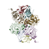
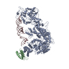
| タイトル | Cryo-EM structure of phosphodiesterase 6 | ||||||||||||
 要素 要素 |
| ||||||||||||
 キーワード キーワード | SIGNALING PROTEIN / GAF domain / phosphohydrolase / G protein-coupled receptor signaling | ||||||||||||
| 機能・相同性 |  機能・相同性情報 機能・相同性情報3',5'-cyclic-GMP phosphodiesterase / positive regulation of G protein-coupled receptor signaling pathway / Inactivation, recovery and regulation of the phototransduction cascade / Activation of the phototransduction cascade / ion binding / response to stimulus / Ca2+ pathway / positive regulation of epidermal growth factor receptor signaling pathway / photoreceptor outer segment membrane / entrainment of circadian clock by photoperiod ...3',5'-cyclic-GMP phosphodiesterase / positive regulation of G protein-coupled receptor signaling pathway / Inactivation, recovery and regulation of the phototransduction cascade / Activation of the phototransduction cascade / ion binding / response to stimulus / Ca2+ pathway / positive regulation of epidermal growth factor receptor signaling pathway / photoreceptor outer segment membrane / entrainment of circadian clock by photoperiod / cGMP binding / 3',5'-cyclic-GMP phosphodiesterase activity / 3',5'-cyclic-AMP phosphodiesterase activity / visual perception / cAMP-mediated signaling / photoreceptor disc membrane / retina development in camera-type eye / molecular adaptor activity / zinc ion binding / metal ion binding 類似検索 - 分子機能 | ||||||||||||
| 生物種 |  | ||||||||||||
| 手法 | 電子顕微鏡法 / 単粒子再構成法 / クライオ電子顕微鏡法 / 解像度: 3.4 Å | ||||||||||||
 データ登録者 データ登録者 | Gulati, S. / Palczewski, K. | ||||||||||||
| 資金援助 |  米国, 3件 米国, 3件
| ||||||||||||
 引用 引用 | ジャーナル: Sci Adv / 年: 2019 タイトル: Cryo-EM structure of phosphodiesterase 6 reveals insights into the allosteric regulation of type I phosphodiesterases. 著者: Sahil Gulati / Krzysztof Palczewski / Andreas Engel / Henning Stahlberg / Lubomir Kovacik /  要旨: Cyclic nucleotide phosphodiesterases (PDEs) work in conjunction with adenylate/guanylate cyclases to regulate the key second messengers of G protein-coupled receptor signaling. Previous attempts to ...Cyclic nucleotide phosphodiesterases (PDEs) work in conjunction with adenylate/guanylate cyclases to regulate the key second messengers of G protein-coupled receptor signaling. Previous attempts to determine the full-length structure of PDE family members at high-resolution have been hindered by structural flexibility, especially in their linker regions and N- and C-terminal ends. Therefore, most structure-activity relationship studies have so far focused on truncated and conserved catalytic domains rather than the regulatory domains that allosterically govern the activity of most PDEs. Here, we used single-particle cryo-electron microscopy to determine the structure of the full-length PDE6αβ2γ complex. The final density map resolved at 3.4 Å reveals several previously unseen structural features, including a coiled N-terminal domain and the interface of PDE6γ subunits with the PDE6αβ heterodimer. Comparison of the PDE6αβ2γ complex with the closed state of PDE2A sheds light on the conformational changes associated with the allosteric activation of type I PDEs. | ||||||||||||
| 履歴 |
|
- 構造の表示
構造の表示
| ムービー |
ムービービューア |
|---|---|
| 構造ビューア | 分子: MolmilJmol/JSmol |
- ダウンロードとリンク
-ダウンロード
| PDBx/mmCIF形式 | 6mzb.cif.gz | 572.6 KB | 表示 | PDBx/mmCIF形式 |
|---|---|---|---|---|
| PDB形式 | pdb6mzb.ent.gz | 489.4 KB | 表示 | PDB形式 |
| PDBx/mmJSON形式 | 6mzb.json.gz | ツリー表示 | PDBx/mmJSON形式 | |
| その他 |  その他のダウンロード その他のダウンロード |
-検証レポート
| 文書・要旨 | 6mzb_validation.pdf.gz | 826.7 KB | 表示 | wwPDB検証レポート |
|---|---|---|---|---|
| 文書・詳細版 | 6mzb_full_validation.pdf.gz | 836.1 KB | 表示 | |
| XML形式データ | 6mzb_validation.xml.gz | 46.3 KB | 表示 | |
| CIF形式データ | 6mzb_validation.cif.gz | 71 KB | 表示 | |
| アーカイブディレクトリ | https://data.pdbj.org/pub/pdb/validation_reports/mz/6mzbftp://data.pdbj.org/pub/pdb/validation_reports/mz/6mzb | HTTPS FTP |
-関連構造データ
| 関連構造データ | 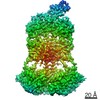 9297MC M: このデータのモデリングに利用したマップデータ C: 同じ文献を引用 ( |
|---|---|
| 類似構造データ | |
| 電子顕微鏡画像生データ | EMPIAR-10228 (タイトル: Cryo-EM structure of phosphodiesterase 6 reveals insights into the allosteric regulation of type I phosphodiesterases Data size: 166.3 Data #1: Aligned micrographs of Phosphodiesterase 6 [micrographs - single frame]) |
-リンク
 PDBj
PDBj


- 集合体
集合体
| 登録構造単位 | 
|
|---|---|
| 1 |
|
-要素
-Rod cGMP-specific 3',5'-cyclic phosphodiesterase subunit ... , 2種, 2分子 BA
| #1: タンパク質 | 分子量: 98449.648 Da / 分子数: 1 / 由来タイプ: 組換発現 / 由来: (組換発現) |
|---|---|
| #2: タンパク質 | 分子量: 99461.789 Da / 分子数: 1 / 由来タイプ: 組換発現 / 由来: (組換発現) |
-タンパク質 , 1種, 2分子 CD
| #3: タンパク質 | 分子量: 9684.229 Da / 分子数: 2 / 由来タイプ: 組換発現 / 由来: (組換発現) |
|---|
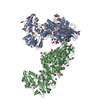
-非ポリマー , 3種, 6分子 

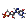
| #4: 化合物 |  分子量: 65.409 Da / 分子数: 2 / 由来タイプ: 合成 / 式: Zn 分子量: 65.409 Da / 分子数: 2 / 由来タイプ: 合成 / 式: Zn#5: 化合物 |  分子量: 24.305 Da / 分子数: 2 / 由来タイプ: 合成 / 式: Mg 分子量: 24.305 Da / 分子数: 2 / 由来タイプ: 合成 / 式: Mg#6: 化合物 |  分子量: 345.205 Da / 分子数: 2 / 由来タイプ: 合成 / 式: C10H12N5O7P 分子量: 345.205 Da / 分子数: 2 / 由来タイプ: 合成 / 式: C10H12N5O7P |
|---|
-実験情報
-実験
| 実験 | 手法: 電子顕微鏡法 |
|---|---|
| EM実験 | 試料の集合状態: PARTICLE / 3次元再構成法: 単粒子再構成法 |
- 試料調製
試料調製
| 構成要素 | 名称: Phosphodiesterase 6 / タイプ: COMPLEX / Entity ID: #1-#3 / 由来: NATURAL |
|---|---|
| 由来(天然) | 生物種: |
| 緩衝液 | pH: 7.5 |
| 試料 | 包埋: NO / シャドウイング: NO / 染色: NO / 凍結: YES |
| 試料支持 | グリッドの材料: COPPER / グリッドのタイプ: Quantifoil R2/2 |
| 急速凍結 | 凍結剤: ETHANE |
- 電子顕微鏡撮影
| 実験機器 |  モデル: Titan Krios / 画像提供: FEI Company |
|---|---|
| 顕微鏡 | モデル: FEI TITAN KRIOS |
| 電子銃 | 電子線源:  FIELD EMISSION GUN / 加速電圧: 300 kV / 照射モード: OTHER FIELD EMISSION GUN / 加速電圧: 300 kV / 照射モード: OTHER |
| 電子レンズ | モード: BRIGHT FIELD |
| 撮影 | 電子線照射量: 80 e/Å2 / 検出モード: SUPER-RESOLUTION フィルム・検出器のモデル: GATAN K2 SUMMIT (4k x 4k) |
- 解析
解析
| ソフトウェア | 名称: PHENIX / バージョン: 1.19.2_4158: / 分類: 精密化 | ||||||||||||||||||||||||
|---|---|---|---|---|---|---|---|---|---|---|---|---|---|---|---|---|---|---|---|---|---|---|---|---|---|
| CTF補正 | タイプ: NONE | ||||||||||||||||||||||||
| 対称性 | 点対称性: C1 (非対称) | ||||||||||||||||||||||||
| 3次元再構成 | 解像度: 3.4 Å / 解像度の算出法: FSC 0.143 CUT-OFF / 粒子像の数: 43597 / 対称性のタイプ: POINT | ||||||||||||||||||||||||
| 拘束条件 |
|
