 Movie
Movie Controller

[English] 日本語
 Yorodumi
- PDB-5zam: Cryo-EM structure of human Dicer and its complexes with a pre-miR... -
+ Open data

- Basic information
| Entry | Database: PDB / ID: 5zam | ||||||
|---|---|---|---|---|---|---|---|
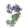
| Title | Cryo-EM structure of human Dicer and its complexes with a pre-miRNA substrate | ||||||
 Components Components |
| ||||||
 Keywords Keywords | HYDROLASE/PROTEIN BINDING/RNA / Dicer / TRBP / Cryo-EM / RNA interference / PROTEIN BINDING / HYDROLASE-PROTEIN BINDING-RNA complex | ||||||
| Function / homology |  Function and homology information Function and homology informationregulation of siRNA processing / regulation of miRNA processing / negative regulation of cytoplasmic pattern recognition receptor signaling pathway / regulation of viral transcription / negative regulation of defense response to virus by host / peripheral nervous system myelin formation / regulation of regulatory ncRNA processing / positive regulation of Schwann cell differentiation / global gene silencing by mRNA cleavage / negative regulation of Schwann cell proliferation ...regulation of siRNA processing / regulation of miRNA processing / negative regulation of cytoplasmic pattern recognition receptor signaling pathway / regulation of viral transcription / negative regulation of defense response to virus by host / peripheral nervous system myelin formation / regulation of regulatory ncRNA processing / positive regulation of Schwann cell differentiation / global gene silencing by mRNA cleavage / negative regulation of Schwann cell proliferation / tRNA-derived small RNA (tsRNA or tRNA-related fragment, tRF) biogenesis / pre-miRNA binding / tRNA decay / ribonuclease III / Small interfering RNA (siRNA) biogenesis / apoptotic DNA fragmentation / deoxyribonuclease I activity / nerve development / positive regulation of myelination / miRNA metabolic process / RISC-loading complex / miRNA processing / ribonuclease III activity / RISC complex assembly / skeletal muscle tissue regeneration / pre-miRNA processing / pre-mRNA binding / siRNA binding / M-decay: degradation of maternal mRNAs by maternally stored factors / siRNA processing / Regulation of MITF-M-dependent genes involved in apoptosis / RISC complex / neural precursor cell proliferation / miRNA binding / positive regulation of muscle cell differentiation / MicroRNA (miRNA) biogenesis / single fertilization / spermatid development / negative regulation of tumor necrosis factor production / negative regulation of tumor necrosis factor-mediated signaling pathway / negative regulation of protein kinase activity / positive regulation of viral genome replication / neuron projection morphogenesis / RNA endonuclease activity / helicase activity / multicellular organism growth / positive regulation of translation / protein sequestering activity / PKR-mediated signaling / double-stranded RNA binding / nuclear body / negative regulation of gene expression / protein domain specific binding / perinuclear region of cytoplasm / enzyme binding / negative regulation of transcription by RNA polymerase II / protein homodimerization activity / RNA binding / extracellular exosome / nucleoplasm / ATP binding / metal ion binding / identical protein binding / nucleus / cytosol / cytoplasm Similarity search - Function | ||||||
| Biological species |  Homo sapiens (human) Homo sapiens (human) | ||||||
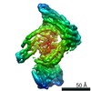
| Method | ELECTRON MICROSCOPY / single particle reconstruction / cryo EM / Resolution: 5.7 Å | ||||||
 Authors Authors | Liu, Z. / Wang, J. / Cheng, H. / Ke, X. / Sun, L. / Zhang, Q.C. / Wang, H.-W. | ||||||
| Funding support |  China, 1items China, 1items
| ||||||
 Citation Citation | Journal: Cell / Year: 2018 Title: Cryo-EM Structure of Human Dicer and Its Complexes with a Pre-miRNA Substrate. Authors: Zhongmin Liu / Jia Wang / Hang Cheng / Xin Ke / Lei Sun / Qiangfeng Cliff Zhang / Hong-Wei Wang / Abstract: Human Dicer (hDicer) is a multi-domain protein belonging to the RNase III family. It plays pivotal roles in small RNA biogenesis during the RNA interference (RNAi) pathway by processing a diverse ...Human Dicer (hDicer) is a multi-domain protein belonging to the RNase III family. It plays pivotal roles in small RNA biogenesis during the RNA interference (RNAi) pathway by processing a diverse range of double-stranded RNA (dsRNA) precursors to generate ∼22 nt microRNA (miRNA) or small interfering RNA (siRNA) products for sequence-directed gene silencing. In this work, we solved the cryoelectron microscopy (cryo-EM) structure of hDicer in complex with its cofactor protein TRBP and revealed the precise spatial arrangement of hDicer's multiple domains. We further solved structures of the hDicer-TRBP complex bound with pre-let-7 RNA in two distinct conformations. In combination with biochemical analysis, these structures reveal a property of the hDicer-TRBP complex to promote the stability of pre-miRNA's stem duplex in a pre-dicing state. These results provide insights into the mechanism of RNA processing by hDicer and illustrate the regulatory role of hDicer's N-terminal helicase domain. | ||||||
| History |
|
- Structure visualization
| Movie |
Movie viewer |
|---|---|
| Structure viewer | Molecule: MolmilJmol/JSmol |
- Downloads & links
-Download
| PDBx/mmCIF format | 5zam.cif.gz | 303.2 KB | Display | PDBx/mmCIF format |
|---|---|---|---|---|
| PDB format | pdb5zam.ent.gz | 226.5 KB | Display | PDB format |
| PDBx/mmJSON format | 5zam.json.gz | Tree view | PDBx/mmJSON format | |
| Others |  Other downloads Other downloads |
-Validation report
| Arichive directory | https://data.pdbj.org/pub/pdb/validation_reports/za/5zamftp://data.pdbj.org/pub/pdb/validation_reports/za/5zam | HTTPS FTP |
|---|
-Related structure data
| Related structure data |  6906MC 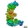 6904C  6905C  5zakC  5zalC M: map data used to model this data C: citing same article ( |
|---|---|
| Similar structure data |


-Links
 PDBj
PDBj


- Assembly
| Deposited unit | 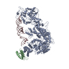
|
|---|---|
| 1 |
|
-Components
| #1: Protein | Mass: 218947.328 Da / Num. of mol.: 1 Source method: isolated from a genetically manipulated source Source: (gene. exp.) Homo sapiens (human) / Gene: DICER1, DICER, HERNA, KIAA0928 / Production host: Homo sapiens (human) / References: UniProt: Q9UPY3, ribonuclease III |
|---|---|
| #2: Protein | Mass: 39085.277 Da / Num. of mol.: 1 Source method: isolated from a genetically manipulated source Source: (gene. exp.) Homo sapiens (human) / Gene: TARBP2, TRBP / Production host: Homo sapiens (human) / References: UniProt: Q15633 |
| #3: RNA chain | Mass: 23375.830 Da / Num. of mol.: 1 Source method: isolated from a genetically manipulated source Source: (gene. exp.) Homo sapiens (human) / Production host: cell-free synthesis (others) |
| Has protein modification | Y |
-Experimental details
-Experiment
| Experiment | Method: ELECTRON MICROSCOPY |
|---|---|
| EM experiment | Aggregation state: PARTICLE / 3D reconstruction method: single particle reconstruction |
- Sample preparation
| Component |
| ||||||||||||||||||
|---|---|---|---|---|---|---|---|---|---|---|---|---|---|---|---|---|---|---|---|
| Molecular weight |
| ||||||||||||||||||
| Source (natural) |
| ||||||||||||||||||
| Source (recombinant) |
| ||||||||||||||||||
| Buffer solution | pH: 8 Details: 50 mM Tris-HCl (pH 8.0), 100 mM NaCl, 1 mM DTT, 2 mM CaCl2 | ||||||||||||||||||
| Specimen | Conc.: 1 mg/ml / Embedding applied: NO / Shadowing applied: NO / Staining applied: NO / Vitrification applied: YES Details: Human Dicer-TRBP with Pre-let-7 complex were distributed homogenenously in ice. | ||||||||||||||||||
| Specimen support | Details: The grid was glow-discharged prior to use. / Grid material: GOLD / Grid mesh size: 400 divisions/in. / Grid type: Quantifoil R1.2/1.3 | ||||||||||||||||||
| Vitrification | Instrument: FEI VITROBOT MARK IV / Cryogen name: ETHANE / Humidity: 100 % / Chamber temperature: 293 K Details: 4 microlitres were applied to do vitrified samples. |
- Electron microscopy imaging
| Experimental equipment |  Model: Titan Krios / Image courtesy: FEI Company |
|---|---|
| Microscopy | Model: FEI TITAN KRIOS |
| Electron gun | Electron source:  FIELD EMISSION GUN / Accelerating voltage: 300 kV / Illumination mode: FLOOD BEAM FIELD EMISSION GUN / Accelerating voltage: 300 kV / Illumination mode: FLOOD BEAM |
| Electron lens | Mode: BRIGHT FIELD / Nominal magnification: 89000 X / Calibrated magnification: 36496 X / Nominal defocus max: 3500 nm / Nominal defocus min: 2500 nm / Calibrated defocus min: 2500 nm / Calibrated defocus max: 3500 nm / Cs: 0.01 mm / C2 aperture diameter: 70 µm / Alignment procedure: COMA FREE |
| Specimen holder | Cryogen: NITROGEN / Specimen holder model: FEI TITAN KRIOS AUTOGRID HOLDER / Temperature (max): 80 K / Temperature (min): 80 K |
| Image recording | Average exposure time: 5.44 sec. / Electron dose: 50 e/Å2 / Detector mode: SUPER-RESOLUTION / Film or detector model: GATAN K2 SUMMIT (4k x 4k) / Num. of grids imaged: 1 / Num. of real images: 2849 |
| EM imaging optics | Phase plate: OTHER |
| Image scans | Width: 3838 / Height: 3710 / Movie frames/image: 32 / Used frames/image: 1-32 |
- Processing
| Software | Name: PHENIX / Version: 1.12_2829: / Classification: refinement | ||||||||||||||||||||||||||||||||
|---|---|---|---|---|---|---|---|---|---|---|---|---|---|---|---|---|---|---|---|---|---|---|---|---|---|---|---|---|---|---|---|---|---|
| EM software |
| ||||||||||||||||||||||||||||||||
| CTF correction | Type: PHASE FLIPPING AND AMPLITUDE CORRECTION | ||||||||||||||||||||||||||||||||
| Symmetry | Point symmetry: C1 (asymmetric) | ||||||||||||||||||||||||||||||||
| 3D reconstruction | Resolution: 5.7 Å / Resolution method: FSC 0.143 CUT-OFF / Num. of particles: 60000 / Algorithm: BACK PROJECTION / Num. of class averages: 1 / Symmetry type: POINT | ||||||||||||||||||||||||||||||||
| Atomic model building | B value: 100 / Protocol: FLEXIBLE FIT / Space: REAL / Target criteria: CC | ||||||||||||||||||||||||||||||||
| Refine LS restraints |
|
