 Movie
Movie Controller

[English] 日本語
 Yorodumi
- PDB-6c1g: High-Resolution Cryo-EM Structures of Actin-bound Myosin States R... -
+ Open data

- Basic information
| Entry | Database: PDB / ID: 6c1g | ||||||
|---|---|---|---|---|---|---|---|
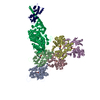
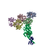
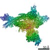
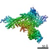
| Title | High-Resolution Cryo-EM Structures of Actin-bound Myosin States Reveal the Mechanism of Myosin Force Sensing | ||||||
 Components Components |
| ||||||
 Keywords Keywords | STRUCTURAL PROTEIN / Mechanochemistry / Mechanobiology / Structural Biology / Cytoskeleton / Molecular Motor / Myosin-I | ||||||
| Function / homology |  Function and homology information Function and homology informationpost-Golgi vesicle-mediated transport / actin filament-based movement / myosin complex / CaM pathway / Cam-PDE 1 activation / Sodium/Calcium exchangers / Calmodulin induced events / Reduction of cytosolic Ca++ levels / cytoskeletal motor activator activity / Activation of Ca-permeable Kainate Receptor ...post-Golgi vesicle-mediated transport / actin filament-based movement / myosin complex / CaM pathway / Cam-PDE 1 activation / Sodium/Calcium exchangers / Calmodulin induced events / Reduction of cytosolic Ca++ levels / cytoskeletal motor activator activity / Activation of Ca-permeable Kainate Receptor / CREB1 phosphorylation through the activation of CaMKII/CaMKK/CaMKIV cascasde / Loss of phosphorylation of MECP2 at T308 / CREB1 phosphorylation through the activation of Adenylate Cyclase / negative regulation of high voltage-gated calcium channel activity / PKA activation / CaMK IV-mediated phosphorylation of CREB / Glycogen breakdown (glycogenolysis) / microfilament motor activity / CLEC7A (Dectin-1) induces NFAT activation / myosin heavy chain binding / negative regulation of ryanodine-sensitive calcium-release channel activity / organelle localization by membrane tethering / Activation of RAC1 downstream of NMDARs / : / autophagosome membrane docking / negative regulation of calcium ion export across plasma membrane / regulation of ryanodine-sensitive calcium-release channel activity / regulation of cardiac muscle cell action potential / tropomyosin binding / presynaptic endocytosis / actin filament bundle / troponin I binding / Synthesis of IP3 and IP4 in the cytosol / filamentous actin / mesenchyme migration / microvillus / Phase 0 - rapid depolarisation / phosphatidylinositol-3,4,5-trisphosphate binding / Negative regulation of NMDA receptor-mediated neuronal transmission / Unblocking of NMDA receptors, glutamate binding and activation / RHO GTPases activate PAKs / calcineurin-mediated signaling / cytoskeletal motor activity / regulation of cell communication by electrical coupling involved in cardiac conduction / skeletal muscle myofibril / brush border / actin filament bundle assembly / Ion transport by P-type ATPases / striated muscle thin filament / Uptake and function of anthrax toxins / protein phosphatase activator activity / skeletal muscle thin filament assembly / Long-term potentiation / actin monomer binding / Calcineurin activates NFAT / Regulation of MECP2 expression and activity / DARPP-32 events / Smooth Muscle Contraction / detection of calcium ion / regulation of cardiac muscle contraction / catalytic complex / RHO GTPases activate IQGAPs / calcium channel inhibitor activity / presynaptic cytosol / Activation of AMPK downstream of NMDARs / skeletal muscle fiber development / cellular response to interferon-beta / regulation of release of sequestered calcium ion into cytosol by sarcoplasmic reticulum / Ion homeostasis / eNOS activation / Tetrahydrobiopterin (BH4) synthesis, recycling, salvage and regulation / stress fiber / Protein methylation / titin binding / actin filament polymerization / phosphatidylinositol-4,5-bisphosphate binding / regulation of cardiac muscle contraction by regulation of the release of sequestered calcium ion / regulation of calcium-mediated signaling / voltage-gated potassium channel complex / FCERI mediated Ca+2 mobilization / calcium channel complex / substantia nigra development / FCGR3A-mediated IL10 synthesis / regulation of heart rate / Antigen activates B Cell Receptor (BCR) leading to generation of second messengers / calyx of Held / Ras activation upon Ca2+ influx through NMDA receptor / adenylate cyclase activator activity / trans-Golgi network membrane / VEGFR2 mediated cell proliferation / VEGFR2 mediated vascular permeability / protein serine/threonine kinase activator activity / actin filament organization / regulation of cytokinesis / cell periphery / spindle microtubule / positive regulation of receptor signaling pathway via JAK-STAT / sarcomere / filopodium / Translocation of SLC2A4 (GLUT4) to the plasma membrane Similarity search - Function | ||||||
| Biological species |  unidentified (others) | ||||||
| Method | ELECTRON MICROSCOPY / helical reconstruction / cryo EM / Resolution: 3.8 Å | ||||||
 Authors Authors | Mentes, A. / Huehn, A. / Liu, X. / Zwolak, A. / Dominguez, R. / Shuman, H. / Ostap, E.M. / Sindelar, C.V. | ||||||
| Funding support |  United States, 1items United States, 1items
| ||||||
 Citation Citation | Journal: Proc Natl Acad Sci U S A / Year: 2018 Title: High-resolution cryo-EM structures of actin-bound myosin states reveal the mechanism of myosin force sensing. Authors: Ahmet Mentes / Andrew Huehn / Xueqi Liu / Adam Zwolak / Roberto Dominguez / Henry Shuman / E Michael Ostap / Charles V Sindelar / Abstract: Myosins adjust their power outputs in response to mechanical loads in an isoform-dependent manner, resulting in their ability to dynamically adapt to a range of motile challenges. Here, we reveal the ...Myosins adjust their power outputs in response to mechanical loads in an isoform-dependent manner, resulting in their ability to dynamically adapt to a range of motile challenges. Here, we reveal the structural basis for force-sensing based on near-atomic resolution structures of one rigor and two ADP-bound states of myosin-IB (myo1b) bound to actin, determined by cryo-electron microscopy. The two ADP-bound states are separated by a 25° rotation of the lever. The lever of the first ADP state is rotated toward the pointed end of the actin filament and forms a previously unidentified interface with the N-terminal subdomain, which constitutes the upper half of the nucleotide-binding cleft. This pointed-end orientation of the lever blocks ADP release by preventing the N-terminal subdomain from the pivoting required to open the nucleotide binding site, thus revealing how myo1b is inhibited by mechanical loads that restrain lever rotation. The lever of the second ADP state adopts a rigor-like orientation, stabilized by class-specific elements of myo1b. We identify a role for this conformation as an intermediate in the ADP release pathway. Moreover, comparison of our structures with other myosins reveals structural diversity in the actomyosin binding site, and we reveal the high-resolution structure of actin-bound phalloidin, a potent stabilizer of filamentous actin. These results provide a framework to understand the spectrum of force-sensing capacities among the myosin superfamily. | ||||||
| History |
|
- Structure visualization
| Movie |
Movie viewer |
|---|---|
| Structure viewer | Molecule: MolmilJmol/JSmol |
- Downloads & links
-Download
| PDBx/mmCIF format | 6c1g.cif.gz | 468.8 KB | Display | PDBx/mmCIF format |
|---|---|---|---|---|
| PDB format | pdb6c1g.ent.gz | 380.5 KB | Display | PDB format |
| PDBx/mmJSON format | 6c1g.json.gz | Tree view | PDBx/mmJSON format | |
| Others |  Other downloads Other downloads |
-Validation report
| Arichive directory | https://data.pdbj.org/pub/pdb/validation_reports/c1/6c1gftp://data.pdbj.org/pub/pdb/validation_reports/c1/6c1g | HTTPS FTP |
|---|
-Related structure data
| Related structure data | 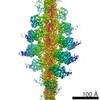 7330MC 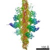 7329C 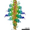 7331C 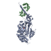 5v7xC 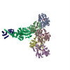 6c1dC 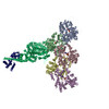 6c1hC M: map data used to model this data C: citing same article ( |
|---|---|
| Similar structure data |
-Links
 PDBj
PDBj


- Assembly
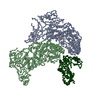
| Deposited unit | 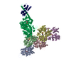
|
|---|---|
| 1 |
|
-Components
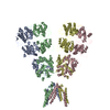
| #1: Protein | Mass: 84143.930 Da / Num. of mol.: 1 / Source method: isolated from a natural source / Source: (natural) | ||||
|---|---|---|---|---|---|
| #2: Protein | Mass: 16721.350 Da / Num. of mol.: 1 / Source method: isolated from a natural source / Source: (natural) unidentified (others) / References: UniProt: P0DP23*PLUS | ||||
| #3: Protein | Mass: 41862.613 Da / Num. of mol.: 5 / Source method: isolated from a natural source / Source: (natural) #4: Chemical | ChemComp-MG /   Mass: 24.305 Da / Num. of mol.: 6 / Source method: obtained synthetically / Formula: Mg Mass: 24.305 Da / Num. of mol.: 6 / Source method: obtained synthetically / Formula: Mg#5: Chemical | ChemComp-ADP /   Mass: 427.201 Da / Num. of mol.: 6 / Source method: obtained synthetically / Formula: C10H15N5O10P2 / Comment: ADP, energy-carrying molecule*YM Mass: 427.201 Da / Num. of mol.: 6 / Source method: obtained synthetically / Formula: C10H15N5O10P2 / Comment: ADP, energy-carrying molecule*YM |
-Experimental details
-Experiment
| Experiment | Method: ELECTRON MICROSCOPY |
|---|---|
| EM experiment | Aggregation state: HELICAL ARRAY / 3D reconstruction method: helical reconstruction |
- Sample preparation
| Component | Name: Complex of actin, myosin-1b, and calmodulin with ADP / Type: COMPLEX / Entity ID: #1-#3 / Source: MULTIPLE SOURCES |
|---|---|
| Molecular weight | Experimental value: NO |
| Source (natural) | Organism: unidentified (others) |
| Buffer solution | pH: 7 |
| Specimen | Embedding applied: NO / Shadowing applied: NO / Staining applied: NO / Vitrification applied: YES |
| Vitrification | Cryogen name: ETHANE |
- Electron microscopy imaging
| Experimental equipment |  Model: Titan Krios / Image courtesy: FEI Company |
|---|---|
| Microscopy | Model: FEI TITAN KRIOS |
| Electron gun | Electron source:  FIELD EMISSION GUN / Accelerating voltage: 300 kV / Illumination mode: SPOT SCAN FIELD EMISSION GUN / Accelerating voltage: 300 kV / Illumination mode: SPOT SCAN |
| Electron lens | Mode: BRIGHT FIELD |
| Image recording | Average exposure time: 11 sec. / Electron dose: 50 e/Å2 / Film or detector model: GATAN K2 SUMMIT (4k x 4k) |
- Processing
| CTF correction | Type: PHASE FLIPPING AND AMPLITUDE CORRECTION |
|---|---|
| Helical symmerty | Angular rotation/subunit: -167.4 ° / Axial rise/subunit: 27.5 Å / Axial symmetry: C1 |
| 3D reconstruction | Resolution: 3.8 Å / Resolution method: FSC 0.143 CUT-OFF / Num. of particles: 7700 Details: Resolution estimated by post-processing in RELION using a mask with soft edges that included only the central subunit. Symmetry type: HELICAL |
| Atomic model building | Protocol: FLEXIBLE FIT / Space: REAL |
