 Movie
Movie Controller

[English] 日本語
 Yorodumi
- PDB-3cnf: 3.88 Angstrom structure of cytoplasmic polyhedrosis virus by cryo... -
+ Open data

- Basic information
| Entry | Database: PDB / ID: 3cnf | ||||||
|---|---|---|---|---|---|---|---|
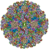
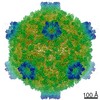
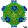
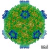
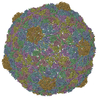
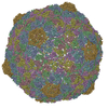
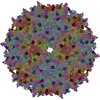
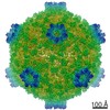
| Title | 3.88 Angstrom structure of cytoplasmic polyhedrosis virus by cryo-electron microscopy | ||||||
 Components Components |
| ||||||
 Keywords Keywords | VIRUS / cytoplasmic polyhedrosis virus / capsid protein / turret protein / polyhedrin-binding domain / guanylyltransferase domain / icosahedral virus | ||||||
| Function / homology |  Function and homology information Function and homology information | ||||||
| Biological species |   Bombyx mori cypovirus 1 Bombyx mori cypovirus 1 | ||||||
| Method | ELECTRON MICROSCOPY / single particle reconstruction / cryo EM / Resolution: 3.88 Å | ||||||
 Authors Authors | Yu, X. / Jin, L. / Zhou, Z.H. | ||||||
 Citation Citation | Journal: Nature / Year: 2008 Title: 3.88 A structure of cytoplasmic polyhedrosis virus by cryo-electron microscopy. Authors: Xuekui Yu / Lei Jin / Z Hong Zhou /  Abstract: Cytoplasmic polyhedrosis virus (CPV) is unique within the Reoviridae family in having a turreted single-layer capsid contained within polyhedrin inclusion bodies, yet being fully capable of cell ...Cytoplasmic polyhedrosis virus (CPV) is unique within the Reoviridae family in having a turreted single-layer capsid contained within polyhedrin inclusion bodies, yet being fully capable of cell entry and endogenous RNA transcription. Biochemical data have shown that the amino-terminal 79 residues of the CPV turret protein (TP) is sufficient to bring CPV or engineered proteins into the polyhedrin matrix for micro-encapsulation. Here we report the three-dimensional structure of CPV at 3.88 A resolution using single-particle cryo-electron microscopy. Our map clearly shows the turns and deep grooves of alpha-helices, the strand separation in beta-sheets, and densities for loops and many bulky side chains; thus permitting atomic model-building effort from cryo-electron microscopy maps. We observed a helix-to-beta-hairpin conformational change between the two conformational states of the capsid shell protein in the region directly interacting with genomic RNA. We have also discovered a messenger RNA release hole coupled with the mRNA capping machinery unique to CPV. Furthermore, we have identified the polyhedrin-binding domain, a structure that has potential in nanobiotechnology applications. | ||||||
| History |
|
- Structure visualization
| Movie |
Movie viewer |
|---|---|
| Structure viewer | Molecule: MolmilJmol/JSmol |
- Downloads & links
-Download
| PDBx/mmCIF format | 3cnf.cif.gz | 112.7 KB | Display | PDBx/mmCIF format |
|---|---|---|---|---|
| PDB format | pdb3cnf.ent.gz | 52.4 KB | Display | PDB format |
| PDBx/mmJSON format | 3cnf.json.gz | Tree view | PDBx/mmJSON format | |
| Others |  Other downloads Other downloads |
-Validation report
| Arichive directory | https://data.pdbj.org/pub/pdb/validation_reports/cn/3cnfftp://data.pdbj.org/pub/pdb/validation_reports/cn/3cnf | HTTPS FTP |
|---|
-Related structure data
| Related structure data | 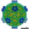 1508MC M: map data used to model this data C: citing same article ( |
|---|---|
| Similar structure data |
-Links
 PDBj
PDBj
- Assembly
| Deposited unit | 
|
|---|---|
| 1 | x 60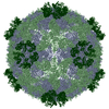
|
| 2 |
|
| 3 | x 5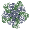
|
| 4 | x 6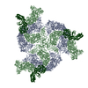
|
| 5 | 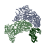
|
| Symmetry | Point symmetry: (Schoenflies symbol: I (icosahedral)) |
-Components
| #1: Protein | Mass: 148560.859 Da / Num. of mol.: 2 / Source method: isolated from a natural source / Source: (natural) Bombyx mori cypovirus 1 / References: UniProt: Q6TS43#2: Protein | | Mass: 119747.117 Da / Num. of mol.: 1 / Source method: isolated from a natural source Details: The cytoplasmic polyhedrosis virus was isolated and purified from infected Bombyx mori larvae. Source: (natural) Bombyx mori cypovirus 1 / References: UniProt: Q9E957 |
|---|
-Experimental details
-Experiment
| Experiment | Method: ELECTRON MICROSCOPY |
|---|---|
| EM experiment | Aggregation state: PARTICLE / 3D reconstruction method: single particle reconstruction |
- Sample preparation
| Component | Name: CYTOPLASMIC POLYHEDROSIS VIRUS / Type: VIRUS |
|---|---|
| Details of virus | Host category: INSECT / Isolate: STRAIN / Type: VIRION |
| Natural host | Organism: Bombyx mori |
| Buffer solution | Name: 10MM PBS / pH: 7.4 / Details: 10MM PBS |
| Specimen | Conc.: 3 mg/ml / Embedding applied: NO / Shadowing applied: NO / Staining applied: NO / Vitrification applied: YES |
| Specimen support | Details: THE VIRIONS WERE EMBEDDED IN A THIN LAYER OF VITREOUS ICE SUSPENDED ACROSS THE HOLES OF HOLEY CARBON FILMS FOR CRYOEM IMAGING. |
| Vitrification | Instrument: HOMEMADE PLUNGER / Cryogen name: ETHANE / Details: PLUNGED INTO LIQUID ETHANE |
- Electron microscopy imaging
| Experimental equipment |  Model: Tecnai Polara / Image courtesy: FEI Company |
|---|---|
| Microscopy | Model: FEI POLARA 300 Details: SAMPLES WERE MAINTAINED AT 100K IN THE ELECTRON MICROSCOPE. |
| Electron gun | Electron source:  FIELD EMISSION GUN / Accelerating voltage: 300 kV / Illumination mode: FLOOD BEAM FIELD EMISSION GUN / Accelerating voltage: 300 kV / Illumination mode: FLOOD BEAM |
| Electron lens | Mode: BRIGHT FIELD / Nominal magnification: 154380 X / Calibrated magnification: 154380 X / Nominal defocus max: 1300 nm / Nominal defocus min: 150 nm / Cs: 2 mm |
| Specimen holder | Temperature: 100 K |
| Image recording | Electron dose: 20 e/Å2 / Film or detector model: GENERIC TVIPS |
| Radiation | Protocol: SINGLE WAVELENGTH / Monochromatic (M) / Laue (L): M / Scattering type: x-ray |
| Radiation wavelength | Relative weight: 1 |
- Processing
| CTF correction | Details: CTF CORRECTION OF EACH PARTICLE | ||||||||||||
|---|---|---|---|---|---|---|---|---|---|---|---|---|---|
| Symmetry | Point symmetry: I (icosahedral) | ||||||||||||
| 3D reconstruction | Method: FOURIER COMMON LINES AND FOURIER- BESSEL SYNTHESIS / Resolution: 3.88 Å / Num. of particles: 12814 / Nominal pixel size: 0.972 Å / Actual pixel size: 0.972 Å Details: PRIOR TO THE MERGING OF PARTICLES FOR 3D RECONSTRUCTION, THE FOURIER TRANSFORM VALUES OF INDIVIDUAL IMAGES WERE CORRECTED FOR THE CTF. Symmetry type: POINT | ||||||||||||
| Refinement | Highest resolution: 3.88 Å | ||||||||||||
| Refinement step | Cycle: LAST / Highest resolution: 3.88 Å
|
