 Movie
Movie Controller

[English] 日本語
 Yorodumi
- PDB-6bt3: High-Resolution Structure Analysis of Antibody V5 Conformational ... -
+ Open data

- Basic information
| Entry | Database: PDB / ID: 6bt3 | ||||||
|---|---|---|---|---|---|---|---|
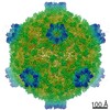
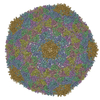
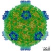
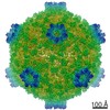
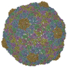
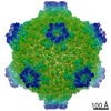
| Title | High-Resolution Structure Analysis of Antibody V5 Conformational Epitope on Human Papillomavirus 16 | ||||||
 Components Components |
| ||||||
 Keywords Keywords | VIRUS / HPV16 / H16.V5 / Fab / VIRUS LIKE PARTICLE-IMMUNE SYSTEM complex | ||||||
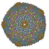
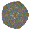
| Function / homology |  Function and homology information Function and homology informationT=7 icosahedral viral capsid / endocytosis involved in viral entry into host cell / virion attachment to host cell / host cell nucleus / structural molecule activity Similarity search - Function | ||||||
| Biological species |   Human papillomavirus type 16 Human papillomavirus type 16 | ||||||
| Method | ELECTRON MICROSCOPY / single particle reconstruction / cryo EM / Resolution: 4.7 Å | ||||||
 Authors Authors | Guan, J. / Bywaters, S.M. / Brendle, S.A. / Ashley, R.E. / Makhov, A.M. / Conway, J.F. / Christensen, N.D. / Hafenstein, S. | ||||||
| Funding support |  United States, 1items United States, 1items
| ||||||
 Citation Citation | Journal: Viruses / Year: 2017 Title: High-Resolution Structure Analysis of Antibody V5 and U4 Conformational Epitopes on Human Papillomavirus 16. Authors: Jian Guan / Stephanie M Bywaters / Sarah A Brendle / Robert E Ashley / Alexander M Makhov / James F Conway / Neil D Christensen / Susan Hafenstein / Abstract: Cancers attributable to human papillomavirus (HPV) place a huge burden on the health of both men and women. The current commercial vaccines are genotype specific and provide little therapeutic ...Cancers attributable to human papillomavirus (HPV) place a huge burden on the health of both men and women. The current commercial vaccines are genotype specific and provide little therapeutic benefit to patients with existing HPV infections. Identifying the conformational epitopes on the virus capsid supports the development of improved recombinant vaccines to maximize long-term protection against multiple types of HPV. Fragments of antibody (Fab) digested from the neutralizing monoclonal antibodies H16.V5 (V5) and H16.U4 (U4) were bound to HPV16 capsids and the structures of the two virus-Fab complexes were solved to near atomic resolution using cryo-electron microscopy. The structures reveal virus conformational changes, the Fab-binding mode to the capsid, the residues comprising the epitope and indicate a potential interaction of U4 with the minor structural protein, L2. Competition enzyme-linked immunosorbent assay (ELISA) showed V5 outcompetes U4 when added sequentially, demonstrating a steric interference even though the footprints do not overlap. Combined with our previously reported immunological and structural results, we propose that the virus may initiate host entry through an interaction between the icosahedral five-fold vertex of the capsid and receptors on the host cell. The highly detailed epitopes identified for the two antibodies provide a framework for continuing biochemical, genetic and biophysical studies. | ||||||
| History |
|
- Structure visualization
| Movie |
Movie viewer |
|---|---|
| Structure viewer | Molecule: MolmilJmol/JSmol |
- Downloads & links
-Download
| PDBx/mmCIF format | 6bt3.cif.gz | 807.6 KB | Display | PDBx/mmCIF format |
|---|---|---|---|---|
| PDB format | pdb6bt3.ent.gz | 666.5 KB | Display | PDB format |
| PDBx/mmJSON format | 6bt3.json.gz | Tree view | PDBx/mmJSON format | |
| Others |  Other downloads Other downloads |
-Validation report
| Arichive directory | https://data.pdbj.org/pub/pdb/validation_reports/bt/6bt3ftp://data.pdbj.org/pub/pdb/validation_reports/bt/6bt3 | HTTPS FTP |
|---|
-Related structure data
| Related structure data |  8243MC 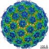 7136C  6bspC M: map data used to model this data C: citing same article ( |
|---|---|
| Similar structure data |
-Links
 PDBj
PDBj


- Assembly
| Deposited unit | 
|
|---|---|
| 1 | x 60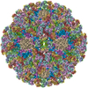
|
| 2 |
|
| 3 | x 5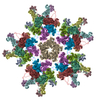
|
| 4 | x 6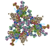
|
| 5 | 
|
| Symmetry | Point symmetry: (Schoenflies symbol: I (icosahedral)) |
-Components
| #1: Antibody | Mass: 23832.492 Da / Num. of mol.: 4 / Source method: isolated from a natural source / Source: (natural) #2: Antibody | Mass: 23739.500 Da / Num. of mol.: 4 / Source method: isolated from a natural source / Source: (natural) #3: Protein | Mass: 56098.617 Da / Num. of mol.: 6 / Source method: isolated from a natural source / Source: (natural) Human papillomavirus type 16 / References: UniProt: P03101Has protein modification | Y | |
|---|
-Experimental details
-Experiment
| Experiment | Method: ELECTRON MICROSCOPY |
|---|---|
| EM experiment | Aggregation state: PARTICLE / 3D reconstruction method: single particle reconstruction |
- Sample preparation
| Component | Name: HPV16 complexed with V5 Fab / Type: COMPLEX / Entity ID: #1-#7 / Source: MULTIPLE SOURCES |
|---|---|
| Buffer solution | pH: 7.4 |
| Specimen | Embedding applied: NO / Shadowing applied: NO / Staining applied: NO / Vitrification applied: YES |
| Vitrification | Cryogen name: ETHANE |
- Electron microscopy imaging
| Experimental equipment |  Model: Tecnai Polara / Image courtesy: FEI Company |
|---|---|
| Microscopy | Model: FEI POLARA 300 |
| Electron gun | Electron source:  FIELD EMISSION GUN / Accelerating voltage: 300 kV / Illumination mode: SPOT SCAN FIELD EMISSION GUN / Accelerating voltage: 300 kV / Illumination mode: SPOT SCAN |
| Electron lens | Mode: BRIGHT FIELD |
| Image recording | Electron dose: 7 e/Å2 / Film or detector model: FEI FALCON II (4k x 4k) |
- Processing
| CTF correction | Type: PHASE FLIPPING AND AMPLITUDE CORRECTION |
|---|---|
| 3D reconstruction | Resolution: 4.7 Å / Resolution method: FSC 0.143 CUT-OFF / Num. of particles: 17612 / Symmetry type: POINT |
