 Movie
Movie Controller

[English] 日本語
 Yorodumi
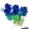
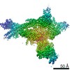
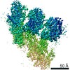
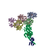
- PDB-6kss: Rat GluD1 receptor(compact conformation) in complex with 7-CKA an... -
+ Open data

- Basic information
| Entry | Database: PDB / ID: 6kss | ||||||
|---|---|---|---|---|---|---|---|
| Title | Rat GluD1 receptor(compact conformation) in complex with 7-CKA and Calcium ions | ||||||
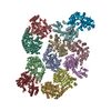
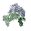
 Components Components | Glutamate receptor ionotropic, delta-1 | ||||||
 Keywords Keywords | MEMBRANE PROTEIN / complex | ||||||
| Function / homology |  Function and homology information Function and homology informationGABA receptor activity / trans-synaptic protein complex / negative regulation of synaptic plasticity / G protein-coupled receptor activity involved in regulation of postsynaptic membrane potential / synaptic signaling via neuropeptide / regulation of postsynapse organization / AMPA glutamate receptor activity / AMPA glutamate receptor complex / social behavior / regulation of postsynaptic membrane neurotransmitter receptor levels ...GABA receptor activity / trans-synaptic protein complex / negative regulation of synaptic plasticity / G protein-coupled receptor activity involved in regulation of postsynaptic membrane potential / synaptic signaling via neuropeptide / regulation of postsynapse organization / AMPA glutamate receptor activity / AMPA glutamate receptor complex / social behavior / regulation of postsynaptic membrane neurotransmitter receptor levels / synaptic transmission, glutamatergic / transmitter-gated monoatomic ion channel activity involved in regulation of postsynaptic membrane potential / postsynaptic density membrane / modulation of chemical synaptic transmission / GABA-ergic synapse / dendritic spine / phospholipase C-activating G protein-coupled receptor signaling pathway / postsynaptic membrane / glutamatergic synapse / metal ion binding / identical protein binding / plasma membrane Similarity search - Function | ||||||
| Biological species |  | ||||||
| Method | ELECTRON MICROSCOPY / single particle reconstruction / cryo EM / Resolution: 8.1 Å | ||||||
 Authors Authors | Burada, A.P. / Kumar, J. | ||||||
| Funding support |  India, 1items India, 1items
| ||||||
 Citation Citation | Journal: Nat Struct Mol Biol / Year: 2020 Title: Cryo-EM structures of the ionotropic glutamate receptor GluD1 reveal a non-swapped architecture. Authors: Ananth Prasad Burada / Rajesh Vinnakota / Janesh Kumar / Abstract: Ionotropic orphan delta (GluD) receptors are not gated by glutamate or any other endogenous ligand but are grouped with ionotropic glutamate receptors (iGluRs) based on sequence similarity. GluD1 ...Ionotropic orphan delta (GluD) receptors are not gated by glutamate or any other endogenous ligand but are grouped with ionotropic glutamate receptors (iGluRs) based on sequence similarity. GluD1 receptors play critical roles in synaptogenesis and synapse maintenance and have been implicated in neuronal disorders, including schizophrenia, cognitive deficits, and cerebral ataxia. Here we report cryo-EM structures of the rat GluD1 receptor complexed with calcium and the ligand 7-chlorokynurenic acid (7-CKA), elucidating molecular architecture and principles of receptor assembly. The structures reveal a non-swapped architecture at the interface of the extracellular amino-terminal domain (ATD) and the ligand-binding domain (LBD). This finding is in contrast with structures of other families of iGluRs, where the dimer partners between the ATD and LBD layers are swapped. Our results demonstrate that principles of architecture and symmetry are not conserved between delta receptors and other iGluRs and provide a molecular blueprint for understanding the functions of the 'orphan' class of iGluRs. | ||||||
| History |
|
- Structure visualization
| Movie |
Movie viewer |
|---|---|
| Structure viewer | Molecule: MolmilJmol/JSmol |
- Downloads & links
-Download
| PDBx/mmCIF format | 6kss.cif.gz | 522.7 KB | Display | PDBx/mmCIF format |
|---|---|---|---|---|
| PDB format | pdb6kss.ent.gz | 430.5 KB | Display | PDB format |
| PDBx/mmJSON format | 6kss.json.gz | Tree view | PDBx/mmJSON format | |
| Others |  Other downloads Other downloads |
-Validation report
| Arichive directory | https://data.pdbj.org/pub/pdb/validation_reports/ks/6kssftp://data.pdbj.org/pub/pdb/validation_reports/ks/6kss | HTTPS FTP |
|---|
-Related structure data
| Related structure data | 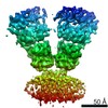 0774MC  0773C  6kspC M: map data used to model this data C: citing same article ( |
|---|---|
| Similar structure data | |
| EM raw data | EMPIAR-10428 (Title: Cryo-EM Structure of GluD1-Orphan Delta Receptor Reveals a Novel Architecture in the Ionotropic Glutamate Receptor Family Data size: 6.5 TB Data #1: ionotropioc Glutamate receptor Delta1 [micrographs - multiframe]) |
-Links
 PDBj
PDBj

- Assembly
| Deposited unit | 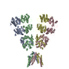
|
|---|---|
| 1 |
|
-Components
| #1: Protein | Mass: 95718.039 Da / Num. of mol.: 4 Source method: isolated from a genetically manipulated source Source: (gene. exp.)  Homo sapiens (human) / References: UniProt: Q62640 Homo sapiens (human) / References: UniProt: Q62640Compound details | The distance between Residue A LYS 527 and Residue A ILE 532 is 20.38 Angstrom. The distance ...The distance between Residue A LYS 527 and Residue A ILE 532 is 20.38 Angstrom. The distance between Residue B LYS 527 and Residue B ILE 532 is 20.46 Angstrom. The distance between Residue C LYS 527 and Residue C ILE 532 is 28.97 Angstrom. The distance between Residue D LYS 527 and Residue D ILE 532 is 19.52 Angstrom. | Has protein modification | Y | Sequence details | Thrombin recognition site | |
|---|
-Experimental details
-Experiment
| Experiment | Method: ELECTRON MICROSCOPY |
|---|---|
| EM experiment | Aggregation state: PARTICLE / 3D reconstruction method: single particle reconstruction |
- Sample preparation
| Component | Name: GluD1 / Type: COMPLEX / Details: Recombinantly expressed protein / Entity ID: all / Source: RECOMBINANT |
|---|---|
| Molecular weight | Value: 0.388 MDa / Experimental value: YES |
| Source (natural) | Organism: |
| Source (recombinant) | Organism: Homo sapiens (human) / Cell: HEK293S GnTI- / Plasmid: pEG BACMAM |
| Buffer solution | pH: 8 / Details: 20mM Tris, 150mM NaCl, 0.75mM DDM, 0.025mM CHS |
| Specimen | Conc.: 0.9 mg/ml / Embedding applied: NO / Shadowing applied: NO / Staining applied: NO / Vitrification applied: YES |
| Specimen support | Grid material: GOLD / Grid mesh size: 300 divisions/in. / Grid type: Quantifoil, UltrAuFoil, R1.2/1.3 |
| Vitrification | Instrument: FEI VITROBOT MARK IV / Cryogen name: ETHANE / Humidity: 95 % / Chamber temperature: 277 K |
- Electron microscopy imaging
| Experimental equipment |  Model: Titan Krios / Image courtesy: FEI Company |
|---|---|
| Microscopy | Model: FEI TITAN KRIOS |
| Electron gun | Electron source:  FIELD EMISSION GUN / Accelerating voltage: 300 kV / Illumination mode: FLOOD BEAM FIELD EMISSION GUN / Accelerating voltage: 300 kV / Illumination mode: FLOOD BEAM |
| Electron lens | Mode: BRIGHT FIELD / Cs: 2.7 mm |
| Specimen holder | Cryogen: NITROGEN |
| Image recording | Average exposure time: 6 sec. / Electron dose: 40.38 e/Å2 / Detector mode: SUPER-RESOLUTION / Film or detector model: GATAN K2 SUMMIT (4k x 4k) / Num. of real images: 4120 |
| Image scans | Movie frames/image: 40 / Used frames/image: 0-40 |
- Processing
| EM software |
| |||||||||||||||||||||||||||||||||||||||||||||
|---|---|---|---|---|---|---|---|---|---|---|---|---|---|---|---|---|---|---|---|---|---|---|---|---|---|---|---|---|---|---|---|---|---|---|---|---|---|---|---|---|---|---|---|---|---|---|
| CTF correction | Type: PHASE FLIPPING AND AMPLITUDE CORRECTION | |||||||||||||||||||||||||||||||||||||||||||||
| Particle selection | Num. of particles selected: 2000 Details: Particles were picked manually, which were used as reference for autopicking. | |||||||||||||||||||||||||||||||||||||||||||||
| Symmetry | Point symmetry: C1 (asymmetric) | |||||||||||||||||||||||||||||||||||||||||||||
| 3D reconstruction | Resolution: 8.1 Å / Resolution method: FSC 0.143 CUT-OFF / Num. of particles: 13422 / Symmetry type: POINT | |||||||||||||||||||||||||||||||||||||||||||||
| Atomic model building | Protocol: RIGID BODY FIT / Space: REAL |
