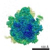
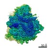
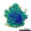
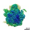
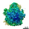
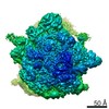
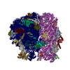
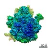
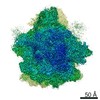
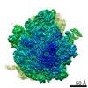
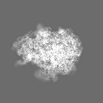
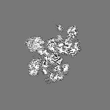
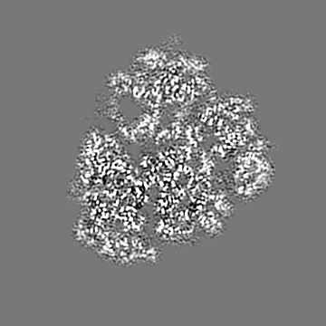
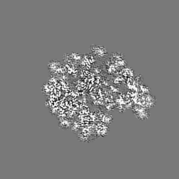
- EMDB-6934: Ribosome Structure bound to ABC-F protein. -
+
Open data
ID or keywords:
Loading...
-
Basic information
Entry
Database: EMDB / ID: EMD-6934
Title
Ribosome Structure bound to ABC-F protein.
Map data
None
Sample
Complex: Bacterial ribosome in complex with MsrE
RNA: x 5 types
Protein or peptide: x 50 types
Protein or peptide: x 1 types
Ligand: x 2 types
Keywords
Ribosome / Antibiotics / Macrolides / ABC-F protein / AZM
Function / homology
Function and homology information
regulation of translation / large ribosomal subunit / transferase activity / ribosomal small subunit assembly / ribosomal small subunit biogenesis / 5S rRNA binding / ribosomal large subunit assembly / small ribosomal subunit / small ribosomal subunit rRNA binding / large ribosomal subunit rRNA binding ...regulation of translation / large ribosomal subunit / transferase activity / ribosomal small subunit assembly / ribosomal small subunit biogenesis / 5S rRNA binding / ribosomal large subunit assembly / small ribosomal subunit / small ribosomal subunit rRNA binding / large ribosomal subunit rRNA binding / cytosolic small ribosomal subunit / cytosolic large ribosomal subunit / cytoplasmic translation / tRNA binding / negative regulation of translation / rRNA binding / structural constituent of ribosome / ribosome / translation / ribonucleoprotein complex / mRNA binding / ATP hydrolysis activity / zinc ion binding / ATP binding / metal ion binding / cytoplasm / cytosol Similarity search - Function
: / ABC-transporter extension domain / ABC transporter / 30S ribosomal protein Thx / 30S ribosomal protein Thx / 30S ribosomal protein / Ribosomal protein L1, bacterial-type / Ribosomal protein L10 / Ribosomal protein L25, long-form / Ribosomal protein L25, beta domain ...: / ABC-transporter extension domain / ABC transporter / 30S ribosomal protein Thx / 30S ribosomal protein Thx / 30S ribosomal protein / Ribosomal protein L1, bacterial-type / Ribosomal protein L10 / Ribosomal protein L25, long-form / Ribosomal protein L25, beta domain / Ribosomal protein L25, C-terminal / Ribosomal protein TL5, C-terminal domain / : / Ribosomal protein S14, type Z / Ribosomal protein L1, conserved site / Ribosomal protein L1 signature. / Ribosomal protein L1 / Ribosomal protein L1, 3-layer alpha/beta-sandwich / Ribosomal protein L1-like / Ribosomal protein L1/ribosomal biogenesis protein / Ribosomal protein L1p/L10e family / Ribosomal protein L11, bacterial-type / Ribosomal protein L31 type A / Ribosomal protein L31 signature. / Ribosomal protein L11, conserved site / Ribosomal protein L11 signature. / Ribosomal protein L31 / Ribosomal protein L31 superfamily / Ribosomal protein L31 / Ribosomal protein L10-like domain superfamily / Ribosomal protein L10P / Ribosomal protein L10 / Ribosomal protein L11, N-terminal / Ribosomal protein L11, N-terminal domain / Ribosomal protein L11/L12 / Ribosomal protein L11, C-terminal / Ribosomal protein L11, C-terminal domain superfamily / Ribosomal protein L11/L12, N-terminal domain superfamily / Ribosomal protein L11/L12 / Ribosomal protein L11, RNA binding domain / Ribosomal L25p family / Ribosomal protein L25 / Ribosomal protein L36 signature. / Ribosomal protein L25/Gln-tRNA synthetase, N-terminal / Ribosomal protein L25/Gln-tRNA synthetase, anti-codon-binding domain superfamily / : / Ribosomal protein L33, conserved site / Ribosomal protein L33 signature. / Ribosomal protein L32p, bacterial type / Ribosomal protein L35, conserved site / Ribosomal protein L35 signature. / Ribosomal protein L35, non-mitochondrial / Ribosomal protein L18, bacterial-type / Ribosomal protein S6, conserved site / Ribosomal protein S6 signature. / Ribosomal protein S3, bacterial-type / Ribosomal protein S13, bacterial-type / Ribosomal protein S19, bacterial-type / : / Ribosomal protein L6, bacterial-type / Ribosomal protein S7, bacterial/organellar-type / Ribosomal protein S11, bacterial-type / Ribosomal protein S20 / Ribosomal protein S20 superfamily / Ribosomal protein S20 / Ribosomal protein S4, bacterial-type / Ribosomal protein L5, bacterial-type / Ribosomal protein S5, bacterial-type / Ribosomal protein L19, conserved site / Ribosomal protein L19 signature. / 30S ribosomal protein S17 / : / Ribosomal protein S6, plastid/chloroplast / Ribosomal protein L36 / Ribosomal protein L36 superfamily / Ribosomal protein L36 / Ribosomal protein L20 signature. / Ribosomal protein L34, conserved site / Ribosomal protein L34 signature. / Ribosomal protein S14/S29 / Ribosomal protein L14P, bacterial-type / Ribosomal protein L27, conserved site / Ribosomal protein L27 signature. / Ribosomal protein S2, bacteria/mitochondria/plastid / Ribosomal protein L35 / Ribosomal protein L35 superfamily / Ribosomal protein L22, bacterial/chloroplast-type / Ribosomal protein L35 / Ribosomal protein L2, bacterial/organellar-type / Ribosomal protein L33 / Ribosomal protein L18 / Ribosomal L18 of archaea, bacteria, mitoch. and chloroplast / Ribosomal protein S18, conserved site / Ribosomal protein S18 signature. / Ribosomal protein L33 / Ribosomal protein S9, bacterial/plastid / Ribosomal protein L33 superfamily / ABC transporter / Ribosomal protein L30, bacterial-type / Ribosomal protein S16 Similarity search - Domain/homology
Macrolide efflux protein / Small ribosomal subunit protein uS14 / Large ribosomal subunit protein uL6 / Small ribosomal subunit protein uS8 / Small ribosomal subunit protein uS7 / Small ribosomal subunit protein uS17 / Large ribosomal subunit protein uL1 / Large ribosomal subunit protein bL33 / Large ribosomal subunit protein uL2 / Large ribosomal subunit protein uL13 ...Macrolide efflux protein / Small ribosomal subunit protein uS14 / Large ribosomal subunit protein uL6 / Small ribosomal subunit protein uS8 / Small ribosomal subunit protein uS7 / Small ribosomal subunit protein uS17 / Large ribosomal subunit protein uL1 / Large ribosomal subunit protein bL33 / Large ribosomal subunit protein uL2 / Large ribosomal subunit protein uL13 / Large ribosomal subunit protein uL16 / Large ribosomal subunit protein bL19 / Large ribosomal subunit protein bL20 / Large ribosomal subunit protein bL21 / Large ribosomal subunit protein bL27 / Large ribosomal subunit protein uL14 / Large ribosomal subunit protein bL32 / Large ribosomal subunit protein bL34 / Small ribosomal subunit protein uS2 / Small ribosomal subunit protein uS3 / Small ribosomal subunit protein uS4 / Small ribosomal subunit protein uS9 / Small ribosomal subunit protein uS11 / Small ribosomal subunit protein uS13 / Small ribosomal subunit protein bS20 / Small ribosomal subunit protein bS18 / Small ribosomal subunit protein uS12 / Small ribosomal subunit protein uS10 / Large ribosomal subunit protein uL3 / Large ribosomal subunit protein uL4 / Large ribosomal subunit protein uL23 / Small ribosomal subunit protein uS19 / Large ribosomal subunit protein uL22 / Large ribosomal subunit protein uL24 / Large ribosomal subunit protein uL5 / Large ribosomal subunit protein uL18 / Small ribosomal subunit protein uS5 / Large ribosomal subunit protein uL30 / Large ribosomal subunit protein uL15 / Large ribosomal subunit protein bL36 / Large ribosomal subunit protein bL25 / Small ribosomal subunit protein bTHX / Small ribosomal subunit protein uS15 / Large ribosomal subunit protein bL31 / Small ribosomal subunit protein bS16 / Large ribosomal subunit protein bL35 / Large ribosomal subunit protein uL11 / Small ribosomal subunit protein bS6 / Large ribosomal subunit protein uL10 / Large ribosomal subunit protein uL29 / Large ribosomal subunit protein bL17 Similarity search - Component
Journal: Proc Natl Acad Sci U S A / Year: 2018 Title: Ribosome protection by antibiotic resistance ATP-binding cassette protein. Authors: Weixin Su / Veerendra Kumar / Yichen Ding / Rya Ero / Aida Serra / Benjamin Sian Teck Lee / Andrew See Weng Wong / Jian Shi / Siu Kwan Sze / Liang Yang / Yong-Gui Gao / Abstract: The ribosome is one of the richest targets for antibiotics. Unfortunately, antibiotic resistance is an urgent issue in clinical practice. Several ATP-binding cassette family proteins confer ...The ribosome is one of the richest targets for antibiotics. Unfortunately, antibiotic resistance is an urgent issue in clinical practice. Several ATP-binding cassette family proteins confer resistance to ribosome-targeting antibiotics through a yet unknown mechanism. Among them, MsrE has been implicated in macrolide resistance. Here, we report the cryo-EM structure of ATP form MsrE bound to the ribosome. Unlike previously characterized ribosomal protection proteins, MsrE is shown to bind to ribosomal exit site. Our structure reveals that the domain linker forms a unique needle-like arrangement with two crossed helices connected by an extended loop projecting into the peptidyl-transferase center and the nascent peptide exit tunnel, where numerous antibiotics bind. In combination with biochemical assays, our structure provides insight into how MsrE binding leads to conformational changes, which results in the release of the drug. This mechanism appears to be universal for the ABC-F type ribosome protection proteins.
History
Deposition
Mar 29, 2018
-
Header (metadata) release
Aug 1, 2018
-
Map release
Aug 1, 2018
-
Update
Sep 17, 2025
-
Current status
Sep 17, 2025
Processing site: PDBj / Status: Released
-
Structure visualization
Movie
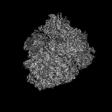
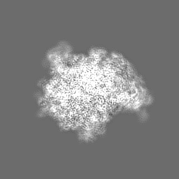
Surface view with section colored by density value
Model: Quantifoil R2/1 / Material: COPPER / Mesh: 300 / Support film - Material: CARBON / Support film - topology: HOLEY / Support film - Film thickness: 0.2 / Pretreatment - Type: GLOW DISCHARGE / Pretreatment - Time: 30 sec.
Vitrification
Cryogen name: ETHANE / Chamber humidity: 100 % / Chamber temperature: 277 K / Instrument: FEI VITROBOT MARK I
-
Electron microscopy
Microscope
FEI TITAN KRIOS
Image recording
Film or detector model: FEI FALCON II (4k x 4k) / Detector mode: INTEGRATING / Average electron dose: 50.0 e/Å2
Electron beam
Acceleration voltage: 300 kV / Electron source: FIELD EMISSION GUN
Electron optics
Illumination mode: OTHER / Imaging mode: BRIGHT FIELD / Cs: 2.7 mm
In the structure databanks used in Yorodumi, some data are registered as the other names, "COVID-19 virus" and "2019-nCoV". Here are the details of the virus and the list of structure data.
Jan 31, 2019. EMDB accession codes are about to change! (news from PDBe EMDB page)
EMDB accession codes are about to change! (news from PDBe EMDB page)
The allocation of 4 digits for EMDB accession codes will soon come to an end. Whilst these codes will remain in use, new EMDB accession codes will include an additional digit and will expand incrementally as the available range of codes is exhausted. The current 4-digit format prefixed with “EMD-” (i.e. EMD-XXXX) will advance to a 5-digit format (i.e. EMD-XXXXX), and so on. It is currently estimated that the 4-digit codes will be depleted around Spring 2019, at which point the 5-digit format will come into force.
The EM Navigator/Yorodumi systems omit the EMD- prefix.
Related info.:Q: What is EMD? / ID/Accession-code notation in Yorodumi/EM Navigator
Yorodumi is a browser for structure data from EMDB, PDB, SASBDB, etc.
This page is also the successor to EM Navigator detail page, and also detail information page/front-end page for Omokage search.
The word "yorodu" (or yorozu) is an old Japanese word meaning "ten thousand". "mi" (miru) is to see.
Related info.:EMDB / PDB / SASBDB / Comparison of 3 databanks / Yorodumi Search / Aug 31, 2016. New EM Navigator & Yorodumi / Yorodumi Papers / Jmol/JSmol / Function and homology information / Changes in new EM Navigator and Yorodumi
 Movie
Movie Controller


 Open data
 Basic information
 Map data
 Sample
 Keywords
 Function and homology information
 Thermus thermophilus (bacteria) /
  Pseudomonas aeruginosa (bacteria)
 Authors
 Singapore, 1 items
  Citation
 Structure visualization
 Downloads & links
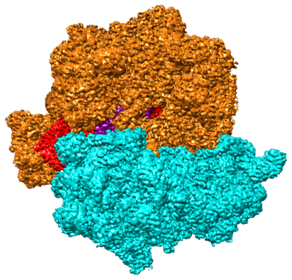 emd_6934.png
 http://ftp.pdbj.org/pub/emdb/structures/EMD-6934


 Z (Sec.)
 Y (Row.)
 X (Col.)


 Sample components
 Processing
 Electron microscopy
 FIELD EMISSION GUN