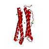
+ データを開く
- 基本情報
基本情報
| 登録情報 | データベース: EMDB / ID: EMD-6800 | |||||||||
|---|---|---|---|---|---|---|---|---|---|---|
| タイトル | Near-atomic resolution reconstruction of under-focused apoferritin | |||||||||
 マップデータ マップデータ | Reconstruction of apoferrintin using under-focused dataset. | |||||||||
 試料 試料 |
| |||||||||
| 生物種 |  Homo sapience (ヒト) Homo sapience (ヒト) | |||||||||
| 手法 | 単粒子再構成法 / クライオ電子顕微鏡法 / 解像度: 2.9 Å | |||||||||
 データ登録者 データ登録者 | Fan X / Zhao LY | |||||||||
| 資金援助 |  中国, 2件 中国, 2件
| |||||||||
 引用 引用 |  ジャーナル: Structure / 年: 2017 ジャーナル: Structure / 年: 2017タイトル: Near-Atomic Resolution Structure Determination in Over-Focus with Volta Phase Plate by Cs-Corrected Cryo-EM. 著者: Xiao Fan / Lingyun Zhao / Chuan Liu / Jin-Can Zhang / Kelong Fan / Xiyun Yan / Hai-Lin Peng / Jianlin Lei / Hong-Wei Wang /  要旨: Volta phase plate (VPP) is a recently developed transmission electron microscope (TEM) apparatus that can significantly enhance the image contrast of biological samples in cryoelectron microscopy, ...Volta phase plate (VPP) is a recently developed transmission electron microscope (TEM) apparatus that can significantly enhance the image contrast of biological samples in cryoelectron microscopy, and therefore provide the possibility to solve structures of relatively small macromolecules at high-resolution. In this work, we performed theoretical analysis and found that using phase plate on objective lens spherical aberration (Cs)-corrected TEM may gain some interesting optical properties, including the over-focus imaging of macromolecules. We subsequently evaluated the imaging strategy of frozen-hydrated apo-ferritin with VPP on a Cs-corrected TEM and obtained the structure of apo-ferritin at near-atomic resolution from both under- and over-focused dataset, illustrating the feasibility and new potential of combining VPP with Cs-corrected TEM for high-resolution cryo-EM. | |||||||||
| 履歴 |
|
- 構造の表示
構造の表示
| ムービー |
 ムービービューア ムービービューア |
|---|---|
| 構造ビューア | EMマップ:  SurfView SurfView Molmil Molmil Jmol/JSmol Jmol/JSmol |
| 添付画像 |
- ダウンロードとリンク
-EMDBアーカイブ
| マップデータ |  emd_6800.map.gz emd_6800.map.gz | 9.8 MB |  EMDBマップデータ形式 EMDBマップデータ形式 | |
|---|---|---|---|---|
| ヘッダ (付随情報) |  emd-6800-v30.xml emd-6800-v30.xml emd-6800.xml emd-6800.xml | 10.5 KB 10.5 KB | 表示 表示 |  EMDBヘッダ EMDBヘッダ |
| FSC (解像度算出) |  emd_6800_fsc.xml emd_6800_fsc.xml | 8.5 KB | 表示 |  FSCデータファイル FSCデータファイル |
| 画像 |  emd_6800.png emd_6800.png | 87.4 KB | ||
| アーカイブディレクトリ |  http://ftp.pdbj.org/pub/emdb/structures/EMD-6800 http://ftp.pdbj.org/pub/emdb/structures/EMD-6800 ftp://ftp.pdbj.org/pub/emdb/structures/EMD-6800 ftp://ftp.pdbj.org/pub/emdb/structures/EMD-6800 | HTTPS FTP |
-検証レポート
| 文書・要旨 |  emd_6800_validation.pdf.gz emd_6800_validation.pdf.gz | 78.4 KB | 表示 |  EMDB検証レポート EMDB検証レポート |
|---|---|---|---|---|
| 文書・詳細版 |  emd_6800_full_validation.pdf.gz emd_6800_full_validation.pdf.gz | 77.6 KB | 表示 | |
| XML形式データ |  emd_6800_validation.xml.gz emd_6800_validation.xml.gz | 493 B | 表示 | |
| アーカイブディレクトリ |  https://ftp.pdbj.org/pub/emdb/validation_reports/EMD-6800 https://ftp.pdbj.org/pub/emdb/validation_reports/EMD-6800 ftp://ftp.pdbj.org/pub/emdb/validation_reports/EMD-6800 ftp://ftp.pdbj.org/pub/emdb/validation_reports/EMD-6800 | HTTPS FTP |
-関連構造データ
- リンク
リンク
| EMDBのページ |  EMDB (EBI/PDBe) / EMDB (EBI/PDBe) /  EMDataResource EMDataResource |
|---|
- マップ
マップ
| ファイル |  ダウンロード / ファイル: emd_6800.map.gz / 形式: CCP4 / 大きさ: 52.7 MB / タイプ: IMAGE STORED AS FLOATING POINT NUMBER (4 BYTES) ダウンロード / ファイル: emd_6800.map.gz / 形式: CCP4 / 大きさ: 52.7 MB / タイプ: IMAGE STORED AS FLOATING POINT NUMBER (4 BYTES) | ||||||||||||||||||||||||||||||||||||||||||||||||||||||||||||||||||||
|---|---|---|---|---|---|---|---|---|---|---|---|---|---|---|---|---|---|---|---|---|---|---|---|---|---|---|---|---|---|---|---|---|---|---|---|---|---|---|---|---|---|---|---|---|---|---|---|---|---|---|---|---|---|---|---|---|---|---|---|---|---|---|---|---|---|---|---|---|---|
| 注釈 | Reconstruction of apoferrintin using under-focused dataset. | ||||||||||||||||||||||||||||||||||||||||||||||||||||||||||||||||||||
| 投影像・断面図 | 画像のコントロール
画像は Spider により作成 | ||||||||||||||||||||||||||||||||||||||||||||||||||||||||||||||||||||
| ボクセルのサイズ | X=Y=Z: 0.88 Å | ||||||||||||||||||||||||||||||||||||||||||||||||||||||||||||||||||||
| 密度 |
| ||||||||||||||||||||||||||||||||||||||||||||||||||||||||||||||||||||
| 対称性 | 空間群: 1 | ||||||||||||||||||||||||||||||||||||||||||||||||||||||||||||||||||||
| 詳細 | EMDB XML:
CCP4マップ ヘッダ情報:
| ||||||||||||||||||||||||||||||||||||||||||||||||||||||||||||||||||||
-添付データ
- 試料の構成要素
-全体 : Near-atomic resolution reconstruction of under-focused apoferritin
| 全体 | 名称: Near-atomic resolution reconstruction of under-focused apoferritin |
|---|---|
| 要素 |
|
-超分子 #1: Near-atomic resolution reconstruction of under-focused apoferritin
| 超分子 | 名称: Near-atomic resolution reconstruction of under-focused apoferritin タイプ: complex / ID: 1 / 親要素: 0 |
|---|---|
| 由来(天然) | 生物種:  Homo sapience (ヒト) Homo sapience (ヒト) |
-実験情報
-構造解析
| 手法 | クライオ電子顕微鏡法 |
|---|---|
 解析 解析 | 単粒子再構成法 |
| 試料の集合状態 | particle |
- 試料調製
試料調製
| 濃度 | 1 mg/mL |
|---|---|
| 緩衝液 | pH: 7.5 |
| グリッド | モデル: Quantifoil / 材質: GOLD / メッシュ: 300 / 支持フィルム - 材質: GRAPHENE / 支持フィルム - トポロジー: CONTINUOUS / 前処理 - タイプ: GLOW DISCHARGE |
| 凍結 | 凍結剤: ETHANE / チャンバー内湿度: 100 % / チャンバー内温度: 283 K / 装置: FEI VITROBOT MARK IV |
- 電子顕微鏡法
| 顕微鏡 | FEI TITAN KRIOS |
|---|---|
| 特殊光学系 | 位相板: VOLTA PHASE PLATE 球面収差補正装置: Microscope was modified with a Cs corrector with two hexapole elements. |
| 撮影 | フィルム・検出器のモデル: FEI FALCON II (4k x 4k) 検出モード: INTEGRATING / デジタル化 - サイズ - 横: 4096 pixel / デジタル化 - サイズ - 縦: 4096 pixel / デジタル化 - サンプリング間隔: 14.0 µm / デジタル化 - 画像ごとのフレーム数: 1-33 / 撮影したグリッド数: 1 / 平均露光時間: 4.0 sec. / 平均電子線量: 25.0 e/Å2 |
| 電子線 | 加速電圧: 300 kV / 電子線源:  FIELD EMISSION GUN FIELD EMISSION GUN |
| 電子光学系 | C2レンズ絞り径: 50.0 µm / 照射モード: FLOOD BEAM / 撮影モード: BRIGHT FIELD / Cs: 0.001 mm |
| 実験機器 |  モデル: Titan Krios / 画像提供: FEI Company |
 ムービー
ムービー コントローラー


 Z (Sec.)
 Y (Row.)
 X (Col.)


