Movie
Movie Controller

[English] 日本語
 Yorodumi
- EMDB-6801: Near-atomic resolution reconstruction of over-focused apoferritin -
+ Open data

- Basic information
| Entry | Database: EMDB / ID: EMD-6801 | |||||||||
|---|---|---|---|---|---|---|---|---|---|---|
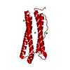
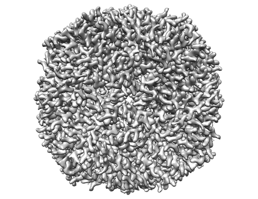
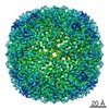
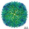
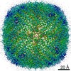
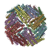
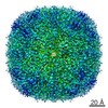
| Title | Near-atomic resolution reconstruction of over-focused apoferritin | |||||||||
 Map data Map data | ||||||||||
 Sample Sample |
| |||||||||
| Function / homology |  Function and homology information Function and homology informationiron ion sequestering activity / ferritin complex / Scavenging by Class A Receptors / Golgi Associated Vesicle Biogenesis / ferroxidase / negative regulation of ferroptosis / autolysosome / ferroxidase activity / negative regulation of fibroblast proliferation / ferric iron binding ...iron ion sequestering activity / ferritin complex / Scavenging by Class A Receptors / Golgi Associated Vesicle Biogenesis / ferroxidase / negative regulation of ferroptosis / autolysosome / ferroxidase activity / negative regulation of fibroblast proliferation / ferric iron binding / autophagosome / iron ion transport / ferrous iron binding / Iron uptake and transport / tertiary granule lumen / ficolin-1-rich granule lumen / intracellular iron ion homeostasis / immune response / iron ion binding / negative regulation of cell population proliferation / Neutrophil degranulation / extracellular exosome / extracellular region / identical protein binding / nucleus / cytoplasm / cytosol Similarity search - Function | |||||||||
| Biological species |  Homo sapiens (human) Homo sapiens (human) | |||||||||
| Method | single particle reconstruction / cryo EM / Resolution: 3.2 Å | |||||||||
 Authors Authors | Fan X / Zhao LY | |||||||||
 Citation Citation | Journal: Structure / Year: 2017 Title: Near-Atomic Resolution Structure Determination in Over-Focus with Volta Phase Plate by Cs-Corrected Cryo-EM. Authors: Xiao Fan / Lingyun Zhao / Chuan Liu / Jin-Can Zhang / Kelong Fan / Xiyun Yan / Hai-Lin Peng / Jianlin Lei / Hong-Wei Wang /  Abstract: Volta phase plate (VPP) is a recently developed transmission electron microscope (TEM) apparatus that can significantly enhance the image contrast of biological samples in cryoelectron microscopy, ...Volta phase plate (VPP) is a recently developed transmission electron microscope (TEM) apparatus that can significantly enhance the image contrast of biological samples in cryoelectron microscopy, and therefore provide the possibility to solve structures of relatively small macromolecules at high-resolution. In this work, we performed theoretical analysis and found that using phase plate on objective lens spherical aberration (Cs)-corrected TEM may gain some interesting optical properties, including the over-focus imaging of macromolecules. We subsequently evaluated the imaging strategy of frozen-hydrated apo-ferritin with VPP on a Cs-corrected TEM and obtained the structure of apo-ferritin at near-atomic resolution from both under- and over-focused dataset, illustrating the feasibility and new potential of combining VPP with Cs-corrected TEM for high-resolution cryo-EM. | |||||||||
| History |
|
- Structure visualization
| Movie |
Movie viewer |
|---|---|
| Structure viewer | EM map: SurfViewMolmilJmol/JSmol |
| Supplemental images |
- Downloads & links
-EMDB archive
| Map data | emd_6801.map.gz | 49.2 MB | EMDB map data format | |
|---|---|---|---|---|
| Header (meta data) | emd-6801-v30.xmlemd-6801.xml | 9.7 KB 9.7 KB | Display Display | EMDB header |
| FSC (resolution estimation) | emd_6801_fsc.xml | 8.3 KB | Display | FSC data file |
| Images |  emd_6801.png emd_6801.png | 113.6 KB | ||
| Archive directory |  http://ftp.pdbj.org/pub/emdb/structures/EMD-6801ftp://ftp.pdbj.org/pub/emdb/structures/EMD-6801 http://ftp.pdbj.org/pub/emdb/structures/EMD-6801ftp://ftp.pdbj.org/pub/emdb/structures/EMD-6801 | HTTPS FTP |
-Related structure data


-Links
| EMDB pages | EMDB (EBI/PDBe) / EMDataResource |
|---|---|
| Related items in Molecule of the Month |


-Map
| File | Download / File: emd_6801.map.gz / Format: CCP4 / Size: 52.7 MB / Type: IMAGE STORED AS FLOATING POINT NUMBER (4 BYTES) | ||||||||||||||||||||||||||||||||||||||||||||||||||||||||||||||||||||
|---|---|---|---|---|---|---|---|---|---|---|---|---|---|---|---|---|---|---|---|---|---|---|---|---|---|---|---|---|---|---|---|---|---|---|---|---|---|---|---|---|---|---|---|---|---|---|---|---|---|---|---|---|---|---|---|---|---|---|---|---|---|---|---|---|---|---|---|---|---|
| Projections & slices | Image control
Images are generated by Spider. | ||||||||||||||||||||||||||||||||||||||||||||||||||||||||||||||||||||
| Voxel size | X=Y=Z: 0.88 Å | ||||||||||||||||||||||||||||||||||||||||||||||||||||||||||||||||||||
| Density |
| ||||||||||||||||||||||||||||||||||||||||||||||||||||||||||||||||||||
| Symmetry | Space group: 1 | ||||||||||||||||||||||||||||||||||||||||||||||||||||||||||||||||||||
| Details | EMDB XML:
CCP4 map header:
| ||||||||||||||||||||||||||||||||||||||||||||||||||||||||||||||||||||
 Z (Sec.)
 Y (Row.)
 X (Col.)


-Supplemental data
- Sample components
-Entire : Near-atomic resolution reconstruction of over-focused apoferritin
| Entire | Name: Near-atomic resolution reconstruction of over-focused apoferritin |
|---|---|
| Components |
|
-Supramolecule #1: Near-atomic resolution reconstruction of over-focused apoferritin
| Supramolecule | Name: Near-atomic resolution reconstruction of over-focused apoferritin type: complex / ID: 1 / Parent: 0 |
|---|---|
| Source (natural) | Organism: Homo sapiens (human) |
-Experimental details
-Structure determination
| Method | cryo EM |
|---|---|
 Processing Processing | single particle reconstruction |
| Aggregation state | particle |
-Sample preparation
| Concentration | 1 mg/mL |
|---|---|
| Buffer | pH: 7.5 |
| Grid | Model: Quantifoil / Material: GOLD / Mesh: 300 / Support film - Material: GRAPHENE / Support film - topology: CONTINUOUS / Pretreatment - Type: GLOW DISCHARGE |
| Vitrification | Cryogen name: ETHANE / Chamber humidity: 100 % / Chamber temperature: 283 K / Instrument: FEI VITROBOT MARK IV |
- Electron microscopy
| Microscope | FEI TITAN KRIOS |
|---|---|
| Specialist optics | Phase plate: VOLTA PHASE PLATE Spherical aberration corrector: Microscope was modified with a Cs corrector with two hexapole elements. |
| Image recording | Film or detector model: FEI FALCON II (4k x 4k) / Detector mode: INTEGRATING / Digitization - Dimensions - Width: 4096 pixel / Digitization - Dimensions - Height: 4096 pixel / Digitization - Sampling interval: 14.0 µm / Digitization - Frames/image: 1-33 / Number grids imaged: 1 / Average exposure time: 4.0 sec. / Average electron dose: 25.0 e/Å2 |
| Electron beam | Acceleration voltage: 300 kV / Electron source:  FIELD EMISSION GUN FIELD EMISSION GUN |
| Electron optics | C2 aperture diameter: 50.0 µm / Illumination mode: FLOOD BEAM / Imaging mode: BRIGHT FIELD / Cs: 0.001 mm |
| Experimental equipment |  Model: Titan Krios / Image courtesy: FEI Company |