 Movie
Movie Controller

[English] 日本語
 Yorodumi
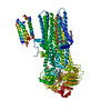
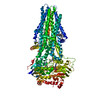
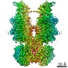
- PDB-5uj9: Cryo-EM structure of bovine multidrug resistance protein 1 (MRP1) -
+ Open data

- Basic information
| Entry | Database: PDB / ID: 5uj9 | ||||||||||||
|---|---|---|---|---|---|---|---|---|---|---|---|---|---|
| Title | Cryo-EM structure of bovine multidrug resistance protein 1 (MRP1) | ||||||||||||
 Components Components | bovine multidrug resistance protein 1 (MRP1),Multidrug resistance-associated protein 1 | ||||||||||||
 Keywords Keywords | TRANSPORT PROTEIN / ABC transporter / multidrug resistance | ||||||||||||
| Function / homology |  Function and homology information Function and homology informationSphingolipid de novo biosynthesis / Heme degradation / Synthesis of Leukotrienes (LT) and Eoxins (EX) / Transport of RCbl within the body / cyclic nucleotide transport / Paracetamol ADME / ABC-family protein mediated transport / glutathione transmembrane transport / leukotriene transport / Cytoprotection by HMOX1 ...Sphingolipid de novo biosynthesis / Heme degradation / Synthesis of Leukotrienes (LT) and Eoxins (EX) / Transport of RCbl within the body / cyclic nucleotide transport / Paracetamol ADME / ABC-family protein mediated transport / glutathione transmembrane transport / leukotriene transport / Cytoprotection by HMOX1 / glutathione transmembrane transporter activity / ABC-type glutathione-S-conjugate transporter / carboxylic acid transmembrane transporter activity / ABC-type glutathione S-conjugate transporter activity / ABC-type xenobiotic transporter / ABC-type xenobiotic transporter activity / lipid transport / xenobiotic transmembrane transporter activity / xenobiotic transport / ABC-type transporter activity / positive regulation of inflammatory response / basolateral plasma membrane / response to xenobiotic stimulus / ATP hydrolysis activity / ATP binding / plasma membrane Similarity search - Function | ||||||||||||
| Biological species |  | ||||||||||||
| Method | ELECTRON MICROSCOPY / single particle reconstruction / cryo EM / Resolution: 3.49 Å | ||||||||||||
 Authors Authors | Johnson, Z.L. / Chen, J. | ||||||||||||
| Funding support |  United States, 3items United States, 3items
| ||||||||||||
 Citation Citation | Journal: Cell / Year: 2017 Title: Structural Basis of Substrate Recognition by the Multidrug Resistance Protein MRP1. Authors: Zachary Lee Johnson / Jue Chen / Abstract: The multidrug resistance protein MRP1 is an ATP-binding cassette (ABC) transporter that confers resistance to many anticancer drugs and plays a role in the disposition and efficacy of several ...The multidrug resistance protein MRP1 is an ATP-binding cassette (ABC) transporter that confers resistance to many anticancer drugs and plays a role in the disposition and efficacy of several opiates, antidepressants, statins, and antibiotics. In addition, MRP1 regulates redox homeostasis, inflammation, and hormone secretion. Using electron cryomicroscopy, we determined the molecular structures of bovine MRP1 in two conformations: an apo form at 3.5 Å without any added substrate and a complex form at 3.3 Å with one of its physiological substrates, leukotriene C. These structures show that by forming a single bipartite binding site, MRP1 can recognize a spectrum of substrates with different chemical structures. We also observed large conformational changes induced by leukotriene C, explaining how substrate binding primes the transporter for ATP hydrolysis. Structural comparison of MRP1 and P-glycoprotein advances our understanding of the common and unique properties of these two important molecules in multidrug resistance to chemotherapy. | ||||||||||||
| History |
|
- Structure visualization
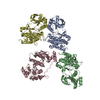
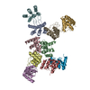
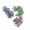
| Movie |
Movie viewer |
|---|---|
| Structure viewer | Molecule: MolmilJmol/JSmol |
- Downloads & links
-Download
| PDBx/mmCIF format | 5uj9.cif.gz | 261.3 KB | Display | PDBx/mmCIF format |
|---|---|---|---|---|
| PDB format | pdb5uj9.ent.gz | 201.8 KB | Display | PDB format |
| PDBx/mmJSON format | 5uj9.json.gz | Tree view | PDBx/mmJSON format | |
| Others |  Other downloads Other downloads |
-Validation report
| Arichive directory | https://data.pdbj.org/pub/pdb/validation_reports/uj/5uj9ftp://data.pdbj.org/pub/pdb/validation_reports/uj/5uj9 | HTTPS FTP |
|---|
-Related structure data
| Related structure data | 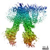 8559MC 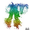 8560C  5ujaC M: map data used to model this data C: citing same article ( |
|---|---|
| Similar structure data | |
| EM raw data | EMPIAR-10802 (Title: Cryo-electron microscopy reconstruction of apo bovine MRP1 Data size: 804.5 Data #1: Unaligned and uncorrected multiframe movies of bovine apo MRP1 [micrographs - multiframe]) |

-Links
 PDBj
PDBj


- Assembly
| Deposited unit | 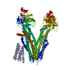
|
|---|---|
| 1 |
|
-Components
| #1: Protein | Mass: 159701.922 Da / Num. of mol.: 1 Source method: isolated from a genetically manipulated source Source: (gene. exp.)  Homo sapiens (human) / References: UniProt: Q8HXQ5 Homo sapiens (human) / References: UniProt: Q8HXQ5 |
|---|---|
| Sequence details | Residues 31-194, corresponding to TMD0, are modeled as poly-alanine. The register in this region ...Residues 31-194, corresponding to TMD0, are modeled as poly-alanine. The register in this region could not be confidently established and thus the numbering assigned to the residues is putative. The poly-alanine regions have been renamed as unknown |
-Experimental details
-Experiment
| Experiment | Method: ELECTRON MICROSCOPY |
|---|---|
| EM experiment | Aggregation state: PARTICLE / 3D reconstruction method: single particle reconstruction |
- Sample preparation
| Component | Name: bovine multidrug resistance protein 1 (MRP1) / Type: COMPLEX / Entity ID: all / Source: RECOMBINANT | ||||||||||||||||||||||||||||
|---|---|---|---|---|---|---|---|---|---|---|---|---|---|---|---|---|---|---|---|---|---|---|---|---|---|---|---|---|---|
| Molecular weight | Value: 0.17 MDa / Experimental value: NO | ||||||||||||||||||||||||||||
| Source (natural) | Organism: | ||||||||||||||||||||||||||||
| Source (recombinant) | Organism: Homo sapiens (human) / Cell: HEK293S GntI- / Plasmid: Baculovirus | ||||||||||||||||||||||||||||
| Buffer solution | pH: 8 | ||||||||||||||||||||||||||||
| Buffer component |
| ||||||||||||||||||||||||||||
| Specimen | Conc.: 4.4 mg/ml / Embedding applied: NO / Shadowing applied: NO / Staining applied: NO / Vitrification applied: YES | ||||||||||||||||||||||||||||
| Specimen support | Grid material: GOLD Grid type: Quantifoil R1.2/1.3 400-mesh Au Holey Carbon Grids | ||||||||||||||||||||||||||||
| Vitrification | Instrument: FEI VITROBOT MARK IV / Cryogen name: ETHANE / Humidity: 100 % / Chamber temperature: 295 K |
- Electron microscopy imaging
| Experimental equipment |  Model: Titan Krios / Image courtesy: FEI Company |
|---|---|
| Microscopy | Model: FEI TITAN KRIOS |
| Electron gun | Electron source:  FIELD EMISSION GUN / Accelerating voltage: 300 kV / Illumination mode: FLOOD BEAM FIELD EMISSION GUN / Accelerating voltage: 300 kV / Illumination mode: FLOOD BEAM |
| Electron lens | Mode: BRIGHT FIELD / Nominal magnification: 37000 X / Nominal defocus max: 2400 nm / Nominal defocus min: 700 nm / Cs: 2.7 mm / C2 aperture diameter: 70 µm |
| Specimen holder | Cryogen: NITROGEN / Specimen holder model: FEI TITAN KRIOS AUTOGRID HOLDER / Temperature (max): 100 K / Temperature (min): 80 K |
| Image recording | Average exposure time: 7 sec. / Electron dose: 84 e/Å2 / Detector mode: SUPER-RESOLUTION / Film or detector model: GATAN K2 SUMMIT (4k x 4k) / Num. of grids imaged: 1 / Num. of real images: 2232 |
| Image scans | Width: 3710 / Height: 3838 / Movie frames/image: 50 / Used frames/image: 1-50 |
- Processing
| Software | Name: REFMAC / Version: 5.8.0158 / Classification: refinement | ||||||||||||||||||||||||||||||||||||||||||||||||||||||||||||||||||||||||||||||||||||||||||||||||||||||||||
|---|---|---|---|---|---|---|---|---|---|---|---|---|---|---|---|---|---|---|---|---|---|---|---|---|---|---|---|---|---|---|---|---|---|---|---|---|---|---|---|---|---|---|---|---|---|---|---|---|---|---|---|---|---|---|---|---|---|---|---|---|---|---|---|---|---|---|---|---|---|---|---|---|---|---|---|---|---|---|---|---|---|---|---|---|---|---|---|---|---|---|---|---|---|---|---|---|---|---|---|---|---|---|---|---|---|---|---|
| EM software |
| ||||||||||||||||||||||||||||||||||||||||||||||||||||||||||||||||||||||||||||||||||||||||||||||||||||||||||
| CTF correction | Type: PHASE FLIPPING AND AMPLITUDE CORRECTION | ||||||||||||||||||||||||||||||||||||||||||||||||||||||||||||||||||||||||||||||||||||||||||||||||||||||||||
| Particle selection | Num. of particles selected: 251986 | ||||||||||||||||||||||||||||||||||||||||||||||||||||||||||||||||||||||||||||||||||||||||||||||||||||||||||
| Symmetry | Point symmetry: C1 (asymmetric) | ||||||||||||||||||||||||||||||||||||||||||||||||||||||||||||||||||||||||||||||||||||||||||||||||||||||||||
| 3D reconstruction | Resolution: 3.49 Å / Resolution method: FSC 0.143 CUT-OFF / Num. of particles: 251986 / Symmetry type: POINT | ||||||||||||||||||||||||||||||||||||||||||||||||||||||||||||||||||||||||||||||||||||||||||||||||||||||||||
| Atomic model building | Protocol: AB INITIO MODEL / Space: RECIPROCAL | ||||||||||||||||||||||||||||||||||||||||||||||||||||||||||||||||||||||||||||||||||||||||||||||||||||||||||
| Refinement | Resolution: 3.49→148 Å / Cor.coef. Fo:Fc: 0.969 / ESU R: 0.448 Stereochemistry target values: MAXIMUM LIKELIHOOD WITH PHASES Details: HYDROGENS HAVE BEEN ADDED IN THE RIDING POSITIONS
| ||||||||||||||||||||||||||||||||||||||||||||||||||||||||||||||||||||||||||||||||||||||||||||||||||||||||||
| Solvent computation | Ion probe radii: 0.8 Å / Shrinkage radii: 0.8 Å / VDW probe radii: 1.2 Å / Solvent model: MASK | ||||||||||||||||||||||||||||||||||||||||||||||||||||||||||||||||||||||||||||||||||||||||||||||||||||||||||
| Displacement parameters | Biso mean: 265.842 Å2
| ||||||||||||||||||||||||||||||||||||||||||||||||||||||||||||||||||||||||||||||||||||||||||||||||||||||||||
| Refinement step | Cycle: 1 / Total: 9857 | ||||||||||||||||||||||||||||||||||||||||||||||||||||||||||||||||||||||||||||||||||||||||||||||||||||||||||
| Refine LS restraints |
|